Interesting case for this week! 64 YO patient with the diagnosis of Stroke (4 separate areas within the brain) no prior echo…. Hello Papillary Fibroelastoma 😍 Got to do the TTE and the TEE with @IjazMalikMD !#echo#echofirst @renujain19 @AmandaGeilfuss@mickeylee52
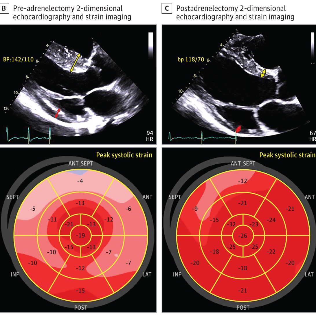
Case of Vanishing HCM, now published https://t.co/VzV6RC2q84
33 yo F p/w labile BP; echo showed marked ASH, mimicking HCM. She was diagnosed with pheochromocytoma & 3 months post bilateral adrenalectomy, she had normalization of VS wall thickness (24mm->12mm) & GLS (-13%->-21%)!
A great find by one of my work colleagues (thanks Simon!). Lovely illustrations of various CHD lesions, created by PD. Dr. med. Daniel Tobler, Kardiologie, Universitätsspital Basel & the New Media Center of the University of Basel.
@DDaniel_Tobler
https://t.co/lPPLyS7AW1
#ASE2019 with some fabulous #Sonographers. So many amazing presentations. Register today for #ASE2020 https://t.co/SdCD3uApvm Gain 44 CMEs at a reduced rate. Great value and still participate in virtual networking with fellow #echonerds 🤓#ASETwitterChampion@ASE360
Some good news to share from my facility. So proud of my team and their dedication during this time @AmandaGeilfuss@EngdahlKirsten@KatieMahalick and all the rest of the amazing staff at Aurora Medical Center Grafton 😁
For my #sonographersunday retweet-really useful suggestions for scanning patients that are ventilated on their stomach. Essential learning during this #COVID19 era. I will also add the great video about why this position is used for optimal ventilation. Great work @cardiacskills