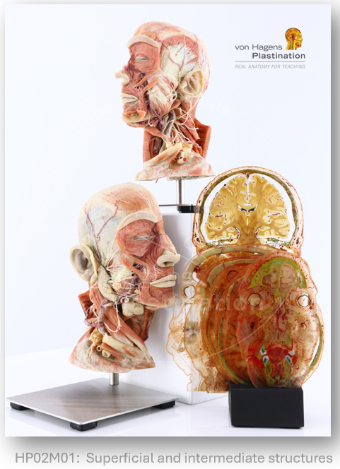
Anatomic Excellence is the exclusive agent for von Hagens Plastination in North America; facilitating the acquisition of ethically-sourced human plastinates.
We are proud to announce our participation as a sponsor at ISCAA 2026 in Vienna!
Join us at the Medical University of Vienna from September 3-6 and visit our booth to explore our anatomical specimens and discover how real human anatomy can enhance medical education.
#Anatomy
Anatomic Excellence is deeply saddened by the passing of Dr. Gunther von Hagens, inventor of Plastination and creator of BODY WORLDS, on July 24th2026, aged 81.
Through Plastination he has transformed anatomy education worldwide, making the complexity and beauty of the human body accessible to students, clinicians and the public alike.
Karen & I are honored to have worked on behalf of a true pioneer and know that his scientific and craftsmanship-based legacy will live on in every anatomy lab it reaches.
Our thoughts are with Dr. Angelina Whalley, Rurik von Hagens, the family, and his close colleagues. His courage, his perseverance and his unwavering belief that education serves humanity will continue to guide us.
https://t.co/lsloJJ4Qt7
Our latest addition to the teaching collection:
The ~13 mm thickness of slices provides a unique combination of cross-sectional and 3D anatomy, bridging the gap between Silicone Plastinates and Sheet Plastinates/Anatomy Glass.
For more detail: https://t.co/RSH1vBUyEU
#Anatomy
This Silicone Plastinate reveals the position and relations of the ventricular system and choroid plexus on one side and the external undissected structure of the cerebrum and cerebellum on the opposite side.
View full description and images https://t.co/U7QlWCQFNd
#Neuroscience
#Bones demonstrate even the most delicate of features and landmarks, including the: wafer-thin #laminapapyracea, delicate #pterygoidhamulus, nutrient and neural #foramina, and grooves for #vessels and #nerves.
Real Anatomy for Teaching (Learning) by von Hagens Plastination
This Cast uniquely reveals the origin, position, and capacity (luminal diameter) of the blood vessels supplying the foot, their points of anastomosis, and their pattern of branching beyond named vessels.
For more details:
https://t.co/LF8TC1Pyzc
#FootAnatomy#VascularSupply
When used alongside Silicone Platinates, Sheet Plastinates provide new ways for learners to visualize and conceptualize anatomy, enhancing their ability to process and mentally manipulate arrangements and morphology.
https://t.co/gKyzvnSwwQ
Our website got a quiet launch — now it’s ready for the spotlight!
Over the past year, we’ve been fine-tuning and improving https://t.co/DW0lL2ATtm based on feedback and user experience.
🔐 Sign up for an account to view prices and save lists
#Anatomy#Plastination#MedicalEd
There are four key principles in Code C:
• Consent – only the individual can donate their body
• Dignity – cadavers must be treated with respect
• Purpose – use must match original consent
• Compliance – facilities must meet HTA standards
🔗 https://t.co/sw0nrsYA4J
von Hagens Plastination teaching specimens stem from the Body Donation Program of the Institute for Plastination in Heidelberg, which is unique in terms of its purpose and accountability.
Transparency and traceability guaranteed: https://t.co/VAu4dlgjgc
This Cast uniquely reveals the origin, position, and capacity (luminal diameter) of the blood vessels supplying the hand, their points of anastomosis, and their pattern of branching beyond named vessels.
See more: https://t.co/Ha5Hqgk6m6
#VascularAnatomy#anatomy
Inflammatory conditions affecting the Parotid gland maycompress the Facial nerve resulting in ipsilateral lowermotor neuron symptoms including weakness of eye andmouth closure, with associated epiphora and escape oforal contents.
#Parotis#Facialisparese#NervusFacialis
This new Plastinate reveals various parts of the fascial system of the thigh including the fascia lata (deep fascia) and associated intermuscular septa dividing the thigh into compartments. Subcutaneous tissues are also revealed alongside paraneural and paravascular sheaths.