Not the official Science Advances cover of this month, but you can still find it in the current issue!
Check our latest paper where we used 3D-CODA to design assembloids that mimic the complexity of human fallopian tubes.
Paper:https://t.co/e3jTJ55iQp
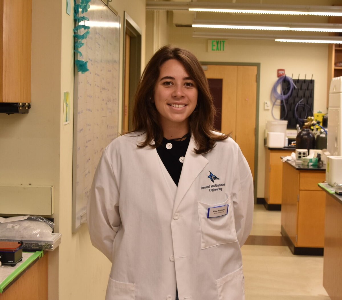
Read about how a joy of discovery in engineering took PhD alum Alice Amitrano from Rome to Johns Hopkins, where she studied how cells adapt to their surroundings, including the physical and chemical ways cancer cells change as they spread through the body. https://t.co/Fm5Lc1kRDg
Elana Fertig, Hartland Jackson, Linghua Wang, Deniz G. Olgun, and Sungsik Kim discussed "Spatial Biology and Actionable Tumor Ecosystems: Beyond the Pretty Picture" to close out the AACR Conference on Pancreatic Cancer.
#AACRpan26@hartjackson@IamLinghua
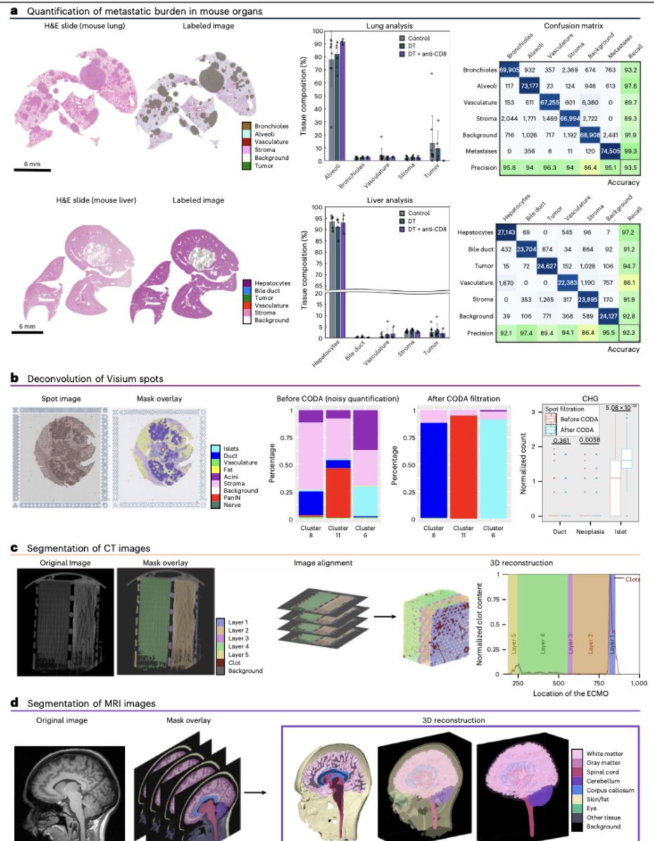
A detailed protocol for CODA to segment tissue components in histological sections for 3D reconstructions.
Led by colleague Prof. Kiemen @JohnsHopkins
Use this protocol here: https://t.co/uz9WAlyw8v
Excited to share our new tool, out today in @NatureProtocols! CODAvision includes a detailed guide on generating high-quality annotations for segmentation and a graphical user interface to make finetuning easy. Huge shoutout to first author @Valen_mat00s! https://t.co/09TjetfmO0
Our weirdly fresh 4D map of hair follicle genesis is finally out in @CellCellPress. It'll make you see spatial more temporally! Brilliant tour-de-force by PhD student Soichiro Asami, with great support from @JEFworks and Luis Garza #devbio#spatialomics
https://t.co/smPWMxm3FV
Great colleague Rong Fan led a team of experts at Yale, Hopkins, and UCSD to write a review on 3D multi-comics of tissues and tumors.
Check it out: https://t.co/5MsXCZT0hx
Immune remodeling during early pancreatic cancer progression.
Individual immunes cells around PanIN lesions are mapped using CODA
More here: https://t.co/7yAKlthbkJ
The new Issue of @cellpressblue is out today with CODA on the cover! Here you see normal pancreatic ducts in blue with tens of precancerous lesions throughout in red. Read more in our recent article here: https://t.co/g23rDAavvY
Currently my favorite video from the lab...
Mapping of blood vessels and nerves in whole embryos of rhesus macaque.
They form scale-free fractals of dimensions 3 and 2, independently of development stage.
More here: https://t.co/LqZrO7Wnfp
The vasculature system as mapped by CODA.
Color coded by radius size and Strahler order.
We found that the vasculature for mouse, rhesus macaque, chicken, and turtle have the same scale-free space-filling fractal geometry (D = 3).
Here: https://t.co/LqZrO7Wnfp
So proud of our lab alumnus, @KaustavBera11 (PhD ’22), on joining @PurdueChemE as an Assistant Professor! Excited to see him launch his lab and continue advancing the frontiers of mechanobiology, biomaterials and tissue engineering. Wishing him every success in this new chapter!