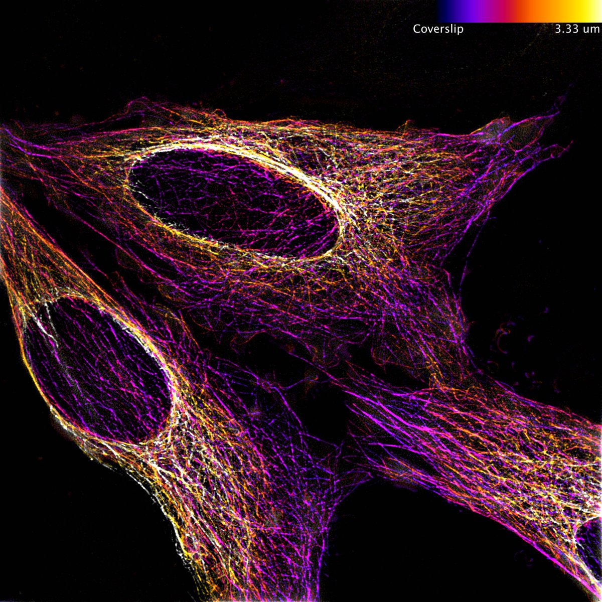
October image of the month is from @Ruby_Peters_ of the
@PaluchLab and shows HeLa cells, labelled with Tubulin-GFP. Taken using our Elyra 7 Lattice SIM and colour coded for depth
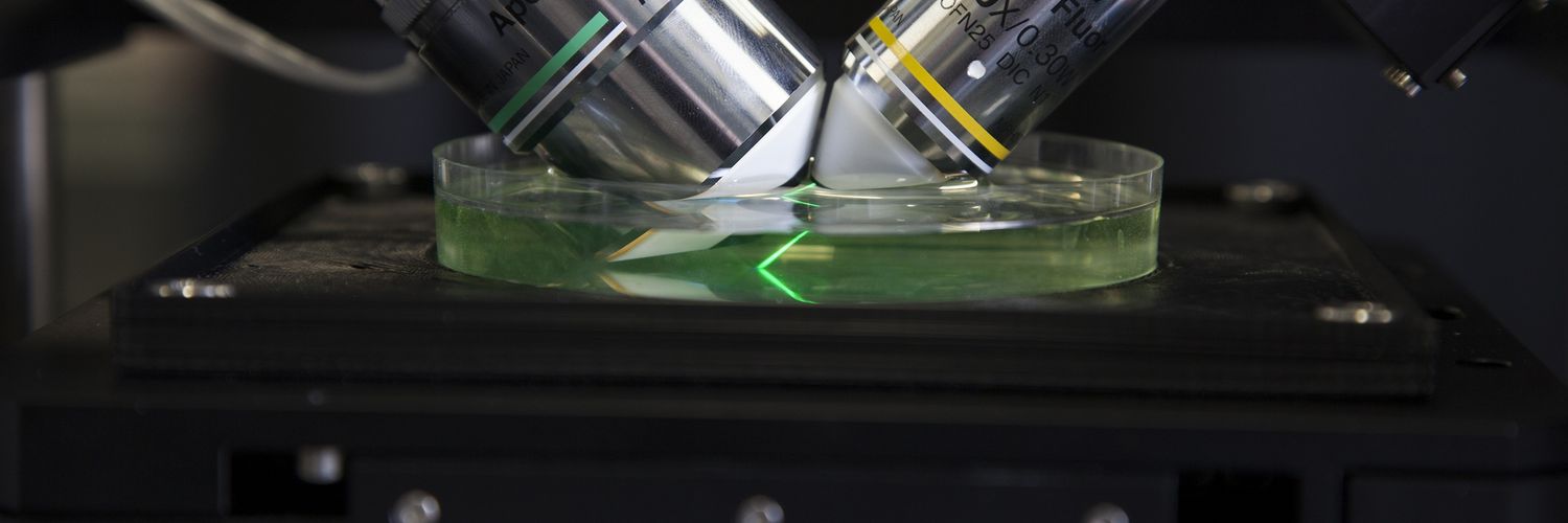
Excited to process and analyse my first nlsABACUS #lightsheet images, even if it is just the negative control😅! Martin Lenz at @CamMicroscopy and @slcuplants has built a brilliant microscope and is an imaging ninja.
how to cover yourself when you are a plant? Just weave your own fabric! Check our collaborative work between @slcuplants@CUBotanicGarden@ChemCambridge@CamMicroscopy on how Dionysia tapetodes extrudes flavones threads from its glandular trichomes. https://t.co/9V8ffw0A2j
August image of the month: Drosophila larvae expressing green and magenta fluorescent proteins in nociceptive (noxious touch), and proprioceptive (body movement) neurons. Image: @Paul_brooks who researches neuron degeneration, captured by confocal microscope in @CamZoology
We're hiring! Research associate position in advanced photo-manipulation microscopy. Come join us in developing technology critical for cutting edge optical trapping, optogenetic and photoablation experiments. https://t.co/wr5GQjt1F5
July image of the month. Puromycin assay on cultured hippocampal neurons showing new proteins that are produced within 15 minutes (yellow/green) and microtuble-associated protein 2 staining (red).
Captured on our laser scanning confocal microscope by Dr Tanja Fuchsberger
The @SarrisLab made incredible use of our two-photon microscope in their recently published research on neutrophil migration to sites of tissue damage https://t.co/YhvZt4Jdec
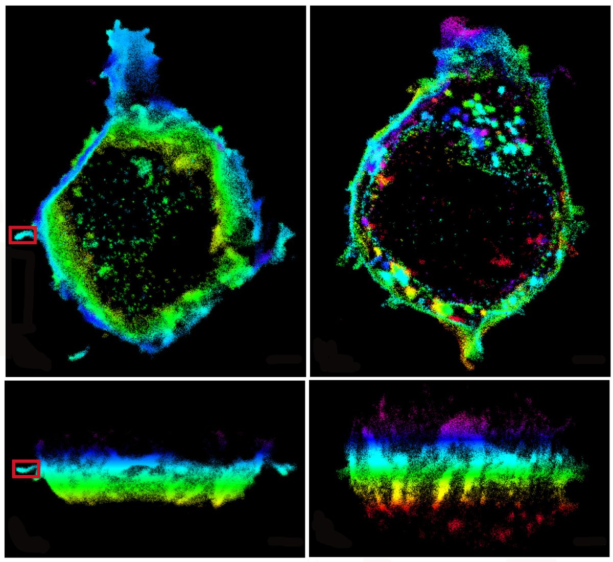
CAIC is starting a monthly series of informal clinics for super-resolution fluorescence microscopy. Come along for advice on getting the most out of our SIM, STED and SMLM systems. First clinic is Friday 21st February - follow link for details https://t.co/K6Y2KAZXBf
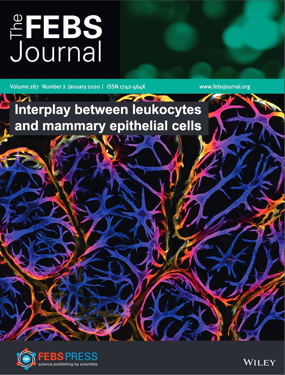
We got the cover! The wonderful @JessieJRH's new paper is out now in @FEBSJournal, with an excellent commentary from @WendyIngman. If you like immune cells and boobs, or nice 3D imaging, this is worth a read. https://t.co/dGYAtEljfg with @katevetpath & @cjwhelix
Fantastic opportunity to join the @EmilianiLab and get involved in advanced optogenetics technologies. You'll also get to work alongside the talented @r_r_sims who developed light sheet and light field microscopes in CAIC.
PhD Position in the #interdisciplinary PhD program #zenith_etn. @EmilianiLab is looking for candidates interested in non-linear optics,wavefront shaping and optogenetics( https://t.co/UBJR4zxlhk ).Send your application soon,deadline is January 5th 2020! #ITN @MSCActions #zenith
Great work by Martin Lenz showcased in his talk about the multi-view light sheet microscope for gentle fast imaging of plants he has developed in the @slcuplants imaging facility.