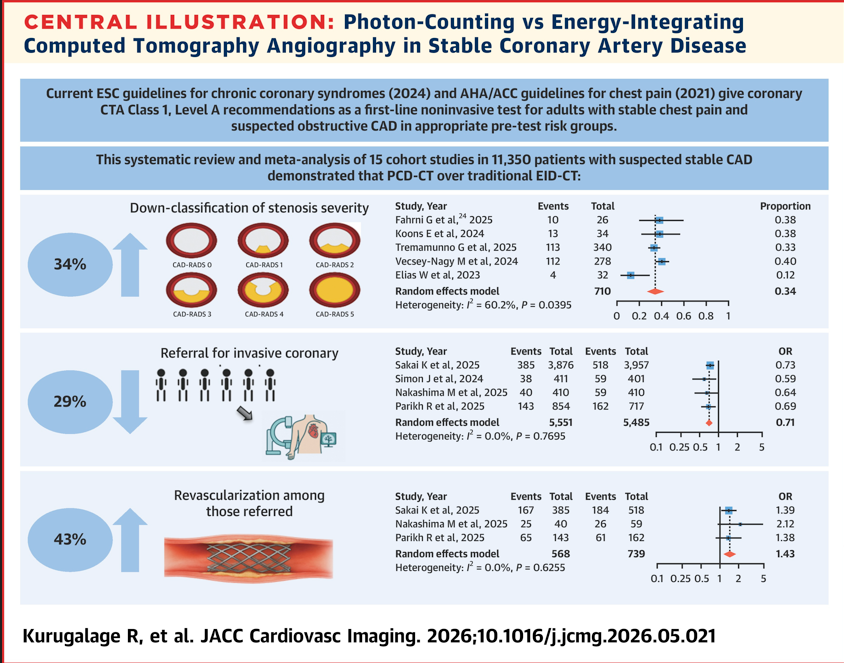
Proud to see our work featured in a new meta-analysis in #JACCImaging!
Even more rewarding to see TWO studies I contributed to- Parikh R et al. and Sakai K et al. included in the central illustration and evidence supporting photon-counting CT from our work
These findings highlight how advances in CT technology may influence not only image quality and diagnostic confidence, but also downstream patient management.
Grateful to my colleagues and collaborators at @StFrancis_LI for their continued dedication to advancing cardiovascular imaging research.
@OKhaliqueMD@DoosupShin@ziadalinyc@ESHLOF
Exciting times for #PCDCT #yesCCT #jaccimaging! 🚀
https://t.co/YRlkFUGEmV
So grateful to work every day with this incredible crew and to run the MKE Half today with our amazing team! ❤️ Huge kudos to Jennee, Olivia, Blair, Pam, Lauren and Julie(full!), and thanks for Caryn & Amanda for cheering us on 🙌 @blairtilkens @lauren_howard31 🏃♀️👟💙
Had such an amazing time at the @WisconsinACC inaugural imaging society dinner with colleagues and friends from across the state!
@renujain19 @jhainesDO@MCWcardiology
@HeartToProve@echo_stepbystep Right but the myocardium does not consistently feel the PVcs either. A sick heart can also increase SV after a PVC but you wouldn’t use that for their CO. Just food for thought. Perhaps an AV calcium score is the answer here (new ESC guidelines).
Excited to share our new publication on Ring-Like Enhancement in @IntJCVImaging
Across heterogeneous cohorts, RLE consistently associated with VA and HF events. May represent a high-risk imaging phenotype rather than a disease-specific marker.
https://t.co/CRCiVvybEO
🧠🫀 Using a large RWD, we studied outcomes of LAAO in patients with cardiac amyloidosis + AF.
1-yr outcomes were comparable to non-CA, but CA mirrors higher-risk cohorts → CHA₂DS₂-VASc may underestimate thromboembolic risk.
👉 https://t.co/3RcEBy32Z2
@crfshj@MCWcardiology