We're pleased to share our latest publication in @SurgEndosc on 𝘙𝘰𝘣𝘰𝘵𝘪𝘤 𝘷𝘦𝘳𝘴𝘶𝘴 𝘓𝘢𝘱𝘢𝘳𝘰𝘴𝘤𝘰𝘱𝘪𝘤 𝘊𝘉𝘋 𝘌𝘹𝘱𝘭𝘰𝘳𝘢𝘵𝘪𝘰𝘯: 𝘖𝘶𝘵𝘤𝘰𝘮𝘦𝘴 𝘧𝘳𝘰𝘮 𝘢 𝘏𝘪𝘨𝘩-𝘝𝘰𝘭𝘶𝘮𝘦 𝘚𝘱𝘦𝘤𝘪𝘢𝘭𝘪𝘴𝘵 𝘗𝘢𝘯𝘤𝘳𝘦𝘢𝘵𝘪𝘤𝘰𝘣𝘪𝘭𝘪𝘢𝘳𝘺 𝘜𝘯𝘪𝘵.
This study reports our experience of 547 consecutive MIS CBDE, demonstrating that RCBDE achieves comporable outcomes to LCBDE, despite preferential use in technically demanding cases.
Following propensity-adjusted analysis, the only significant difference was a modest increase in operative duration with the robotic approach.
Our findings support the integration of robotic surgery within a structured minimally invasive biliary pathway in dedicated specialist centres.
Link to paper:
https://t.co/hFKqksipRv
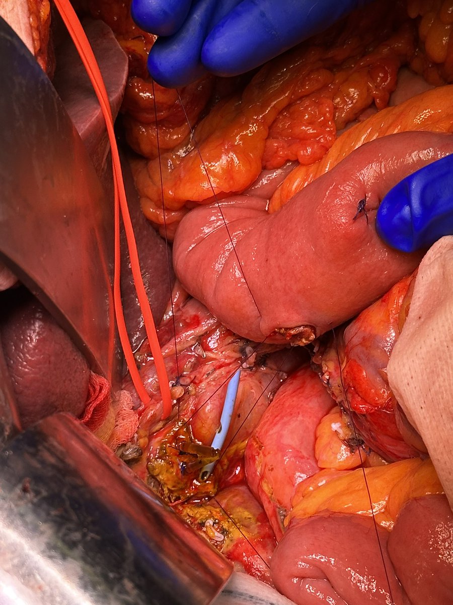
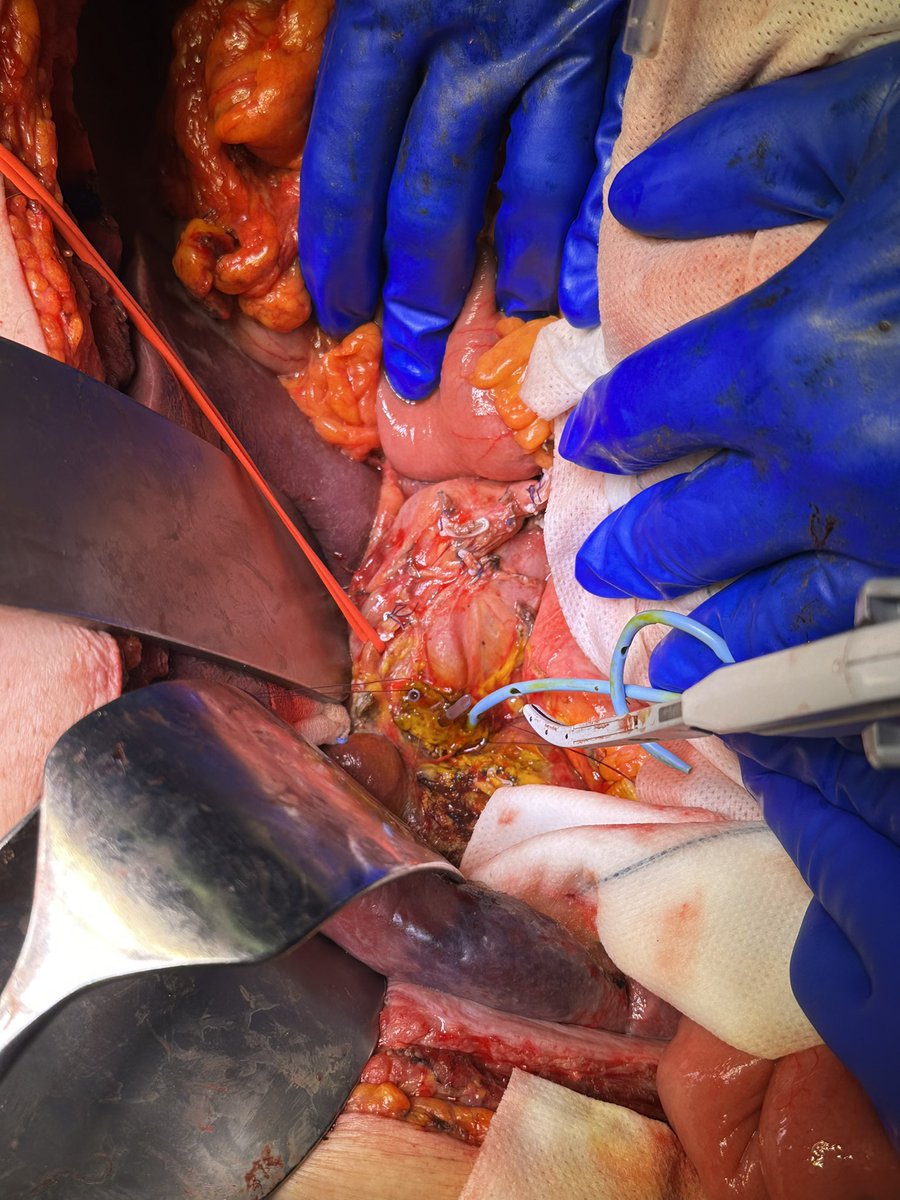
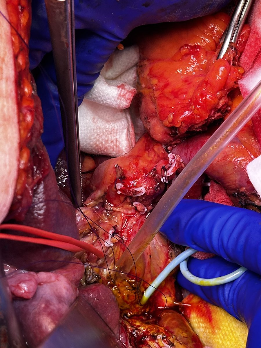
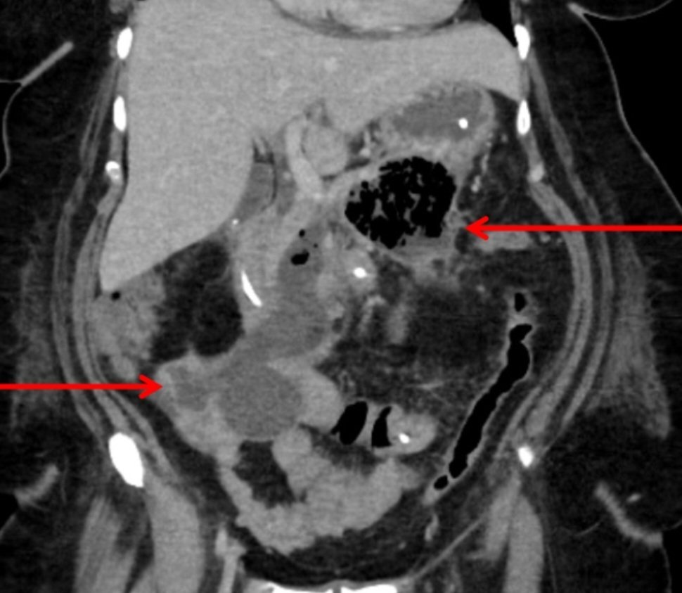
𝗔𝗻𝘁𝗲𝗿𝗶𝗼𝗿 𝗚𝗮𝘀𝘁𝗿𝗼𝘀𝘁𝗼𝗺𝘆 𝗮𝗻𝗱 𝗥𝗲𝘁𝗿𝗶𝗲𝘃𝗮𝗹 𝗼𝗳 𝗜𝗺𝗽𝗮𝗰𝘁𝗲𝗱 𝗛𝗼𝘁 𝗔𝘅𝗶𝗼𝘀 𝗦𝘁𝗲𝗻𝘁
🔴Anterior gastrostomy just at level of palpable bulge and proximal to pylorus
➡️Correlated with Hot Axios position on CT
🔴3/0 PDS used as stay sutures to improve visibility
➡️Just anterior leaf of gastrostomy
➡️To falciform using slip knot
🔴Despite manoeuvres described difficult to visualise Hot Axios stent
🔴Ligaclips applied to mucosa where stent could be palpated
🔴Intraoperative XR screening used to orientate the stent in relation to ligaclips
🔴Hook diathermy used to divide stomach mucosa to the right of the ligaclips to identify the stent which proved to be unsuccesful
🔴Real time IOUS utilised
➡️Critical step
➡️Key in identifying location of Hot Axios stent
🔴Once identified the stent which buried beneath the mucosa was removed with careful traction
🔴Bleeding encountered upon stent retrieval
➡️Controlled with pressure, judicious use of diathermy and placement of Veriset
🔴Anterior gastrostomy sutured close in 2 layers with 3/0 PDS
🔴Postoperatively patient started on IV PPI and initiated on oral fluids and diet in stepwise manner
#FOAMed #GITwitter #EUS #HPB #SoMe4Surgery #surgery
𝗥𝗼𝗯𝗼𝘁𝗶𝗰 𝗧𝗿𝗮𝗻𝘀𝗴𝗮𝘀𝘁𝗿𝗶𝗰 𝗡𝗲𝗰𝗿𝗼𝘀𝗲𝗰𝘁𝗼𝗺𝘆 𝗮𝗻𝗱 𝗖𝗼𝗻𝗰𝗼𝗺𝗶𝘁𝗮𝗻𝘁 𝗖𝗵𝗼𝗹𝗲𝗰𝘆𝘀𝘁𝗲𝗰𝘁𝗼𝗺𝘆 𝘄𝗶𝘁𝗵 𝗜𝗖𝗚
Preoperative Workup:
🔴Preoperative CT imaging
➡️Retrogastric WON
➡️Portal HTN with evidence of portocavernous transformation
🔴Preoperative MRCP
➡️Help distinguish % of necrosis vs. liquid similar to EUS
🔴Symptomatic pt with no evidence of ongoing infection
➡️Suitable for single stage MIS
Surgical Approach:
🟢IOUS essential to identify locality of WON
🟢Anterior gastrostomy
🟢Necrosectomy performed
➡️Single stage clearance of WON
➡️EUS requires multiple sessions with recurrent hits of sepsis between interventions
➡️Visual feedback critical in 🤖 due to lack of haptic feedback
🟢Haemostatic agents applied into cavity
🟢Cystgastrostomy maturation
➡️0 Vicryl suture in figure of 8
➡️Prefer over stapler due to haemostatic benefit of suturing
🟢Two layer closure of anterior gastrostomy
🟢Subtotal cholecystectomy
➡️Fused hepatocystic triangle
➡️Increased difficulty in performing cholecystectomy early in severe acute pancreatitis
Benefit of 🤖:
🔵Improved views of WON cavity
➡️3D enhancement
🔵Wristed instruments
➡️Better articulation for suturing of cystgastrostomy
#FOAMed #GITwitter #MedEd #SoMe4Surgery #Robotics #Pancreatitis #surgery
“Three duct” biliary-jejunal anastomosis at the level of Hilar Plate after a difficult whipple + biliary duct resection for extra hepatic cholangiocarcinoma
Pleased to share my 7th publication from my TPIAT thesis with @HpbLeicester in @JournalofGISurg.
We found lower islet yields in genetic CP (48k vs 149k IEQ), with comparable pain outcomes.
Early genetic assessment and referral key.
Link to paper:
https://t.co/VRCoJVFrTT
Robotic Longitudinal Pancreaticojejunostomy
🔵Chronic pancreatitis secondary to ETOH excess - now teetotal
🔵Multiple admissions with chronic pain
🔵Significant impairment on QoL
🔵2 emergency admissions
➡️Acute pancreatitis with side branch disruption
🔵Normal LFTs
🔵CT/MRCP
➡️Dilated PD with "chain of lakes"
➡️Non-obstructing stone at HOP
🔵MDT
➡️Decision to proceed to surgery
🔴Adjuncts such as IOUS, ICG and intraoperative pancreatoscopy
➡️Improves operative safety
➡️Promotes learning and training in complex benign PB conditions
🔴Benefit of ICG
➡️Visual confirmation of splenic artery and proximity to PD/pancreatic parenchyma
#FOAMed #MedEd #surgery #hpb
Camera Flip (Image Inversion) during Robotic Ventral Hernia Repair
Image Inversion Process
🔴Involves a three-step process: ➡️Unlocking the display and reassigning robotic arms via the “manual command” button
➡️Flipping the 30° endoscope lens in the opposite direction.
🔴This reorients the surgical field so that movements in the upper space (“ceiling”) of the display are transformed into movements in the lower field (“floor”)
Benefits
🔴Improved Ergonomics
➡️Reorients the surgical field for intuitive forehand suturing and reduces physical strain during ventral hernia defect closure
🔴Enhanced Precision
➡️Facilitates accurate suturing and assists with tension-free fascial closure
Technical Requirement
🔴Requires expertise with the robotic platform and proper execution of the image inversion process
𝙀𝙢𝙚𝙧𝙜𝙚𝙣𝙩 𝙇𝙖𝙥𝙖𝙧𝙤𝙨𝙘𝙤𝙥𝙞𝙘 𝙄𝙣𝙛𝙧𝙖𝙘𝙤𝙡𝙞𝙘 𝙉𝙚𝙘𝙧𝙤𝙨𝙚𝙘𝙩𝙤𝙢𝙮
Patient Selection:
🔵Central, infracolic collections that are dominantly solid (WON) often not amenable to perc. drainage and/or narrow window of access for transgastric approach
🔵Too unwell for concomitant cystjejunostomy
🔵Septic
🔵3-4 weeks into acute attack of SAP
Approach:
🔴Initial identification of bulge in transverse mesocolon
🔴Location confirmed with IOUS
🔴Window created into infracolic compartment
🔴Necrosectomy and washout performed
🔴2 x 18F Robinsons drains placed adjacent to cyst
Key Points:
🟢Timing of intervention critical
🟢Surgical approach allows 𝘀𝗶𝗻𝗴𝗹𝗲 𝘀𝘁𝗮𝗴𝗲 𝗰𝗹𝗲𝗮𝗿𝗮𝗻𝗰𝗲 of WON which avoids multiple hits of sepsis
🟢Concomitant cholecystectomy can be undertaken in select cases
🟢Patient selection and discussion in benign specialist MDT key
Link to our paper in @JournalofGISurg below:
https://t.co/kcdwWFwSjz
#FOAMed #GITwitter #surgery #SoMe4Surgery #MedEd #hpb
𝙇𝙖𝙥𝙖𝙧𝙤𝙨𝙘𝙤𝙥𝙞𝙘 𝙍𝙚𝙥𝙖𝙞𝙧 𝙤𝙛 𝙋𝙚𝙧𝙛𝙤𝙧𝙖𝙩𝙚𝙙 𝘿𝙪𝙤𝙙𝙚𝙣𝙖𝙡 𝙐𝙡𝙘𝙚𝙧 𝙬𝙞𝙩𝙝 𝙋𝙧𝙞𝙢𝙖𝙧𝙮 𝘾𝙡𝙤𝙨𝙪𝙧𝙚 & 𝙊𝙢𝙚𝙣𝙩𝙖𝙡 𝙋𝙖𝙩𝙘𝙝
Presentation:
🔵1 day history of sever upper abdominal pain
🔵Smoker and excess ETOH
🔵CXR
➡️Pneumoperitoneum
🔵CT
➡️Perforated duodenal ulcer
🔵Time from ED➡️theatre 3 hours
Operative Approach:
🔴Thorough peritoneal lavage
➡️Lap washout allows for thorough removal of intra-abdominal contamination
➡️Modern OR table tilting can provide better view then open in select cases
🔴If fragile tissue then omental patch only
➡️This approach is a modified version of Grahams patch repair
➡️When suitable, we prefer primary closure with patch after
🔴For large perforations kocherisation of duodenum may be indicated
🔴Convert to open if:
➡️Large ulcer
➡️Associated ulcer bleed
🔴H. Pylori eradication therapy on discharge
🔴OGD in 6-8 weeks to confirm healing
Key Points:
🟢Timely intervention critical
🟢Lap approach in EGS should be encouraged
🟢Compared to open, advantages of lap are:
➡️Reduced pain
➡️Reduced LOS
➡️Reduced incisional hernia rate
🟢Trainees should be exposed to lap EGS early to develop skills and confidence
#FOAMed #GITwitter #MedEd #SurgEd #EGS #SoMe4Surgery
𝗘𝗺𝗲𝗿𝗴𝗲𝗻𝗰𝘆 𝗥𝗼𝗯𝗼𝘁𝗶𝗰 𝗥𝗲𝗽𝗮𝗶𝗿 𝗼𝗳 𝗟𝗲𝗳𝘁 𝗜𝗻𝗴𝘂𝗶𝗻𝗮𝗹 𝗛𝗲𝗿𝗻𝗶𝗮 𝘄𝗶𝘁𝗵 𝗜𝗖𝗚
Surgical Approach:
🔴Initial assessment of small bowel with ICG
🔴Small bowel placed in pelvis with warm wet swabs overlying
🔴TAPP approach to hernia repair
➡️Critical view of MPO
➡️ICG to help delineate key structures
➡️Progrip mesh
➡️3-0 absorbable V-lock to close peritoneum
🔴Reassessment of small bowel after hernia repair
➡️ICG present in bruised area demonstrating viability
➡️Aided decision making to not resect or oversew
Robotics in EGS:
🟢Case selection key
🟢MDT approach with anaesthetic and scrub teams to coordinate
🟢Minimally invasive approaches in EGS should be encouraged
#FOAMed #GITwitter #MedEd #Surgery #Hernia #EGS
Emergency Laparoscopic Repair of Strangulated Femoral Hernia with Small Bowel Resection & ICG
🔴Reduction of small bowel
➡️Await perfusion recovery
➡️Allows for demarcation of ischaemic segment
🔴Undertake hernia repair
➡️TAPP approach
➡️Opted for suture repair for this case
➡️Biologic mesh alternative option
➡️Synthetic mesh can be considered if no bowel resection
🔴ICG to assess bowel perfusion and delineate length of resection
➡️Given intraop at time of assessment
➡️Immediate assessment within 30 seconds key
➡️Excellent tool for teaching trainees
🔴Advanced lap skills crucial
🔴Lap EGS recommended
➡️Underutilisation in the UK
Laparoscopic Infracolic Necrosectomy and Drainage of RIF Abscess
🔴Septic patient but no organ failure
🔴Narrow window of access for EUS or IR drainage
🔴Single stage clearance of WON and drainage of RIF abscess
➡️Prevents hits of sepsis between multiple procedures
➡️Address both collections at once
➡️Select cases
➡️Benign PB MDT
➡️"One size does not fit all"
Our paper on infracolic approach in @JournalofGISurg:
https://t.co/kcdwWFwSjz
Our collaboration with @BehindTheKnife on MIS for complicated SAP:
https://t.co/Ihu684TBjg
#Surgery #HPB #Pancreatitis
𝗥𝗼𝗯𝗼𝘁𝗶𝗰 𝗖𝗼𝗺𝗺𝗼𝗻 𝗕𝗶𝗹𝗲 𝗗𝘂𝗰𝘁 𝗘𝘅𝗽𝗹𝗼𝗿𝗮𝘁𝗶𝗼𝗻 𝗣𝗼𝘀𝘁 𝗖𝗵𝗼𝗹𝗲𝗰𝘆𝘀𝘁𝗲𝗰𝘁𝗼𝗺𝘆
Patient Presentation:
🔵Previous cholecystectomy
🔵2 previous ERCPs
🔵Recurrent large stone
➡️1.7 x 1.1 cm
➡️Failed endoscopic clearance
➡️+/- requirement for EHL
🔵MDT decision to proceed to bile duct exploration
Surgical Approach:
🔴Careful dissection of adhesions in the GB fossa to expose the CBD
🔴ICG injected immediately following induction of anaesthesia
🔴Firefly mode used to confirm the CBD
🔴Vertical choledochotomy made with robotic shears
🔴Large stone extracted by milking it gently through the choledochotomy
🔴Check choledochoscopy to confirm clearance of the CBD
🔴Closure of the CBD with 4/0 Vicryl
Benefit of Robotic Platform:
🟢Wristed instruments which improves ergonomics for suturing
🟢May reduce the learning curve for transductal CBDE when compared with laparoscopy
Multiple CBD stones are not, in themselves, a contraindication to LCBDE.
With appropriate laparoscopic expertise, choledochoscopy and access to EHL/laser for larger or impacted stones, most cases can be cleared with primary duct closure, avoiding routine T-tubes or biliary stenting.
Success depends on training, experience, and commitment to an LCBDE programme.
𝗘𝗺𝗲𝗿𝗴𝗲𝗻𝘁 𝗟𝗮𝗽𝗮𝗿𝗼𝘀𝗰𝗼𝗽𝗶𝗰 𝗖𝗵𝗼𝗹𝗲𝗰𝘆𝘀𝘁𝗲𝗰𝘁𝗼𝗺𝘆 𝗮𝗻𝗱 𝗧𝗿𝗮𝗻𝘀𝗰𝘆𝘀𝘁𝗶𝗰 𝗟𝗖𝗕𝗗𝗘
Patient Presentation:
🟢Acute cholecystitis with deranged LFTs
🟢Surgery performed <24 hours from admission
Surgical Approach:
🔴Timing critical in acute cases
🔴Retraction of duodenum improves view
🔴Cystic artery clipped prior to IOC in this case due to confidence in anatomy
🔴3mm choledochoscope for stone retrieval
➡️Remove scope over wire if CBD stone is larger than cystic duct
🔴Check choledochoscopy
#FOAMed #MedEd #Surgery #SoMe4Surgery #gallstones