Need a DURABLE way to remember what causes DURAL enhancement?
Feeling left behind when it comes to leptomeningeal disease?
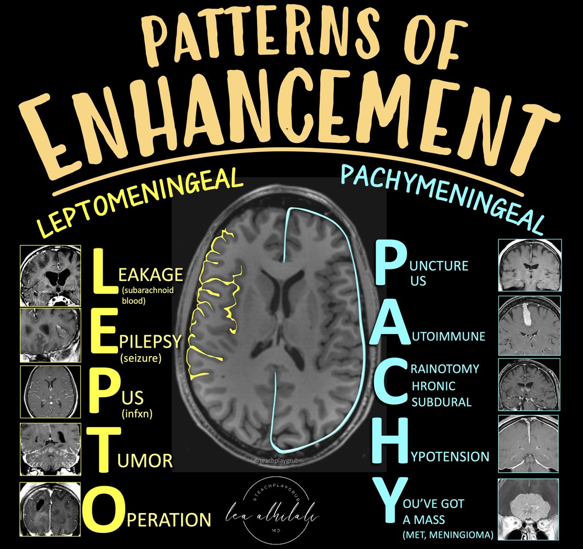
Here’s an easy way to remember the differential diagnoses for the two types of meningeal enhancement:
LEPTOMENINGEAL
Remember:
L = Leakage (subarachnoid hemorrhage)
E = Epilepsy (recent seizure)
P = Pus (infection, either bacterial, viral or granulomatous/fungal)
T = Tumor (leptomeningeal metastatic dz)
O = Operation (post operative)
PACHYMENINGEAL
Remember
P = Puncture (recent LP), Pus (subdural empyema)
A = Autoimmune (sarcoidosis, IgG4)
C = Craniotomy (post-op), Chronic subdural
H = Hypotension (intracranial hypotension)
Y = You’ve got a mass (mets, usually from direct bony extension, meningioma)
Now you’ve got a lot information PACH-ed into an easy & memorable mnemonic!
Facet joints are the only synovial joints in the spine & can be involved in a large number of pathological processe . F J Arthritis could be due to aging,repetitive stress on the joints, poor posture,obesity,...
FJs help in load transmission, flexion& extensionin of spine,..etc
1/Hate it when one radiologist called the stenosis mild, the next one said moderate--but it was unchanged?!
How do you grade it? Do you estimate? Measure? Guess???
Let this thread take the load off your back w/a lumbar grading system that’s easy, reproducible & evidence-based!
1/Diagnosing discitis can be a pain in the backside!
When it’s easy, it’s easy--but when it’s hard, it’s REALLY HARD!
Only one of these is discitis—can you tell which one?
This #tweetorial will help show you how!
#meded#medtwitter#FOAMed#spine#radtwitter#radres
1/“I love memorizing classification systems!”—said no one in clinical practice, ever
Overwhelmed by all the RAD reporting systems?
Here’s a #tweetorial on 1 that’s helpful in head & neck imaging, that you can understand instead of memorize—NI-RADS!
#meded#medtwitter#radres
Can't remember the types of craniotomies off the top of your head?
Do you need another thing to memorize like you need a hole in your head?
Here's a quick review figure of common craniotomies to help you out!
#meded#FOAMed#medtwitter#Neurosurgery#neurotwitter#radres
Within the last week in US.
MR tech on-duty, not in the magnet room.
MR table was out of the magnet room.
Nurse and tech-aide brought patient-on-gurney into magnet room.
Patient thrown (patient relatively unharmed).
Nurse struck reported fract. femur and pelvis.
#MRIsafety#MRI
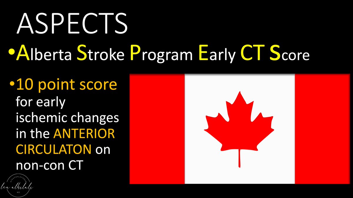
2/Many know anterior circulation ASPECTS. It uses a 10 point scoring system to semi-quantitate the amount of the MCA territory infarcted on non-contrast head CT.
If you need a review: here’s my tweetorial on ASPECTS: https://t.co/urxRZBpSEc
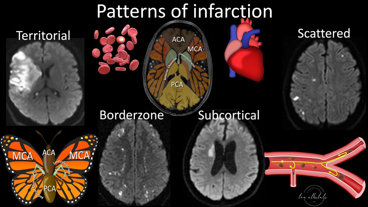
1/I always tell my fellows, “Anyone can see the bright spot on diffusion—what sets you apart is if you can tell them why it’s there!”
Can you tell a stroke’s etiology from its appearance on MRI?
Here’s a #tweetorial to show you how!
#medtwitter#neurotwitter#stroke#neurorad
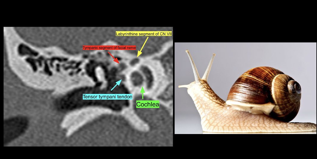
Use the snail eyes sign to locate the facial nerve on a coronal CT of the temporal bone. Start with finding the snail shell/cochlea. The eyes reflect two segments of the facial nerve and the nose is the tendon of the tensor tympani muscle. #Neurorad#anatomy#radres#MedTwitter
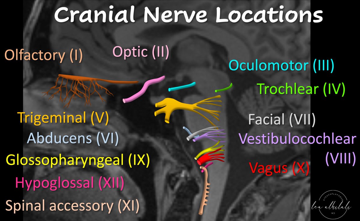
My attending once said, "Just b/c you don't see it, doesn't mean it's not there."
Just b/c you don't see all cranial nerves on all MR exams doesn't mean they aren't there
You must know their #anatomy so you know when pathology will affect them
#medtwitter#meded#neurotwitter