“In examining disease, we gain wisdom about anatomy and physiology and biology. In examining the person with disease, we gain wisdom about life.”
—Oliver Sacks
The key is to enjoy hanging out on the edge. That is, you find it interesting to attempt things one step beyond where you are right now. It could be the edge of your ability or the edge of your knowledge or the edge of your network.
If you reach — but just a little — and you do it every week, then you'll take on challenges that are manageable enough that you win most of the time, but meaningful enough that you improve as well.
How I still try to keep up to date with the literature!
1. Every week browse new articles in major journals for my field (Blood, JCO etc)
2. I still read/skim the UpToDate article for all new patients I see regarding their diagnosis
3. Follow key people on X
4. Call my friends
It’s interview season! Here’s an article I wrote with advice for applicants, based on 15+ years of doing interviews at every level from med school to residency to fellowship. Give it a read and let me know what you think. https://t.co/97FBg96Xj0
One ordinary moment in a doctor's mind:
Overthinking yourself into despair trying to figure out whether your headache is from dehydration, a migraine, muscle tension, malnutrition, stress, lack of sleep, bad posture, or a brain tumor.
Are you right when it’s bright?
Bright cortical signal on diffusion images is classically associated w/hypoxic-ischemic injury.
But there are many mimics!
Do you know how to recognize the different patterns of cortical restricted diffusion? Here’s a figure & some pearls to help!
Cortical restricted diffusion many seen in a variety of conditions—many with VERY different pathophysiology & prognosis.
There are 6 main patterns:
(1) Cortex & deep gray
(2) Diffuse Cortex
(3) Focal Cortex
(4) Limbic
(5) Deep gray
(6) White matter
Here are some steps to help differentiate:
(1) Is the presentation acute or chronic?
(2) If it’s acute, toxic-metabolic & hypoxic-ischemic events account for the majority of cases. Seizure & encephalitis are less frequent and typically more focal
(3) In the chronic setting, isolated cortical involvement is almost diagnostic of CJD
Hopefully, this will clear up any confusion about the diffusion!
Even if you’re in it for the LONG haul, sometimes it’s good to come up SHORT!
If you can’t summarize, you’ll never memorize!
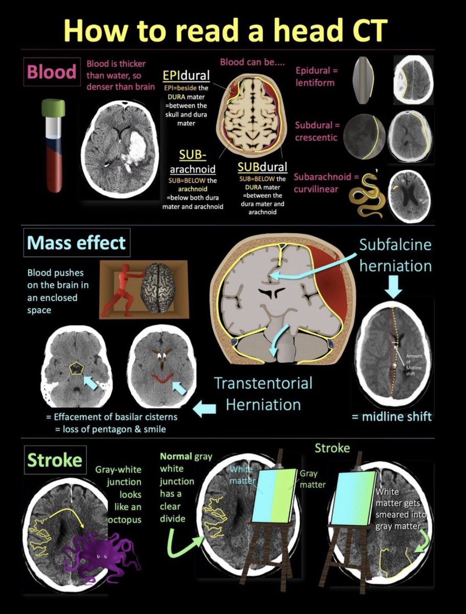
Here's a cheat sheet for a truly critical skill in imaging: How to read a head CT!
If you're looking at head CTs, this is what you NEED to know!
➡️The 3 main things to look for on head CTs are:
🔸Blood
🔸Mass Effect
🔸Stroke
➡️Blood
🔸CT is just a measure of density. Blood is thicker than water, so blood is denser than brain (70% water) on head CT
Blood can be:
🔸EPIdural: looks lentiform
🔸SUBdural: looks crescentic
🔸SUBarachnoid: looks snake-like
➡️Mass effect
🔸You won’t bleed out in your head. Instead, calvarium is a closed space & brain gets compressed
Look for:
🔸Subfalcine herniation = midline shift, ventricles shift to the other side
🔸Transtentorial herniation = Loss of the basilar cisterns--normally look like a pentagon & smiley face
➡️Stroke
🔸Normal gray-white differentiation looks like an octopus & is crispy & well-defined
🔸Stroke looks like someone smeared white matter paint into gray matter paint
You can see the full thread here:
https://t.co/NyrzkWz0Q2
So now you know what to look for on a head CT. Hopefully this short version stays with you for the long haul!
@AbdullahHanjraa If October doesn't work, We will sing "November never disappoints" and "December is the month of miracles". 🤣 ✨But have to keep going!