#Susac's syndrome is a rare immune-mediated endotheliopathy characterized by a classic triad of #hearing loss, #retinal vaso-occlusive disease, and #encephalopathy.
It was first described by American neurologist John O. Susac, who in 1975 saw a young woman with a previously unreported clinical triad of encephalopathy (“personality changes and paranoid psychosis”), branch retinal artery occlusions, and deafness. This patient was presented to him at a conference in Albany, New York. Shortly thereafter, John Selhorst referred a 40-year-old woman with an identical set of clinical findings. They published these cases in 1979 as #microangiopathy of the #brain and #retina and since then there have been many additional patients reported.
📸 John O. Susac, MD (1940–2012) holding a cut-away model of the human brain in which he discovered Susac’s syndrome.
📸 Susac JO. Susac's syndrome: the triad of microangiopathy of the brain and retina with hearing loss in young women. Neurology. 1994;44(4):591-593. https://t.co/d1trbb0A9V
#WorldHearingDay #WorldHearingDay2025
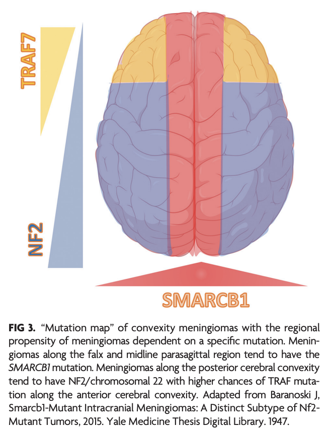
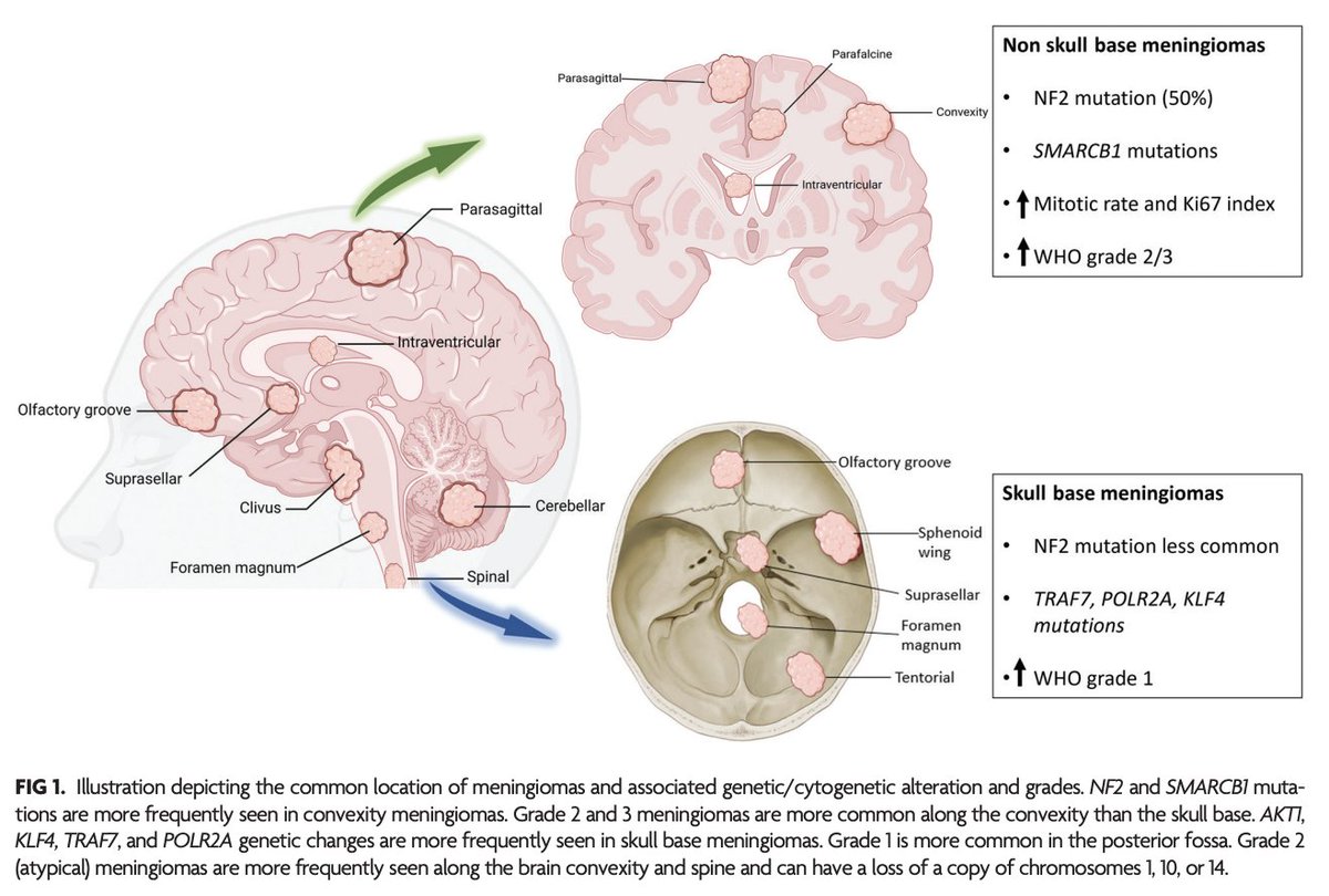
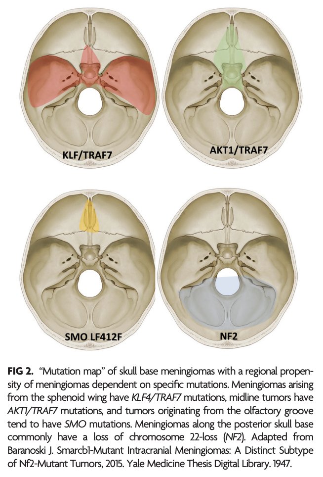
"Meningioma: Molecular Updates from the 2021 World Health Organization Classification of CNS Tumors and Imaging Correlates"
https://t.co/8h33Pwm0dR
@NeetuSo27437480
The MRI Neuroanatomy Post 🗞️
The parieto-occipital fissure (yellow) divides the occipital lobe from the parietal lobe and intersects the calcarine sulcus (pink) - resembling a tilted "Y".
The primary visual cortex lies along the calcarine sulcus.
Above the calcarine sulcus lies the cuneus (green) - the site of basic visual processing.
Below the calcarine sulcus lies the lingual gyrus (blue) - needed for face & landmark perception and recognition, and visual memory.
#MRI #Neuroanatomy #neuroscience #radiology #anatomy #neurotwitter #neurosurgery #MedEd #education #MedTwitter #neuro #ArtificialInteligence @futureradres