Even in early/intermediate AMD, structural retinal changes on OCT are linked to reduced visual function under bright & low light. These findings highlight key imaging markers of functional decline before advanced disease.
#Ophthalmology#OpenAccess
https://t.co/cKXV8xeS8g
@JAMAOphth@ApellisPharma The work of the original report & commentary would of course not be possible without the visionary leadership of @KonBalaskas
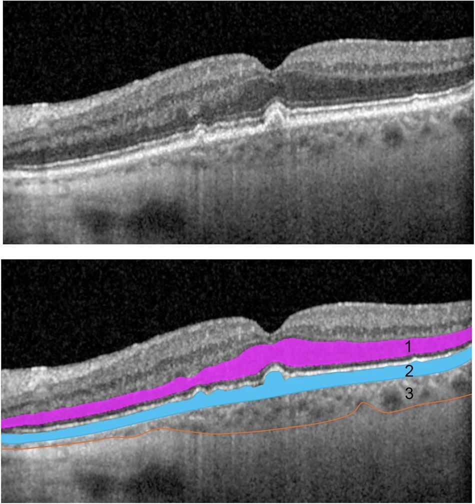
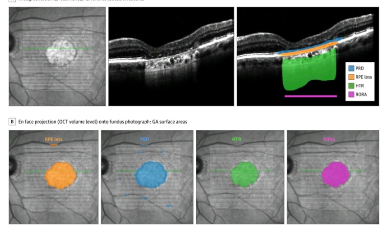
Pegcetacoplan associated with slower growth of photoreceptor degeneration (PRD), RPE-loss, PRD in isolation, and RPE and Outer Retinal Atrophy.
Our analysis of automatically-segmented quantitative OCT biomarkers out in @JAMAOphth based on OAKS & DERBY @ApellisPharma data.
Deep-learning spectral-domain optical coherence tomography analysis from DERBY and OAKS trials showed association with reduction of photoreceptor degeneration, RPE-loss, and RPE and outer retinal atrophy over 24 months. https://t.co/n4ID9x169u
@JAMAOphth@ApellisPharma https://t.co/zVF6on9v9Y
Extremely honoured to have eminent academics offer commentary on our work !
We thank and share their commitment to an open dialogue on the role of complement-inhibition in GA.
However, some of the authors' points, perplexed us ...
@JAMAOphth@ApellisPharma All patients already had foveal RPE-loss and PRD prior to starting any treatment, with a mean occupancy of 57% RORA, 60% RPE-loss, and 86% PRD.
This limits ability to draw any association between the treatment and GA features with the foveal region or foveal function.
@JAMAOphth@ApellisPharma Slower growth of PRD in isolation with Pegcetacoplan is relevant because:
1. Area of PRD in isolation is an independent predictor of GA Growth
2. It suggests that there a greater protective effect on photoreceptors than with RPE
Hot off the press 👇‼️ https://t.co/MpHeVxBfpl
Very proud to see the first AI-enabled analysis of Geographic Atrophy OCT feature changes over 24 months from the phase III DERBY & OAKS studies published by our team in @JAMAOphth 😀@MoorfieldsBRC@UCLeye 🙌
"Sharing data for [medical] research is highly beneficial to the scientific community and beyond, but cannot come at the expense of patient privacy"
Unfortunately, digital masks don't do away with this expense.
Great paper by @yvesalexandre
https://t.co/lkkddAA0P3
Up to 40% of patients with DMO do NOT respond to 1st-line anti-VEGF and thereby eligible for alternative treatment like intravitreal dexamethasone.
How well does it work? And for how long?
Answered in this new paper https://t.co/4gI8bl3GyT @JCM_MDPI@Moorfields
🧵1/5
@JCM_MDPI@Moorfields ... in our cohort, the median time to retreatment was 10.4 months.
So now that we can approximate the timeline for a positive response, we can time retreatment !
🧵4/5
@JCM_MDPI@Moorfields ... following a positive visual outcome, there is a 50% chance of sustaining this response beyond 4 months.
This makes sense as good things don't last forever, the dexamethasone delivery system we studied is designed to last 6 months.
... so that's when we retreat right?
🧵3/5
@JCM_MDPI@Moorfields Following a single intravitreal 700 µg dexamethasone injection, there is:
- >75% chance of gaining ≥5 ETDRS letters (or 1 Snellen line)
- >50% chance of gaining ≥10 ETDRS letters (2 Snellen lines)
... good times, but for how long can patients sustain this? ...
🧵2/5
Delighted to share our new paper authored by @jmnunezdorio & ORNATE team!
Link: https://t.co/ixwnWLVuyA
We developed novel deep learning systems (DLS) to curate then detect referable diabetic retinopathy in a community-based setting.
Need an answer and reference for the prevalence of diabetic retinopathy and vision-threatening diabetic retinopathy in India ? 🤓
@SivaprasadSobha and colleagues have the answer here: 👀
https://t.co/no1VOblo9U @TheLancet@LancetGH#diabetes#DiabetesAwareness
A real-world, #EMR-based cost analysis of UK NHS #glaucoma clinic patients found the vast majority were mild/low risk for prog. to #blindness & may be more appropriately managed w/ alternative, more affordable models of care. #ophthalmology https://t.co/dGTsuQJ24b


![MaximeTaquet's tweet photo. "Sharing data for [medical] research is highly beneficial to the scientific community and beyond, but cannot come at the expense of patient privacy"
Unfortunately, digital masks don't do away with this expense.
Great paper by @yvesalexandre
https://t.co/lkkddAA0P3 https://t.co/yqnp6KAMV7](https://pbs.twimg.com/media/F1WRIduXgAAhfn6.jpg)


