Our goal is to make ECG interpretation simple, practical, and fun for acute providers; and we believe that mastery of this simple skill will save lives.
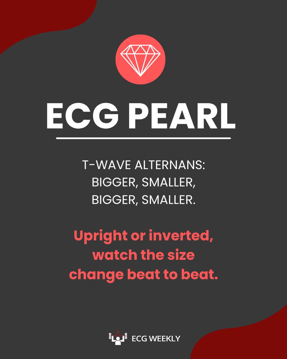
T-wave alternans: bigger, smaller, bigger, smaller. 🔎
Upright or inverted doesn’t matter—it’s the beat-to-beat change in size.
Spot it, and you’ve spotted a patient at very high risk of torsades.
📷 The T-wave size.
Bigger, smaller, bigger, smaller, beat to beat. Upright or inverted doesn’t matter.
It’s different from electrical alternans, and it flags a patient at very high risk of torsades. 📷
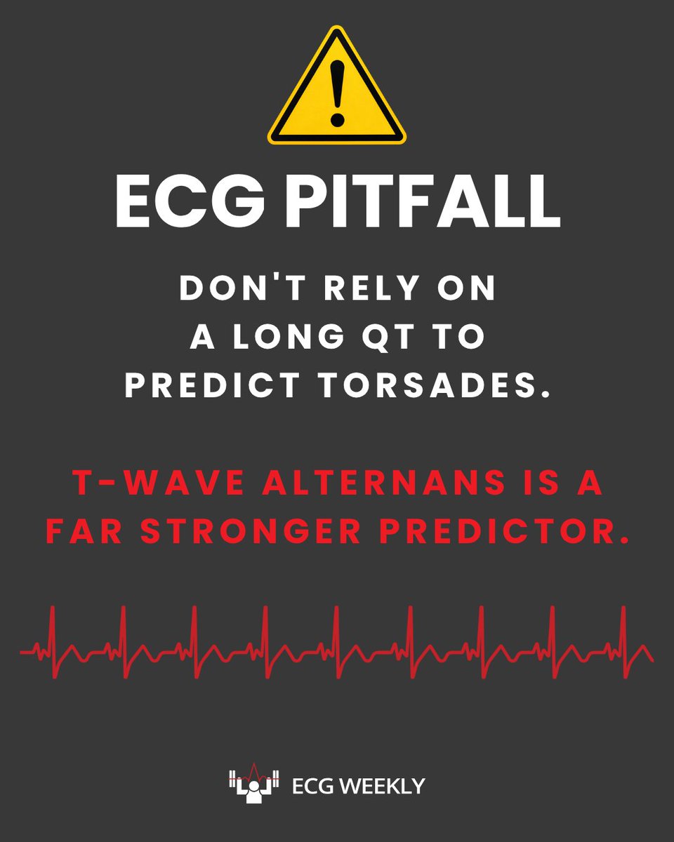
Don’t rely on a long QT to predict torsades. ⚠️
The QT is often prolonged, but T-wave alternans is a far stronger predictor.
Long QT: fix the electrolytes and watch closely.
T-wave alternans: torsades may be minutes away.
T-wave alternans can be an ominous warning: bigger, smaller, bigger, smaller from beat to beat. ⚠️
Dr. Mattu explains why this pattern deserves serious attention when you’re worried about impending torsades.
Would you recognize it in time?
https://t.co/lSsocVXJ8E
The Aslanger pattern isn’t in the formal ACC/AHA guidelines. It’s been described in the interventional cardiology literature.
Dr. Mattu’s tip: if you’re worried, share that literature with your cardiologist so they know what you’re talking about. 🔎
A 45 yo m presents to the emergency department with palpitations. He has no known history of coronary artery disease. He is alert, with a systolic blood pressure of approximately 120 mm Hg and a heart rate near 180 beats/min. The following ECG shows.......https://t.co/oRwvrVBmci
✅ It’s looking for ST elevation in two contiguous leads. Lead III alone won’t meet that rule.
But isolated ST elevation in III can be part of the Aslanger pattern, linked to acute coronary occlusion. Don’t let “nonspecific” end the read. 🔎
Don’t dismiss isolated ST elevation in lead III. ⚠️
Your computer is looking for two contiguous leads—so check V1 vs V2 and the lateral leads yourself.
It may be the Aslanger pattern: acute RCA or circumflex occlusion without traditional STEMI criteria.
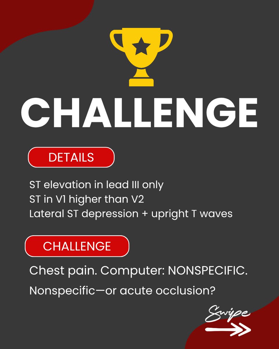
Chest pain. ST elevation in lead III only. ST in V1 higher than V2. Lateral ST depression with upright T waves. 🔎
Computer: nonspecific.
Nonspecific—or acute occlusion?
71M with chest pain. Lead III has ST elevation—but II and aVF don’t. 👀
The computer called it nonspecific. Cardiology: not a STEMI.
But the ED physician recognized a subtle high-risk pattern.
What did he see?
https://t.co/zyMixwlgAw
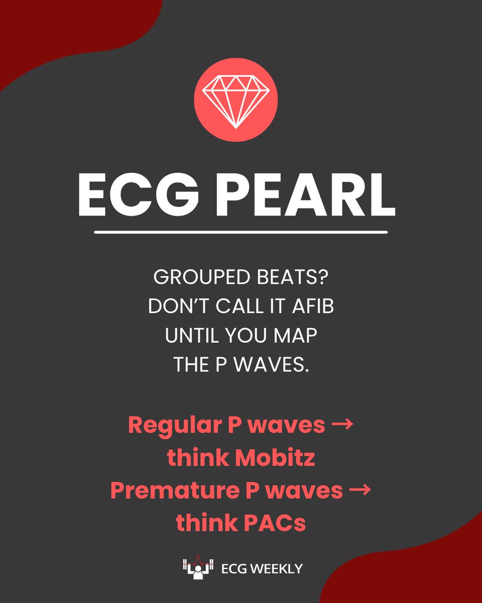
MYTH ❌Before calling AFib, confirm:
No clear P waves marching through
The rhythm is irregularly irregular
Don’t let “irregular” end the interpretation.
A 76-year-old man presents with chest pain suggestive of acute coronary syndrome. An initial 12-lead ECG is obtained and shown below, but does not meet conventional STEMI criteria......https://t.co/v1GpwHsZhC
📚 Last day of #PrimeDay!
Looking to build your ECG library? We've put together our recommended ECG books for every stage of learning—from beginners to advanced clinicians.
Learn why we recommend each one:
https://t.co/Hz9vcly59s
#AmazonAssociate#ECGWeekly#EmergencyMedicine