Today we will learn about...
Category: Hypertension > Endocrine Hypertension Syndromes > Primary Hyperaldosteronism
A 59-year-old man is referred for evaluation of hypertension that had remained above 165/100 mm Hg while he was taking amlodipine 10 mg daily, hydrochlorothiazide 25 mg daily, and metoprolol succinate 100 mg daily. Five years earlier, he required emergency department treatment for lip and tongue swelling after his first lisinopril dose. He was instructed never to take an angiotensin-converting-enzyme inhibitor again. His father had hypertension and ischemic stroke at age 71 years.
He has osteoarthritis treated intermittently with naproxen, rheumatoid arthritis treated with prednisone 5 mg daily, and gastroesophageal reflux disease treated with omeprazole. He does not smoke and drinks one glass of wine nightly. He reports no other drug allergies. Four weeks before repeat endocrine testing, hydrochlorothiazide and metoprolol were replaced with verapamil sustained release and doxazosin; naproxen was stopped.
At this visit, blood pressure is 156/94 mm Hg in both arms, pulse 74/min, respiratory rate 14/min, temperature 37.0 °C, and oxygen saturation 97% on room air. He has no edema, pulmonary crackles, or cardiac murmur. Mild proximal leg weakness is present without synovitis or focal neurologic deficits.
Laboratory studies show sodium 145 mmol/L (normal: 136-145 mmol/L), potassium 4.1 mmol/L (normal: 3.5-5.0 mmol/L), bicarbonate 31 mmol/L (normal: 22-29 mmol/L), creatinine 1.0 mg/dL (normal: 0.7-1.3 mg/dL), and fasting glucose 94 mg/dL (normal: 70-99 mg/dL). Urine microscopy is bland. Plasma aldosterone concentration is 18 ng/dL (laboratory reference: 4-31 ng/dL), plasma renin activity is 0.3 ng/mL/h (laboratory reference: 0.2-1.6 ng/mL/h), and the aldosterone-to-renin ratio is 60. Adrenal CT shows a 1.6-cm left adrenal nodule without a right-sided mass.
The endocrinologist plans confirmatory testing before considering adrenal venous sampling.
Which planned confirmatory test is contraindicated for this patient?
🎬 Watch the video explanation: https://t.co/YdU0AH9v0S
Visit https://t.co/iqlyRShjul for the answer and detailed explanation. Access requires a valid US NPI (licensed healthcare providers only).
#Hypertension #MedicalEducation #PrimaryHyperaldosteronism #EndlessMedical #MultipleChoiceQuestions #EndlessMedicalcom #EndocrineHypertensionSyndromes #EndlessMedicalAcademy #Medical #MCQ
Dig deeper on this topic at https://t.co/ofNUUCifgT, where multiple AI models debate, cross-check references, and verify calculations to give you the most updated and accurate medical information with minimal hallucinations and errors.
Further reading:
1. Primary Aldosteronism: An Endocrine Society Clinical Practice Guideline - (2025) - by Adler GK, Stowasser ...
Confidence: 100%
Educational value: Supports understanding of the medical concepts in the question.
https://t.co/Gl9eRAn9nz
Links to sources are provided for optional further reading only.
Verified, approved and authored using AI by Lukasz Kiljanek MD, board-certified in Internal Medicine, Nephrology and Hypertension
*For medical education. May contain errors. Intended for licensed healthcare professionals & educators. Not medical advice. Full disclaimer: https://t.co/d57C8xdFf2
📿 Medical Pearl
Category: Hypertension > Hypertension in Chronic Kidney Disease (CKD)
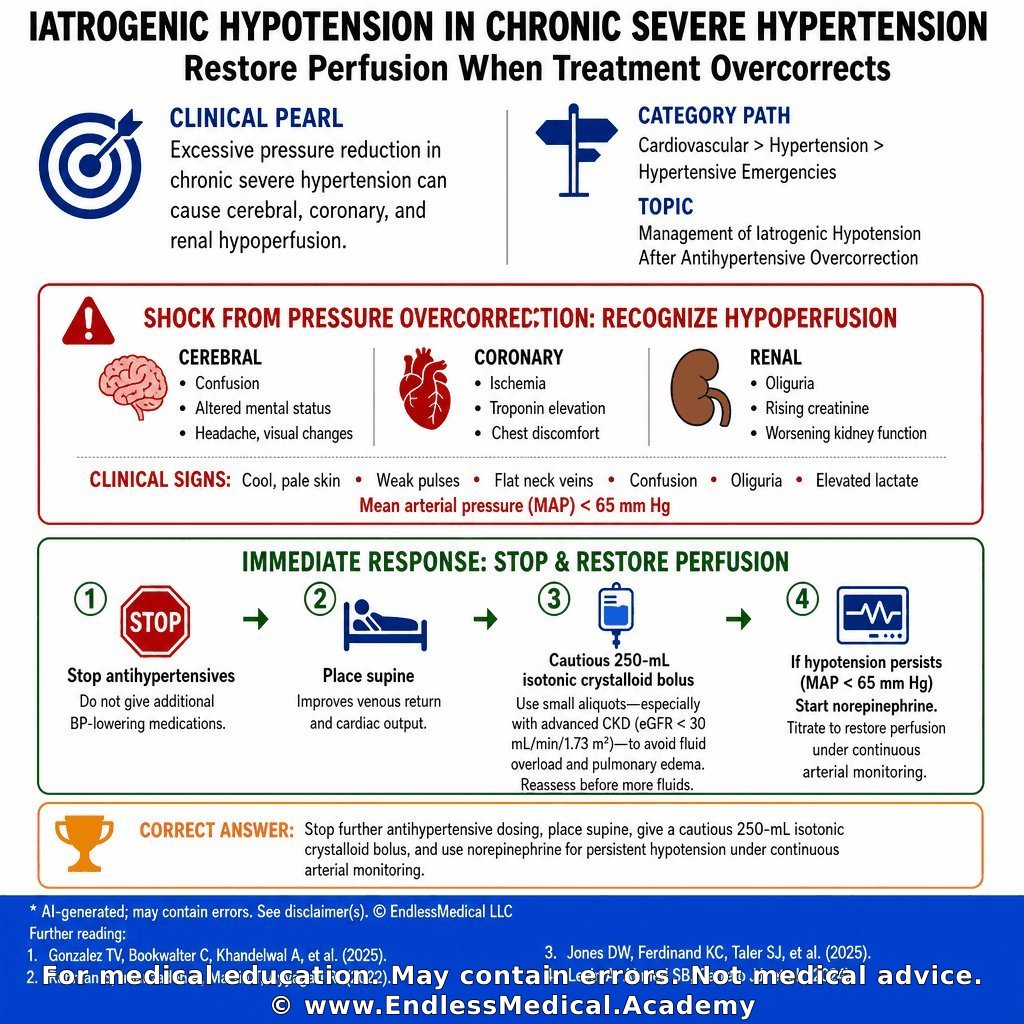
Excessive pressure reduction in chronic severe hypertension can cause cerebral, coronary, and renal hypoperfusion.
Clinically, cool skin, confusion, oliguria, elevated lactate, and a mean arterial pressure below 65 mm Hg indicate shock requiring immediate reversal of the treatment strategy.
At a basic level, stop antihypertensives and restore perfusion.
At an advanced level, use small crystalloid aliquots in advanced chronic kidney disease and initiate norepinephrine when hypotension persists, because large empiric fluid loads may cause pulmonary edema.
🔗 Learn more: https://t.co/fKSiLzcdFS
#MedicalEducation #ClinicalMedicine #ClinicalPearl
@MedTwitter @MedPedsJournal
Dig deeper on this topic at https://t.co/ofNUUCifgT, where multiple AI models debate, cross-check references, and verify calculations to give you the most updated and accurate medical information with minimal hallucinations and errors.
Further reading:
1. Gonzalez TV, Bookwalter C, Khandelwal A, François CJ, Takahashi EA, Rajiah PS. Imaging in Renovascular Hypertension: State of the Art. RadioGraphics. 2025;45(12):e240211. - RadioGraphics - (2025) - by Gonzalez TV, Bookwal...
Confidence: 100%
Educational value: Supports understanding of the medical concepts in the question.
https://t.co/y4mpeoZuwk
2. 2025 AHA/ACC/AANP/AAPA/ABC/ACCP/ACPM/AGS/AMA/ASPC/NMA/PCNA/SGIM Guideline for the Prevention, Detection, Evaluation and Management of High Blood Pressure in Adults - (2025) - by Jones DW, Ferdinand ...
Confidence: 100%
Educational value: Supports understanding of the medical concepts in the question.
https://t.co/hwUnAJ0wIz
3. KDIGO 2024 Clinical Practice Guideline for the Evaluation and Management of Chronic Kidney Disease: Executive Summary - (2024) - by Levin A, Ahmed SB, C...
Confidence: 100%
Educational value: Supports understanding of the medical concepts in the question.
https://t.co/G9vsO1pkXs
Links to sources are provided for optional further reading only.
Verified, approved and authored using AI by Lukasz Kiljanek MD, board-certified in Internal Medicine, Nephrology and Hypertension
*For medical education. May contain errors. Intended for licensed healthcare professionals & educators. Not medical advice. Full disclaimer: https://t.co/d57C8xdFf2
Let's study together! Can you solve this question?
Category: Nephrology: Kidney Disease Diagnosis and Management > Glomerulonephritis: Causes, Diagnosis, and Management > Drug-Induced Glomerulonephritis
A 21-year-old woman is admitted with 3 weeks of fatigue, ankle swelling, tea-colored urine, and progressive exertional dyspnea. During the past 2 days, she has developed blood-streaked sputum. Her military problem list says “giant cell arteritis,” but subsequent rheumatology records document Takayasu arteritis with renal artery involvement and hypertension. She also has protein S deficiency and had an unprovoked deep venous thrombosis at age 17. Current medications are hydralazine, amlodipine, low-dose prednisone, and warfarin. She has taken hydralazine continuously for 18 months. She has no known allergies. She previously used methamphetamine but stopped 2 years ago and denies cocaine use. She follows a vegan diet, lives with her spouse, and completed a brief period of military service. Her father had a deep venous thrombosis, but there is no family history of kidney disease or systemic autoimmune disease.
Prior records show that fluorescein angiography demonstrated delayed retinal arterial filling and peripheral nonperfusion. During an earlier arteritis flare, the erythrocyte sedimentation rate was 74 mm/h (normal: 0–20 mm/h) and C-reactive protein was 6.1 mg/dL (normal: <0.5 mg/dL). Next-generation sequencing identified a pathogenic PROS1 variant. An indium-111 leukocyte scan obtained during a previous fever evaluation showed no focal uptake, and a lipid panel was within its laboratory reference intervals.
Her pulse is 107/min, temperature is 37.6°C, respiratory rate is 20/min, blood pressure is 156/88 mm Hg, and oxygen saturation is **91% on room air**. She has pale conjunctivae, **fine bilateral inspiratory crackles**, **nonblanching palpable purpura over both shins**, and mild bilateral ankle edema. There is no sinus tenderness, nasal ulceration, oral ulceration, or active synovitis. Mild comedonal acne is present on her forehead.
The hemoglobin is **8.9 g/dL (normal: 12.0��16.0 g/dL)**, white blood cell count is 10.6 K/µL (normal: 4.0–11.0 K/µL), and blood urea nitrogen is **44 mg/dL (normal: 7–20 mg/dL)**. Serum creatinine has increased from 0.8 mg/dL (normal: 0.5–1.1 mg/dL) 2 months ago to **3.1 mg/dL (normal: 0.5–1.1 mg/dL)**. The international normalized ratio is 2.4 (normal without anticoagulation: 0.8–1.1; therapeutic goal: 2.0–3.0). Urinalysis shows **3+ blood (normal: negative)** and **2+ protein (normal: negative)**. Microscopy shows **70 erythrocytes/high-power field (normal: 0–2/high-power field), dysmorphic erythrocytes, and erythrocyte casts (normal: absent)**. The urine protein-to-creatinine ratio is 1.8 g/g (normal: <0.2 g/g).
Serologic testing shows a perinuclear ANCA titer greater than 1:640 (normal: <1:20), **MPO-ANCA greater than 8.0 AI (normal: <1.0 AI)**, and **PR3-ANCA 2.3 AI (normal: <1.0 AI)**. The antinuclear antibody titer is 1:640 (normal: <1:80), anti-histone antibody is 4.2 U (normal: <1.0 U), C3 is 62 mg/dL (normal: 90–180 mg/dL), and C4 is 8 mg/dL (normal: 10–40 mg/dL). Anti-double-stranded DNA antibody is 4 IU/mL (normal: <10 IU/mL), and anti-glomerular basement membrane antibody is 0.2 AI (normal: <1.0 AI). A chest radiograph shows bilateral patchy airspace opacities without cardiomegaly. Kidney ultrasonography shows normal-sized kidneys without hydronephrosis. Warfarin is held, and after the international normalized ratio reaches 1.1 (normal: 0.8–1.1), an ultrasound-guided biopsy of the left native kidney shows **necrotizing and crescentic glomerulonephritis**. Immunofluorescence shows only sparse mesangial IgG and C3 staining, without linear IgG or a full-house pattern.
Which diagnosis best explains her acute kidney and pulmonary findings?
🎬 Watch the video explanation: https://t.co/8jeLYn6k2j
Visit https://t.co/IEmahwcKpW for the answer and detailed explanation. Access requires a valid US NPI (licensed healthcare providers only).
Dig deeper on this topic at https://t.co/ofNUUCifgT, where multiple AI models debate, cross-check references, and verify calculations to give you the most updated and accurate medical information with minimal hallucinations and errors.
Further reading:
1. KDOQI US Commentary on the KDIGO 2024 Clinical Practice Guideline for the Management of Antineutrophil Cytoplasmic Antibody-Associated Vasculitis - (2026) - by Salem J. Almaani, Is...
Confidence: 100%
Educational value: Supports understanding of the medical concepts in the question.
https://t.co/CHgSFfsYgI
2. Anti-neutrophil cytoplasmic antibody associated glomerulonephritis complicating treatment with hydralazine - Kidney International - (2021) - by Dominick Santoriello...
Confidence: 100%
Educational value: Supports understanding of the medical concepts in the question.
https://t.co/Ynmv9Wj0ZL
Links to sources are provided for optional further reading only.
Verified, approved and authored using AI by Lukasz Kiljanek MD, board-certified in Internal Medicine, Nephrology and Hypertension
*For medical education. May contain errors. Intended for licensed healthcare professionals & educators. Not medical advice. Full disclaimer: https://t.co/d57C8xdFf2
This question is for you!
Category: Hypertension > Management of Resistant Hypertension
A 31-year-old woman is evaluated at a rural outpatient clinic for blood pressure that remains above goal despite confirmed adherence to three antihypertensive agents. Pharmacy refill records and pill counts at two prior visits verify daily use of lisinopril 20 mg, amlodipine 10 mg, and chlorthalidone 25 mg. She also takes naproxen 440 mg as needed for osteoarthritis several times per week and methimazole 5 mg daily for recurrent Graves disease. She does not smoke or drink alcohol.
Over the last 4 months she has developed muscle cramps, intermittent headaches, and increased nocturia. Temperature is 36.3 °C, pulse 85/min, respirations 17/min, blood pressure 148/80 mm Hg in both arms while seated, oxygen saturation 97% on room air, and BMI 28 kg/m². Cardiopulmonary examination is normal. No abdominal bruits are heard. There is 1+ bilateral pretibial edema. The thyroid is normal in size without tenderness, and the skin shows no striae or easy bruising.
Laboratory studies show sodium 146 mmol/L, potassium 2.9 mmol/L, chloride 103 mmol/L, bicarbonate 30 mmol/L, blood urea nitrogen 20 mg/dL, creatinine 1.4 mg/dL, glucose 92 mg/dL, and 24-hour urinary potassium excretion 72 mmol. Urinalysis shows specific gravity 1.014 and no protein. Morning cortisol after an overnight 1-mg dexamethasone suppression test is 0.6 μg/dL, and thyroid-stimulating hormone is 1.8 μIU/mL. Abdominal ultrasonography performed locally last month showed normal-sized kidneys without hydronephrosis and no adrenal mass. Computed tomographic angiography was avoided because she develops flushing after iodinated contrast. A 12-lead electrocardiogram and chest radiograph are unremarkable.
Advanced imaging and subspecialty services are not easily accessible in this rural setting. Which single diagnostic test is most appropriate now to identify the specific secondary cause of her resistant hypertension?
🎬 Watch the video explanation: https://t.co/kyCfqysmOU
Visit https://t.co/S0zujDuNBX for the answer and detailed explanation. Access requires a valid US NPI (licensed healthcare providers only).
#MedicalEducation #MCQ #EndlessMedical #ManagementofResistantHypertension #MultipleChoiceQuestions #EndlessMedicalcom #EndlessMedicalAcademy #Medical #Hypertension
Dig deeper on this topic at https://t.co/ofNUUCifgT, where multiple AI models debate, cross-check references, and verify calculations to give you the most updated and accurate medical information with minimal hallucinations and errors.
Further reading:
1. Resistant Hypertension: Detection, Evaluation, and Management: A Scientific Statement From the American Heart Association - (2018) - by Carey RM, Calhoun DA...
Confidence: 100%
Educational value: Supports understanding of the medical concepts in the question.
https://t.co/c6yWCQSyIa
2. Primary Aldosteronism: An Endocrine Society Clinical Practice Guideline - (2025) - by Adler GK, Stowasser ...
Confidence: 100%
Educational value: Supports understanding of the medical concepts in the question.
https://t.co/jCrGo9q6DI
Links to sources are provided for optional further reading only.
Verified, approved and authored using AI by Lukasz Kiljanek MD, board-certified in Internal Medicine, Nephrology and Hypertension
*For medical education. May contain errors. Intended for licensed healthcare professionals & educators. Not medical advice. Full disclaimer: https://t.co/d57C8xdFf2
Let's study together! Can you solve this question?
Category: Hypertension > Management of Resistant Hypertension
A 20-year-old woman is seen in the nephrology clinic for routine follow-up of long-standing resistant hypertension. Three years ago, angiography demonstrated multifocal fibromuscular dysplasia of the right renal artery that was treated with percutaneous angioplasty, but home blood-pressure logs have remained in the 150s/90s.
Her current antihypertensive regimen at maximally tolerated doses is lisinopril 40 mg once daily, amlodipine 10 mg once daily, and chlorthalidone 25 mg once daily. She also takes allopurinol 300 mg once daily for gout, colchicine 0.6 mg as needed for flares, sertraline 100 mg once daily for depression, and vitamin D 1000 IU daily. She denies NSAID use, licorice, and over-the-counter supplements.
At today’s visit she feels well. After 5 minutes of seated rest, temperature is 36.8 °C, pulse 88/min, respirations 14/min, blood pressure 152/96 mm Hg, oxygen saturation 98% on room air, and BMI 31 kg/m². Cardiopulmonary and neurologic examinations are unremarkable. There is 1+ bilateral ankle edema.
Laboratory studies obtained this morning show sodium 139 mmol/L, potassium 5.3 mmol/L, creatinine 1.9 mg/dL, blood urea nitrogen 30 mg/dL, uric acid 9.8 mg/dL, calcium 9.1 mg/dL, hemoglobin 13.4 g/dL, platelets 248 × 10⁹/L, leukocytes 6.7 × 10⁹/L, urine albumin-to-creatinine ratio 310 mg/g, plasma aldosterone 4 ng/dL, and plasma renin activity 1.4 ng/mL/h measured while she remains on lisinopril.
The nephrologist plans to add a fourth antihypertensive agent. Which of the following medications should be avoided because of a specific contraindication or major precaution in this patient?
🎬 Watch the video explanation: https://t.co/JumfqU8cx1
Visit https://t.co/WwAmzbUXlX for the answer and detailed explanation. Access requires a valid US NPI (licensed healthcare providers only).
#EndlessMedical #MultipleChoiceQuestions #Hypertension #ManagementofResistantHypertension #Medical #MCQ #MedicalEducation #EndlessMedicalAcademy #EndlessMedicalcom
Dig deeper on this topic at https://t.co/ofNUUCifgT, where multiple AI models debate, cross-check references, and verify calculations to give you the most updated and accurate medical information with minimal hallucinations and errors.
Further reading:
1. Resistant Hypertension: Detection, Evaluation, and Management: A Scientific Statement From the American Heart Association - (2018) - by Carey RM, Calhoun DA...
Confidence: 100%
Educational value: Supports understanding of the medical concepts in the question.
https://t.co/c6yWCQSyIa
Links to sources are provided for optional further reading only.
Verified, approved and authored using AI by Lukasz Kiljanek MD, board-certified in Internal Medicine, Nephrology and Hypertension
*For medical education. May contain errors. Intended for licensed healthcare professionals & educators. Not medical advice. Full disclaimer: https://t.co/d57C8xdFf2
📿 Medical Pearl
Category: Nephrology: Kidney Disease Diagnosis and Management > Electrolyte Disorders > Hyponatremia
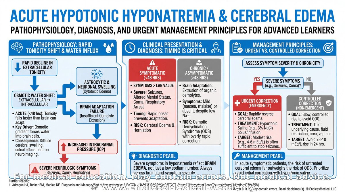
Acute hypotonic hyponatremia can cause rapid cerebral edema because the brain has not yet adapted to the fall in extracellular tonicity.
The most dangerous early manifestations are headache, nausea, vomiting, confusion, seizure, and respiratory compromise.
Low serum osmolality means water shifts into brain cells; high serum osmolality means water shifts out of them.
Osmotic demyelination syndrome is a different problem that usually follows overly rapid correction of chronic hyponatremia rather than the initial hypo-osmolar insult.
🎬 Watch the video explanation: https://t.co/A9HKDbziOW
🔗 Learn more: https://t.co/L6BcAnQrgu
#MedicalEducation #ClinicalPearl #NursesOfTwitter
@MedStudentChat @MedTwitter
Dig deeper on this topic at https://t.co/ofNUUCifgT, where multiple AI models debate, cross-check references, and verify calculations to give you the most updated and accurate medical information with minimal hallucinations and errors.
Further reading:
1. Adrogué HJ, Tucker BM, Madias NE. Diagnosis and Management of Hyponatremia: A Review. JAMA. 2022;328(3):280-291. - JAMA - (2022) - by Adrogué, H. J., Tuck...
Confidence: 100%
Educational value: Supports understanding of the medical concepts in the question.
https://t.co/5OQKONmYiD
Links to sources are provided for optional further reading only.
Verified, approved and authored using AI by Lukasz Kiljanek MD, board-certified in Internal Medicine, Nephrology and Hypertension
*For medical education. May contain errors. Intended for licensed healthcare professionals & educators. Not medical advice. Full disclaimer: https://t.co/d57C8xdFf2
Are you as smart as you think you are? Solve it!
Category: Nephrology: Kidney Disease Diagnosis and Management > Glomerulonephritis: Causes, Diagnosis, and Management > Drug-Induced Glomerulonephritis
A 57-year-old woman presents to a critical access hospital in rural Wyoming with 10 days of fatigue, migratory arthralgias, dark urine, and decreasing urine output. Four days ago, she noticed nonblanching spots over both lower legs. She has no fever, cough, hemoptysis, sinus symptoms, abdominal pain, hematochezia, or recent diarrheal illness. Her Crohn disease has been clinically inactive for more than 1 year. Dense wildfire smoke has grounded rotor-wing transport, and closure of the only mountain highway is expected to delay ground transfer for at least 12 hours. The hospital has basic laboratory, microscopy, radiography, and telemedicine services but no kidney-biopsy service or on-site nephrologist.
Her history includes colonic Crohn disease, Graves disease, rosacea, contact dermatitis, and pyridoxine-responsive sideroblastic anemia. Methimazole previously caused cholestatic liver injury, and she declined radioactive iodine and thyroid surgery. She has taken propylthiouracil continuously for 5 years. Current medications are propylthiouracil, mesalamine, pyridoxine, topical metronidazole, topical triamcinolone as needed, and ibuprofen taken twice daily for the past 6 days for arthralgias. She has no known allergies. She smoked 1.5 packs/day for 20 years, quit 5 years ago, and resumed smoking 0.5 pack/day 6 months ago. Her mother had Graves disease, but there is no family history of glomerular disease or systemic vasculitis.
During recent preventive care, a screening mammogram was negative, hemoglobin A1c was 5.4% (normal: 4.0–5.6%), and a DXA scan documented low bone mass without osteoporosis. At an urgent-care visit 3 days ago, a urine culture showed no growth. The prothrombin time was 12.4 seconds (normal: 11.0–13.5 seconds), international normalized ratio was 1.0 (normal: 0.8–1.1), and activated partial thromboplastin time was 30 seconds (normal: 25–35 seconds).
Her pulse is 105/min, temperature is 36.8°C, respiratory rate is 16/min, blood pressure is 131/72 mm Hg, and oxygen saturation is 98% on room air. She has **palpable purpura over both lower legs**, trace ankle edema, and mild tenderness of the wrists without synovitis. Her lungs are clear. Stable centrofacial erythema from rosacea and a small lichenified patch at the belt line are also present.
Serum creatinine has increased from 0.9 mg/dL (normal: 0.5–1.1 mg/dL) 4 months ago to **2.6 mg/dL (normal: 0.5–1.1 mg/dL)**. Blood urea nitrogen is **38 mg/dL (normal: 7–20 mg/dL)**. Hemoglobin is 11.4 g/dL (normal: 12.0–16.0 g/dL), white blood cell count is 8.2 K/µL (normal: 4.0–11.0 K/µL), and eosinophils are 1.8% (normal: 0–5%). Potassium is 4.7 mEq/L (normal: 3.5–5.0 mEq/L). Urinalysis shows **3+ blood (normal: negative)** and **2+ protein (normal: negative)**. Urine microscopy shows **more than 50 erythrocytes/high-power field (normal: 0–2/high-power field), 2 leukocytes/high-power field (normal: 0–5/high-power field), dysmorphic erythrocytes, and erythrocyte casts (normal: absent)**. The urine protein-to-creatinine ratio is 1.3 g/g (normal: <0.2 g/g).
A serology specimen collected at the urgent-care visit has just resulted from the regional laboratory. The perinuclear ANCA titer is greater than 1:640 (normal: <1:20), **MPO-ANCA is greater than 8.0 AI (normal: <1.0 AI)**, and **PR3-ANCA is 1.8 AI (normal: <1.0 AI)**. The antinuclear antibody titer is 1:160 (normal: <1:80), C3 is 112 mg/dL (normal: 90–180 mg/dL), and C4 is 24 mg/dL (normal: 10–40 mg/dL). Antigen-specific repeat testing, anti-glomerular basement membrane testing, and kidney biopsy cannot be completed before transfer.
Which diagnosis is most likely?
🎬 Watch the video explanation: https://t.co/3CjXLHGeVz
Visit https://t.co/CC8uo9pgW6 for the answer and detailed explanation. Access requires a valid US NPI (licensed healthcare providers only).
#MedicalEducation #Medical #EndlessMedicalAcademy #GlomerulonephritisCausesDiagnosisandManagement #EndlessMedicalcom #EndlessMedical #MCQ #DrugInducedGlomerulonephritis #NephrologyKidneyDiseaseDiagnosisandManagement #MultipleChoiceQuestions
Dig deeper on this topic at https://t.co/ofNUUCifgT, where multiple AI models debate, cross-check references, and verify calculations to give you the most updated and accurate medical information with minimal hallucinations and errors.
Further reading:
1. KDOQI US Commentary on the KDIGO 2024 Clinical Practice Guideline for the Management of Antineutrophil Cytoplasmic Antibody (ANCA)-Associated Vasculitis - (2026) - by Salem J. Almaani, Is...
Confidence: 100%
Educational value: Supports understanding of the medical concepts in the question.
https://t.co/CHgSFfsYgI
2. Long-term outcomes and clinicopathological characteristics of propylthiouracil-induced ANCA-associated glomerulonephritis: a case-control study - Clinical and Experimental Rheumatology - (2026) - by Di Wu, Ruifeng Zhang...
Confidence: 100%
Educational value: Supports understanding of the medical concepts in the question.
https://t.co/8vwTx9jVfj
Links to sources are provided for optional further reading only.
Verified, approved and authored using AI by Lukasz Kiljanek MD, board-certified in Internal Medicine, Nephrology and Hypertension
*For medical education. May contain errors. Intended for licensed healthcare professionals & educators. Not medical advice. Full disclaimer: https://t.co/d57C8xdFf2
So you think you're smart? Prove it!
Category: Nephrology: Kidney Disease Diagnosis and Management > Glomerulonephritis: Causes, Diagnosis, and Management > Drug-Induced Glomerulonephritis
A 21-year-old woman is admitted with 3 weeks of fatigue, ankle swelling, tea-colored urine, and progressive exertional dyspnea. During the past 2 days, she has developed blood-streaked sputum. Her military problem list says “giant cell arteritis,” but subsequent rheumatology records document Takayasu arteritis with renal artery involvement and hypertension. She also has protein S deficiency and had an unprovoked deep venous thrombosis at age 17. Current medications are hydralazine, amlodipine, low-dose prednisone, and warfarin. She has taken hydralazine continuously for 18 months. She has no known allergies. She previously used methamphetamine but stopped 2 years ago and denies cocaine use. She follows a vegan diet, lives with her spouse, and completed a brief period of military service. Her father had a deep venous thrombosis, but there is no family history of kidney disease or systemic autoimmune disease.
Prior records show that fluorescein angiography demonstrated delayed retinal arterial filling and peripheral nonperfusion. During an earlier arteritis flare, the erythrocyte sedimentation rate was 74 mm/h (normal: 0–20 mm/h) and C-reactive protein was 6.1 mg/dL (normal: <0.5 mg/dL). Next-generation sequencing identified a pathogenic PROS1 variant. An indium-111 leukocyte scan obtained during a previous fever evaluation showed no focal uptake, and a lipid panel was within its laboratory reference intervals.
Her pulse is 107/min, temperature is 37.6°C, respiratory rate is 20/min, blood pressure is 156/88 mm Hg, and oxygen saturation is **91% on room air**. She has pale conjunctivae, **fine bilateral inspiratory crackles**, **nonblanching palpable purpura over both shins**, and mild bilateral ankle edema. There is no sinus tenderness, nasal ulceration, oral ulceration, or active synovitis. Mild comedonal acne is present on her forehead.
The hemoglobin is **8.9 g/dL (normal: 12.0–16.0 g/dL)**, white blood cell count is 10.6 K/µL (normal: 4.0–11.0 K/µL), and blood urea nitrogen is **44 mg/dL (normal: 7–20 mg/dL)**. Serum creatinine has increased from 0.8 mg/dL (normal: 0.5–1.1 mg/dL) 2 months ago to **3.1 mg/dL (normal: 0.5–1.1 mg/dL)**. The international normalized ratio is 2.4 (normal without anticoagulation: 0.8–1.1; therapeutic goal: 2.0–3.0). Urinalysis shows **3+ blood (normal: negative)** and **2+ protein (normal: negative)**. Microscopy shows **70 erythrocytes/high-power field (normal: 0–2/high-power field), dysmorphic erythrocytes, and erythrocyte casts (normal: absent)**. The urine protein-to-creatinine ratio is 1.8 g/g (normal: <0.2 g/g).
Serologic testing shows a perinuclear ANCA titer greater than 1:640 (normal: <1:20), **MPO-ANCA greater than 8.0 AI (normal: <1.0 AI)**, and **PR3-ANCA 2.3 AI (normal: <1.0 AI)**. The antinuclear antibody titer is 1:640 (normal: <1:80), anti-histone antibody is 4.2 U (normal: <1.0 U), C3 is 62 mg/dL (normal: 90–180 mg/dL), and C4 is 8 mg/dL (normal: 10–40 mg/dL). Anti-double-stranded DNA antibody is 4 IU/mL (normal: <10 IU/mL), and anti-glomerular basement membrane antibody is 0.2 AI (normal: <1.0 AI). A chest radiograph shows bilateral patchy airspace opacities without cardiomegaly. Kidney ultrasonography shows normal-sized kidneys without hydronephrosis. Warfarin is held, and after the international normalized ratio reaches 1.1 (normal: 0.8–1.1), an ultrasound-guided biopsy of the left native kidney shows **necrotizing and crescentic glomerulonephritis**. Immunofluorescence shows only sparse mesangial IgG and C3 staining, without linear IgG or a full-house pattern.
Which diagnosis best explains her acute kidney and pulmonary findings?
🎬 Watch the video explanation: https://t.co/8jeLYn6k2j
Visit https://t.co/IEmahwcKpW for the answer and detailed explanation. Access requires a valid US NPI (licensed healthcare providers only).
#NephrologyKidneyDiseaseDiagnosisandManagement #GlomerulonephritisCausesDiagnosisandManagement #MCQ #DrugInducedGlomerulonephritis #MultipleChoiceQuestions #MedicalEducation #EndlessMedical #EndlessMedicalcom #EndlessMedicalAcademy #Medical
Dig deeper on this topic at https://t.co/ofNUUCifgT, where multiple AI models debate, cross-check references, and verify calculations to give you the most updated and accurate medical information with minimal hallucinations and errors.
Further reading:
1. KDOQI US Commentary on the KDIGO 2024 Clinical Practice Guideline for the Management of Antineutrophil Cytoplasmic Antibody-Associated Vasculitis - (2026) - by Salem J. Almaani, Is...
Confidence: 100%
Educational value: Supports understanding of the medical concepts in the question.
https://t.co/CHgSFfsYgI
2. Anti-neutrophil cytoplasmic antibody associated glomerulonephritis complicating treatment with hydralazine - Kidney International - (2021) - by Dominick Santoriello...
Confidence: 100%
Educational value: Supports understanding of the medical concepts in the question.
https://t.co/Ynmv9Wj0ZL
Links to sources are provided for optional further reading only.
Verified, approved and authored using AI by Lukasz Kiljanek MD, board-certified in Internal Medicine, Nephrology and Hypertension
*For medical education. May contain errors. Intended for licensed healthcare professionals & educators. Not medical advice. Full disclaimer: https://t.co/d57C8xdFf2
Let's study together! Can you solve this question?
Category: Hypertension > Management of Resistant Hypertension
A 51-year-old man is evaluated at a routine nephrology visit for persistently elevated blood pressure. Medical history includes Crohn disease status post ileocolic resection, asthma, obstructive sleep apnea treated with nocturnal continuous positive airway pressure, coronary artery disease treated with a drug-eluting stent 2 years ago, and chronic musculoskeletal pain. Medications are lisinopril 40 mg daily, amlodipine 10 mg daily, chlorthalidone 25 mg every morning, aspirin 81 mg daily, budesonide-formoterol inhaler twice daily, and acetaminophen as needed. He reports daily marijuana use and at least five alcoholic drinks each evening. He does not use NSAIDs or sympathomimetics.
Seated vital signs are blood pressure 176/98 mm Hg in both arms, pulse 84/min, respiratory rate 14/min, temperature 36.3 °C, oxygen saturation 98% on room air, and BMI 32 kg/m². Cardiovascular, pulmonary, abdominal, thyroid, and peripheral vascular examinations are otherwise unremarkable. There are no abdominal bruits and no peripheral edema.
Recent laboratory studies show sodium 140 mmol/L, potassium 4.1 mmol/L, creatinine 1.0 mg/dL, calcium 9.3 mg/dL, and fasting glucose 97 mg/dL. Morning plasma aldosterone is 6 ng/dL and plasma renin activity is 0.6 ng/mL/h, giving an aldosterone-to-renin ratio below the screening threshold for primary aldosteronism. A 24-hour urine collection shows sodium excretion 180 mmol and metanephrines within the reference range. Ambulatory blood-pressure monitoring recorded a 24-hour average of 168/90 mm Hg. Pharmacy refill data, pill counts, and serum drug levels confirm adherence.
Which of the following statements best characterizes the epidemiologic phenotype represented by this patient after objective exclusion of pseudoresistance?
🎬 Watch the video explanation: https://t.co/G9HnI4StUQ
Visit https://t.co/e43soRIiP2 for the answer and detailed explanation. Access requires a valid US NPI (licensed healthcare providers only).
#Hypertension #MedicalEducation #ManagementofResistantHypertension #MultipleChoiceQuestions #Medical #MCQ #EndlessMedical #EndlessMedicalAcademy #EndlessMedicalcom
Dig deeper on this topic at https://t.co/ofNUUCifgT, where multiple AI models debate, cross-check references, and verify calculations to give you the most updated and accurate medical information with minimal hallucinations and errors.
Further reading:
1. Resistant Hypertension: Detection, Evaluation, and Management: A Scientific Statement From the American Heart Association - (2018) - by Carey RM, Calhoun DA...
Confidence: 100%
Educational value: Supports understanding of the medical concepts in the question.
https://t.co/c6yWCQSyIa
2. Azizi M, Vongpatanasin W, Fisher NDL, et al. Diagnosis and Management of Resistant Hypertension: A Review. - JAMA - (2026) - by Azizi M, Vongpatanas...
Confidence: 100%
Educational value: Supports understanding of the medical concepts in the question.
https://t.co/Xa4gY79wYz
Links to sources are provided for optional further reading only.
Verified, approved and authored using AI by Lukasz Kiljanek MD, board-certified in Internal Medicine, Nephrology and Hypertension
*For medical education. May contain errors. Intended for licensed healthcare professionals & educators. Not medical advice. Full disclaimer: https://t.co/d57C8xdFf2
📿 Medical Pearl
Category: Nephrology: Kidney Disease Diagnosis and Management > Electrolyte Disorders > Hyponatremia
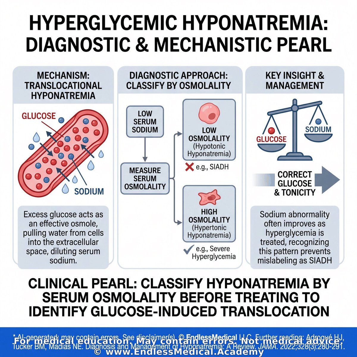
A low sodium with a high serum osmolality usually means another effective osmole is pulling water into the extracellular space.
Recognizing that pattern prevents mislabeling hyperglycemic emergencies as SIADH.
The basic takeaway is to classify hyponatremia by serum osmolality before treating it.
An advanced insight is that hyperglycemia produces translocational hyponatremia, so the sodium abnormality often improves as the glucose and tonicity are corrected.
🎬 Watch the video explanation: https://t.co/GmGUnzYrmO
🔗 Learn more: https://t.co/bLZtWDSDGg
#DoctorsOfTwitter #MedicalEducation #MedStudent
@MedTwitter @MedEdChat
Dig deeper on this topic at https://t.co/ofNUUCifgT, where multiple AI models debate, cross-check references, and verify calculations to give you the most updated and accurate medical information with minimal hallucinations and errors.
Verified, approved and authored using AI by Lukasz Kiljanek MD, board-certified in Internal Medicine, Nephrology and Hypertension
*For medical education. May contain errors. Intended for licensed healthcare professionals & educators. Not medical advice. Full disclaimer: https://t.co/d57C8xdFf2
I challenge you today to solve this question!
Category: Hypertension > Renovascular Hypertension and Kidney Artery Disorders > Renal Artery Stenosis
A 33-year-old woman presents to a U.S. emergency department with 1 day of throbbing right-sided headache, palpitations, malaise, and home blood pressure readings that have remained elevated for 6 months despite adherence. She reports an audible abdominal whooshing when supine and intermittent orthostatic lightheadedness without syncope. She denies chest pain and exertional dyspnea. Her history includes systemic lupus erythematosus diagnosed at age 22 without current nephritis, anterior uveitis in remission, and bilateral carpal tunnel syndrome. Her mother had carotid artery dissection in her 40s. She follows a vegan diet, is a former smoker who quit 5 years ago, and uses no alcohol or illicit drugs. She has regular menses and no known pregnancy.
She has no medication allergies and reports fragrance-triggered headaches. Home medications taken this morning include amlodipine 10 mg PO daily, carvedilol 12.5 mg PO twice daily, hydrochlorothiazide 25 mg PO daily, lisinopril 10 mg PO daily started 3 weeks ago, ibuprofen 400 mg PO up to three times daily as needed for headaches used twice this week, prednisone 5 mg PO daily, hydroxychloroquine 200 mg PO twice daily, vitamin B12 1000 mcg PO daily, depot medroxyprogesterone acetate 150 mg IM every 3 months with the last injection 7 weeks ago, and cetirizine 10 mg PO daily as needed for seasonal symptoms.
Triage vital signs are tympanic temperature 38.4 °C, repeat oral temperature 37.8 °C, manual radial pulse 138/min and regular, respiratory rate 14/min, blood pressure 107/53 mm Hg, and oxygen saturation 91% on room air that improves to 97% with 2 L/min by nasal cannula. She is alert, oriented, communicates choices coherently, and repeats risks and benefits. Funduscopic examination shows mild arteriolar narrowing with arteriovenous nicking but no cotton-wool spots or papilledema. Cardiopulmonary examination shows no murmur or rales. The abdomen is soft with a prominent systolic bruit maximal in the right periumbilical and flank region. Peripheral pulses are symmetric without bruits, and there is no lower-extremity edema. Tinel sign is present at both wrists; slit-lamp examination shows no active anterior chamber cells.
Initial ED results show regular narrow-complex tachycardia at 138/min on ECG and no infiltrate or effusion on chest radiograph. Sodium is 138 mEq/L (reference: 135–145 mEq/L), potassium 5.3 mEq/L (reference: 3.5–5.0 mEq/L), bicarbonate 23 mEq/L (reference: 22–29 mEq/L), glucose 96 mg/dL (reference: 70–140 mg/dL), and creatinine 1.4 mg/dL (reference: 0.5–1.1 mg/dL), increased from 1.2 mg/dL one week earlier (reference: 0.5–1.1 mg/dL). Estimated GFR is approximately 51 mL/min/1.73 m² today (reference: at least 90 mL/min/1.73 m²) versus 61 mL/min/1.73 m² previously (reference: at least 90 mL/min/1.73 m²). White blood cell count is 9.2 × 10^3/µL (reference: 4.0–11.0 × 10^3/µL) with neutrophils 72% (reference: 40%–70%), hemoglobin is 12.8 g/dL (reference: 12.0–16.0 g/dL), platelets are 274 × 10^3/µL (reference: 150–450 × 10^3/µL), and lactate is 1.2 mmol/L (reference: 0.5��2.2 mmol/L). Urinalysis shows specific gravity 1.018 (reference: 1.005–1.030), protein 1+ (reference: negative), 0–2 RBC/hpf (reference: 0–2 RBC/hpf), and negative nitrites and leukocyte esterase (reference: negative). Two peripheral blood-culture sets are pending. Bedside renal ultrasound shows a right kidney length of 8.8 cm and a left kidney length of 10.6 cm, preserved cortical echogenicity, and no hydronephrosis.
She declines contrast-enhanced imaging, and formal vascular-laboratory duplex ultrasound shows right proximal renal artery peak systolic velocity 395 cm/s, aortic peak systolic velocity 92 cm/s, renal-aortic ratio 4.29, delayed intrarenal upstroke with acceleration time 0.12 s, and a tardus-parvus waveform. Left renal artery velocities and waveforms are within reference ranges. Outpatient testing last week showed plasma renin activity 8.2 ng/mL/h (laboratory reference: 0.2–3.3 ng/mL/h), plasma aldosterone 24 ng/dL (laboratory reference: 4–31 ng/dL), aldosterone-renin ratio 2.9 (screening reference: less than 20), morning cortisol 16 µg/dL (reference: 5–25 µg/dL), TSH 2.0 mIU/L (reference: 0.4–4.0 mIU/L), and 24-hour urinary fractionated metanephrines within laboratory reference ranges.
After oral acetaminophen and 1 L isotonic IV crystalloid, her oral temperature is 37.8 °C, pulse is 108/min, blood pressure is 118/66 mm Hg, and oxygen saturation is 97% on room air. Cognition remains intact. The senior resident proposes immediate CT angiography of the renal arteries using low-osmolality iodinated contrast and, if a tight stenosis is confirmed, same-day revascularization. The patient explicitly states, “I do not want any contrast study until I speak with my partner. I’m okay with ultrasound today.” A stat serum hCG has been sent and is pending.
Which single course of action is most appropriate from a legal and ethical standpoint right now, given this clinical context?
🎬 Watch the video explanation: https://t.co/tfOnQCWUmj
Visit https://t.co/6LICT2vdrm for the answer and detailed explanation. Access requires a valid US NPI (licensed healthcare providers only).
Dig deeper on this topic at https://t.co/ofNUUCifgT, where multiple AI models debate, cross-check references, and verify calculations to give you the most updated and accurate medical information with minimal hallucinations and errors.
Further reading:
1. ACR Manual on Contrast Media – Administration of Contrast Media to Pregnant or Potentially Pregnant Patients - (2025) - by American College of ...
Confidence: 100%
Educational value: Supports understanding of the medical concepts in the question.
https://t.co/Vir6dBgloN
Links to sources are provided for optional further reading only.
Verified, approved and authored using AI by Lukasz Kiljanek MD, board-certified in Internal Medicine, Nephrology and Hypertension
*For medical education. May contain errors. Intended for licensed healthcare professionals & educators. Not medical advice. Full disclaimer: https://t.co/d57C8xdFf2
Get better every day and solve this!
Category: Nephrology: Kidney Disease Diagnosis and Management > Glomerulonephritis: Causes, Diagnosis, and Management > Drug-Induced Glomerulonephritis
A 57-year-old woman presents to a critical access hospital in rural Wyoming with 10 days of fatigue, migratory arthralgias, dark urine, and decreasing urine output. Four days ago, she noticed nonblanching spots over both lower legs. She has no fever, cough, hemoptysis, sinus symptoms, abdominal pain, hematochezia, or recent diarrheal illness. Her Crohn disease has been clinically inactive for more than 1 year. Dense wildfire smoke has grounded rotor-wing transport, and closure of the only mountain highway is expected to delay ground transfer for at least 12 hours. The hospital has basic laboratory, microscopy, radiography, and telemedicine services but no kidney-biopsy service or on-site nephrologist.
Her history includes colonic Crohn disease, Graves disease, rosacea, contact dermatitis, and pyridoxine-responsive sideroblastic anemia. Methimazole previously caused cholestatic liver injury, and she declined radioactive iodine and thyroid surgery. She has taken propylthiouracil continuously for 5 years. Current medications are propylthiouracil, mesalamine, pyridoxine, topical metronidazole, topical triamcinolone as needed, and ibuprofen taken twice daily for the past 6 days for arthralgias. She has no known allergies. She smoked 1.5 packs/day for 20 years, quit 5 years ago, and resumed smoking 0.5 pack/day 6 months ago. Her mother had Graves disease, but there is no family history of glomerular disease or systemic vasculitis.
During recent preventive care, a screening mammogram was negative, hemoglobin A1c was 5.4% (normal: 4.0–5.6%), and a DXA scan documented low bone mass without osteoporosis. At an urgent-care visit 3 days ago, a urine culture showed no growth. The prothrombin time was 12.4 seconds (normal: 11.0–13.5 seconds), international normalized ratio was 1.0 (normal: 0.8–1.1), and activated partial thromboplastin time was 30 seconds (normal: 25–35 seconds).
Her pulse is 105/min, temperature is 36.8°C, respiratory rate is 16/min, blood pressure is 131/72 mm Hg, and oxygen saturation is 98% on room air. She has **palpable purpura over both lower legs**, trace ankle edema, and mild tenderness of the wrists without synovitis. Her lungs are clear. Stable centrofacial erythema from rosacea and a small lichenified patch at the belt line are also present.
Serum creatinine has increased from 0.9 mg/dL (normal: 0.5–1.1 mg/dL) 4 months ago to **2.6 mg/dL (normal: 0.5–1.1 mg/dL)**. Blood urea nitrogen is **38 mg/dL (normal: 7–20 mg/dL)**. Hemoglobin is 11.4 g/dL (normal: 12.0–16.0 g/dL), white blood cell count is 8.2 K/µL (normal: 4.0–11.0 K/µL), and eosinophils are 1.8% (normal: 0–5%). Potassium is 4.7 mEq/L (normal: 3.5–5.0 mEq/L). Urinalysis shows **3+ blood (normal: negative)** and **2+ protein (normal: negative)**. Urine microscopy shows **more than 50 erythrocytes/high-power field (normal: 0–2/high-power field), 2 leukocytes/high-power field (normal: 0–5/high-power field), dysmorphic erythrocytes, and erythrocyte casts (normal: absent)**. The urine protein-to-creatinine ratio is 1.3 g/g (normal: <0.2 g/g).
A serology specimen collected at the urgent-care visit has just resulted from the regional laboratory. The perinuclear ANCA titer is greater than 1:640 (normal: <1:20), **MPO-ANCA is greater than 8.0 AI (normal: <1.0 AI)**, and **PR3-ANCA is 1.8 AI (normal: <1.0 AI)**. The antinuclear antibody titer is 1:160 (normal: <1:80), C3 is 112 mg/dL (normal: 90–180 mg/dL), and C4 is 24 mg/dL (normal: 10–40 mg/dL). Antigen-specific repeat testing, anti-glomerular basement membrane testing, and kidney biopsy cannot be completed before transfer.
Which diagnosis is most likely?
🎬 Watch the video explanation: https://t.co/3CjXLHGeVz
Visit https://t.co/CC8uo9pgW6 for the answer and detailed explanation. Access requires a valid US NPI (licensed healthcare providers only).
#EndlessMedical #NephrologyKidneyDiseaseDiagnosisandManagement #MultipleChoiceQuestions #DrugInducedGlomerulonephritis #MCQ #EndlessMedicalcom #MedicalEducation #GlomerulonephritisCausesDiagnosisandManagement #Medical #EndlessMedicalAcademy
Dig deeper on this topic at https://t.co/ofNUUCifgT, where multiple AI models debate, cross-check references, and verify calculations to give you the most updated and accurate medical information with minimal hallucinations and errors.
Further reading:
1. KDOQI US Commentary on the KDIGO 2024 Clinical Practice Guideline for the Management of Antineutrophil Cytoplasmic Antibody (ANCA)-Associated Vasculitis - (2026) - by Salem J. Almaani, Is...
Confidence: 100%
Educational value: Supports understanding of the medical concepts in the question.
https://t.co/CHgSFfsYgI
2. Long-term outcomes and clinicopathological characteristics of propylthiouracil-induced ANCA-associated glomerulonephritis: a case-control study - Clinical and Experimental Rheumatology - (2026) - by Di Wu, Ruifeng Zhang...
Confidence: 100%
Educational value: Supports understanding of the medical concepts in the question.
https://t.co/8vwTx9jVfj
Links to sources are provided for optional further reading only.
Verified, approved and authored using AI by Lukasz Kiljanek MD, board-certified in Internal Medicine, Nephrology and Hypertension
*For medical education. May contain errors. Intended for licensed healthcare professionals & educators. Not medical advice. Full disclaimer: https://t.co/d57C8xdFf2
This question is for you!
Category: Nephrology: Kidney Disease Diagnosis and Management > Glomerulonephritis: Causes, Diagnosis, and Management > Drug-Induced Glomerulonephritis
A 21-year-old woman is admitted with 3 weeks of fatigue, ankle swelling, tea-colored urine, and progressive exertional dyspnea. During the past 2 days, she has developed blood-streaked sputum. Her military problem list says “giant cell arteritis,” but subsequent rheumatology records document Takayasu arteritis with renal artery involvement and hypertension. She also has protein S deficiency and had an unprovoked deep venous thrombosis at age 17. Current medications are hydralazine, amlodipine, low-dose prednisone, and warfarin. She has taken hydralazine continuously for 18 months. She has no known allergies. She previously used methamphetamine but stopped 2 years ago and denies cocaine use. She follows a vegan diet, lives with her spouse, and completed a brief period of military service. Her father had a deep venous thrombosis, but there is no family history of kidney disease or systemic autoimmune disease.
Prior records show that fluorescein angiography demonstrated delayed retinal arterial filling and peripheral nonperfusion. During an earlier arteritis flare, the erythrocyte sedimentation rate was 74 mm/h (normal: 0–20 mm/h) and C-reactive protein was 6.1 mg/dL (normal: <0.5 mg/dL). Next-generation sequencing identified a pathogenic PROS1 variant. An indium-111 leukocyte scan obtained during a previous fever evaluation showed no focal uptake, and a lipid panel was within its laboratory reference intervals.
Her pulse is 107/min, temperature is 37.6°C, respiratory rate is 20/min, blood pressure is 156/88 mm Hg, and oxygen saturation is **91% on room air**. She has pale conjunctivae, **fine bilateral inspiratory crackles**, **nonblanching palpable purpura over both shins**, and mild bilateral ankle edema. There is no sinus tenderness, nasal ulceration, oral ulceration, or active synovitis. Mild comedonal acne is present on her forehead.
The hemoglobin is **8.9 g/dL (normal: 12.0–16.0 g/dL)**, white blood cell count is 10.6 K/µL (normal: 4.0–11.0 K/µL), and blood urea nitrogen is **44 mg/dL (normal: 7–20 mg/dL)**. Serum creatinine has increased from 0.8 mg/dL (normal: 0.5–1.1 mg/dL) 2 months ago to **3.1 mg/dL (normal: 0.5–1.1 mg/dL)**. The international normalized ratio is 2.4 (normal without anticoagulation: 0.8–1.1; therapeutic goal: 2.0–3.0). Urinalysis shows **3+ blood (normal: negative)** and **2+ protein (normal: negative)**. Microscopy shows **70 erythrocytes/high-power field (normal: 0–2/high-power field), dysmorphic erythrocytes, and erythrocyte casts (normal: absent)**. The urine protein-to-creatinine ratio is 1.8 g/g (normal: <0.2 g/g).
Serologic testing shows a perinuclear ANCA titer greater than 1:640 (normal: <1:20), **MPO-ANCA greater than 8.0 AI (normal: <1.0 AI)**, and **PR3-ANCA 2.3 AI (normal: <1.0 AI)**. The antinuclear antibody titer is 1:640 (normal: <1:80), anti-histone antibody is 4.2 U (normal: <1.0 U), C3 is 62 mg/dL (normal: 90–180 mg/dL), and C4 is 8 mg/dL (normal: 10–40 mg/dL). Anti-double-stranded DNA antibody is 4 IU/mL (normal: <10 IU/mL), and anti-glomerular basement membrane antibody is 0.2 AI (normal: <1.0 AI). A chest radiograph shows bilateral patchy airspace opacities without cardiomegaly. Kidney ultrasonography shows normal-sized kidneys without hydronephrosis. Warfarin is held, and after the international normalized ratio reaches 1.1 (normal: 0.8–1.1), an ultrasound-guided biopsy of the left native kidney shows **necrotizing and crescentic glomerulonephritis**. Immunofluorescence shows only sparse mesangial IgG and C3 staining, without linear IgG or a full-house pattern.
Which diagnosis best explains her acute kidney and pulmonary findings?
🎬 Watch the video explanation: https://t.co/8jeLYn6k2j
Visit https://t.co/IEmahwcKpW for the answer and detailed explanation. Access requires a valid US NPI (licensed healthcare providers only).
#MultipleChoiceQuestions #MCQ #MedicalEducation #DrugInducedGlomerulonephritis #GlomerulonephritisCausesDiagnosisandManagement #EndlessMedical #NephrologyKidneyDiseaseDiagnosisandManagement #EndlessMedicalcom #EndlessMedicalAcademy #Medical
Dig deeper on this topic at https://t.co/ofNUUCifgT, where multiple AI models debate, cross-check references, and verify calculations to give you the most updated and accurate medical information with minimal hallucinations and errors.
Further reading:
1. KDOQI US Commentary on the KDIGO 2024 Clinical Practice Guideline for the Management of Antineutrophil Cytoplasmic Antibody-Associated Vasculitis - (2026) - by Salem J. Almaani, Is...
Confidence: 100%
Educational value: Supports understanding of the medical concepts in the question.
https://t.co/CHgSFfsYgI
2. Anti-neutrophil cytoplasmic antibody associated glomerulonephritis complicating treatment with hydralazine - Kidney International - (2021) - by Dominick Santoriello...
Confidence: 100%
Educational value: Supports understanding of the medical concepts in the question.
https://t.co/Ynmv9Wj0ZL
Links to sources are provided for optional further reading only.
Verified, approved and authored using AI by Lukasz Kiljanek MD, board-certified in Internal Medicine, Nephrology and Hypertension
*For medical education. May contain errors. Intended for licensed healthcare professionals & educators. Not medical advice. Full disclaimer: https://t.co/d57C8xdFf2
📿 Medical Pearl
Category: Nephrology: Kidney Disease Diagnosis and Management > Electrolyte Disorders > Hyponatremia
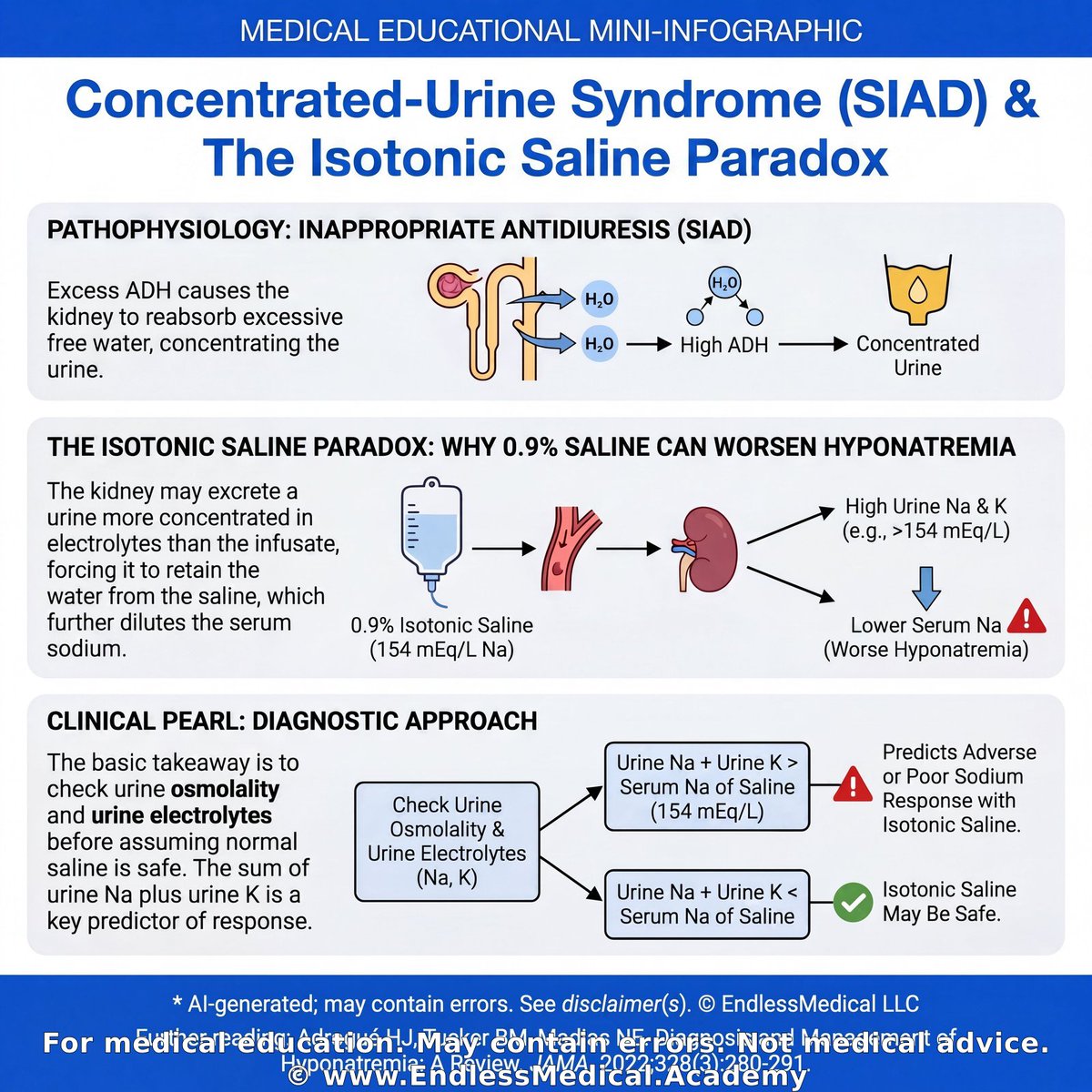
Isotonic saline can worsen hyponatremia when antidiuresis forces the kidney to excrete a urine more concentrated in electrolytes than the infusate.
That paradox is clinically important because the wrong fluid can deepen neurologic injury.
The basic takeaway is to check urine osmolality and urine electrolytes before assuming normal saline is safe.
An advanced clue is that urine sodium plus urine potassium greater than the electrolyte concentration of 0.9% saline predicts poor or adverse sodium response in SIAD physiology.
🎬 Watch the video explanation: https://t.co/53J2LDlb7A
🔗 Learn more: https://t.co/YPkM0mayWr
#MedEdTwitter #MedTwitter #NursesOfTwitter
@MedTwitJC @MedTwitter
Dig deeper on this topic at https://t.co/ofNUUCifgT, where multiple AI models debate, cross-check references, and verify calculations to give you the most updated and accurate medical information with minimal hallucinations and errors.
Further reading:
1. Adrogué HJ, Tucker BM, Madias NE. Diagnosis and Management of Hyponatremia: A Review. JAMA. 2022;328(3):280-291. - JAMA - (2022) - by Adrogué, H. J., Tuck...
Confidence: 100%
Educational value: Supports understanding of the medical concepts in the question.
https://t.co/5OQKONmYiD
Links to sources are provided for optional further reading only.
Verified, approved and authored using AI by Lukasz Kiljanek MD, board-certified in Internal Medicine, Nephrology and Hypertension
*For medical education. May contain errors. Intended for licensed healthcare professionals & educators. Not medical advice. Full disclaimer: https://t.co/d57C8xdFf2
Learn about...
Category: Hypertension > Management of Resistant Hypertension
During a resistant-hypertension multidisciplinary conference, four patients are reviewed for treatment failure after maximally tolerated triple therapy with an angiotensin-converting enzyme inhibitor or angiotensin receptor blocker, a long-acting dihydropyridine calcium-channel blocker, and chlorthalidone.
Patient A is a 46-year-old woman with type 2 diabetes and obesity. Home measurements and a 24-hour ambulatory study average 166/100 mm Hg while she takes lisinopril 40 mg daily, amlodipine 10 mg daily, and chlorthalidone 25 mg daily. Serum potassium is 3.8 mmol/L, estimated GFR is 81 mL/min/1.73 m², urinary metanephrines are normal, renal artery duplex ultrasonography is normal, and drug assays confirm adherence. She has not yet received spironolactone or eplerenone.
Patient B is a 68-year-old man with long-standing diabetes complicated by autonomic neuropathy. Ambulatory blood pressure averages 164/96 mm Hg on losartan 100 mg daily, amlodipine 10 mg daily, and chlorthalidone 25 mg daily. He previously stopped clonidine because of somnolence. Potassium is 4.2 mmol/L and estimated GFR is 77 mL/min/1.73 m². Seated blood pressure is 170/94 mm Hg, but after standing for 2 minutes it falls to 142/82 mm Hg with light-headedness.
Patient C is a 42-year-old man with World Health Organization group 1 pulmonary arterial hypertension attributed to remote methamphetamine exposure who is followed for systemic blood-pressure control. Home measurements and a 24-hour ambulatory study average 165/102 mm Hg despite lisinopril 40 mg daily, amlodipine 10 mg daily, and chlorthalidone 25 mg daily. Spironolactone 50 mg daily and later eplerenone 50 mg twice daily were each discontinued after recurrent serum potassium values above 5.5 mmol/L. A prior clonidine trial caused severe fatigue. Current estimated GFR is 74 mL/min/1.73 m², potassium is 4.9 mmol/L, screening for primary aldosteronism and pheochromocytoma is unrevealing, and there are no orthostatic symptoms.
Patient D is a 61-year-old man with obesity and obstructive sleep apnea treated with nocturnal continuous positive airway pressure. Ambulatory blood pressure averages 164/98 mm Hg while he takes valsartan 320 mg daily, amlodipine 10 mg daily, chlorthalidone 25 mg daily, and doxazosin 8 mg nightly. Potassium is 4.4 mmol/L, estimated GFR is 69 mL/min/1.73 m², renal artery imaging is normal, and adherence is confirmed.
According to the 2020 International Society of Hypertension guidance, which patient is the best candidate for doxazosin 4 mg orally at bedtime as the next pharmacologic step?
🎬 Watch the video explanation: https://t.co/6RuCufOySB
Visit https://t.co/UsSgGDgVDV for the answer and detailed explanation. Access requires a valid US NPI (licensed healthcare providers only).
Dig deeper on this topic at https://t.co/ofNUUCifgT, where multiple AI models debate, cross-check references, and verify calculations to give you the most updated and accurate medical information with minimal hallucinations and errors.
Further reading:
1. Resistant Hypertension: Detection, Evaluation, and Management: A Scientific Statement From the American Heart Association - (2018) - by Carey RM, Calhoun DA...
Confidence: 100%
Educational value: Supports understanding of the medical concepts in the question.
https://t.co/c6yWCQSyIa
2. 2020 International Society of Hypertension Global Hypertension Practice Guidelines - (2020) - by Unger T, Borghi C, C...
Confidence: 100%
Educational value: Supports understanding of the medical concepts in the question.
https://t.co/p8GYypVNcq
Links to sources are provided for optional further reading only.
Verified, approved and authored using AI by Lukasz Kiljanek MD, board-certified in Internal Medicine, Nephrology and Hypertension
*For medical education. May contain errors. Intended for licensed healthcare professionals & educators. Not medical advice. Full disclaimer: https://t.co/d57C8xdFf2
I challenge you today to solve this question!
Category: Hypertension > Renovascular Hypertension and Kidney Artery Disorders > Medical Management Strategies for Renovascular Hypertension
A 43-year-old man returns to nephrology clinic for follow-up of apparent treatment-resistant hypertension diagnosed 4 months ago. Home readings remain 165–180/95–110 mm Hg despite reported adherence. His father developed hypertension in his 40s and had myocardial infarction at age 68. The patient has schizophrenia, compensated metabolic dysfunction-associated steatotic liver disease cirrhosis (Child–Pugh A), idiopathic pulmonary fibrosis, chronic plaque psoriasis, bilateral high-frequency sensorineural hearing loss, and migraine without aura. He does not smoke, use illicit substances, take nonsteroidal anti-inflammatory drugs, or use herbal products. He reports no episodic palpitations, flushing, orthostatic symptoms, exertional chest pain, transient neurologic symptoms, or prior flash pulmonary edema.
Current medications are amlodipine 10 mg orally daily, metoprolol tartrate 100 mg orally twice daily, hydrochlorothiazide 25 mg orally daily, lisinopril 10 mg orally daily started 2 weeks ago, risperidone 2 mg orally nightly, and nintedanib 100 mg orally twice daily. He reports adequate oral intake and no recent diarrhea, vomiting, or diuretic overuse.
Vital signs are temperature 36.8 °C, pulse 92/min, respiratory rate 14/min, blood pressure 182/106 mm Hg in the right arm while seated, and oxygen saturation 94% on room air. Repeat automated readings are concordant in both arms. Funduscopic examination shows no hemorrhages, exudates, or papilledema. A loud continuous right paraumbilical bruit radiates posteriorly. There is no peripheral edema, carotid bruit, pulse asymmetry, livedo reticularis, or abdominal tenderness. Fine bibasilar inspiratory crackles and stable psoriatic plaques are present.
Laboratory results show serum creatinine before lisinopril of 1.02 mg/dL (normal: 0.7–1.3 mg/dL) and serum creatinine today of 1.49 mg/dL (normal: 0.7–1.3 mg/dL). Serum potassium is 4.2 mmol/L (normal: 3.5–5.0 mmol/L), bicarbonate is 24 mmol/L (normal: 22–28 mmol/L), fasting glucose is 96 mg/dL (normal: 70–99 mg/dL), and LDL cholesterol is 118 mg/dL (optimal: <100 mg/dL). Urinalysis shows no blood or cellular casts (normal: absent), and urine protein-to-creatinine ratio is 0.08 g/g (normal: <0.20 g/g). A 24-hour ambulatory blood-pressure study last month confirmed sustained daytime hypertension. Electrocardiography shows voltage criteria for left ventricular hypertrophy. Six weeks ago, renal ultrasound showed a right kidney measuring 8.2 cm with cortical thinning and a left kidney measuring 12.1 cm with preserved cortical thickness.
Which individual finding most strongly supports a hemodynamically important unilateral renal-artery lesion and warrants dedicated renal vascular imaging rather than simple escalation of antihypertensive therapy?
🎬 Watch the video explanation: https://t.co/yYiwRhuL7H
Visit https://t.co/2LttR1LdgD for the answer and detailed explanation. Access requires a valid US NPI (licensed healthcare providers only).
#Hypertension #Medical #MCQ #EndlessMedicalAcademy #MedicalManagementStrategiesforRenovascularHypertension #EndlessMedicalcom #EndlessMedical #MedicalEducation #MultipleChoiceQuestions #RenovascularHypertensionandKidneyArteryDisorders
Dig deeper on this topic at https://t.co/ofNUUCifgT, where multiple AI models debate, cross-check references, and verify calculations to give you the most updated and accurate medical information with minimal hallucinations and errors.
Further reading:
1. 2025 AHA/ACC/AANP/AAPA/ABC/ACCP/ACPM/AGS/AMA/ASPC/NMA/PCNA/SGIM Guideline for the Prevention, Detection, Evaluation and Management of High Blood Pressure in Adults - (2025) - by Jones DW, Ferdinand ...
Confidence: 100%
Educational value: Supports understanding of the medical concepts in the question.
https://t.co/WSuIJeObOZ
2. Revascularization for Renovascular Disease: A Scientific Statement From the American Heart Association - (2022) - by Bhalla V, Textor SC,...
Confidence: 100%
Educational value: Supports understanding of the medical concepts in the question.
https://t.co/wB8aUPMzSq
Links to sources are provided for optional further reading only.
Verified, approved and authored using AI by Lukasz Kiljanek MD, board-certified in Internal Medicine, Nephrology and Hypertension
*For medical education. May contain errors. Intended for licensed healthcare professionals & educators. Not medical advice. Full disclaimer: https://t.co/d57C8xdFf2
Are you as smart as you think you are? Solve it!
Category: Hypertension > Renovascular Hypertension and Kidney Artery Disorders > Renal Artery Stenosis
A 33-year-old woman presents to a U.S. emergency department with 1 day of throbbing right-sided headache, palpitations, malaise, and home blood pressure readings that have remained elevated for 6 months despite adherence. She reports an audible abdominal whooshing when supine and intermittent orthostatic lightheadedness without syncope. She denies chest pain and exertional dyspnea. Her history includes systemic lupus erythematosus diagnosed at age 22 without current nephritis, anterior uveitis in remission, and bilateral carpal tunnel syndrome. Her mother had carotid artery dissection in her 40s. She follows a vegan diet, is a former smoker who quit 5 years ago, and uses no alcohol or illicit drugs. She has regular menses and no known pregnancy.
She has no medication allergies and reports fragrance-triggered headaches. Home medications taken this morning include amlodipine 10 mg PO daily, carvedilol 12.5 mg PO twice daily, hydrochlorothiazide 25 mg PO daily, lisinopril 10 mg PO daily started 3 weeks ago, ibuprofen 400 mg PO up to three times daily as needed for headaches used twice this week, prednisone 5 mg PO daily, hydroxychloroquine 200 mg PO twice daily, vitamin B12 1000 mcg PO daily, depot medroxyprogesterone acetate 150 mg IM every 3 months with the last injection 7 weeks ago, and cetirizine 10 mg PO daily as needed for seasonal symptoms.
Triage vital signs are tympanic temperature 38.4 °C, repeat oral temperature 37.8 °C, manual radial pulse 138/min and regular, respiratory rate 14/min, blood pressure 107/53 mm Hg, and oxygen saturation 91% on room air that improves to 97% with 2 L/min by nasal cannula. She is alert, oriented, communicates choices coherently, and repeats risks and benefits. Funduscopic examination shows mild arteriolar narrowing with arteriovenous nicking but no cotton-wool spots or papilledema. Cardiopulmonary examination shows no murmur or rales. The abdomen is soft with a prominent systolic bruit maximal in the right periumbilical and flank region. Peripheral pulses are symmetric without bruits, and there is no lower-extremity edema. Tinel sign is present at both wrists; slit-lamp examination shows no active anterior chamber cells.
Initial ED results show regular narrow-complex tachycardia at 138/min on ECG and no infiltrate or effusion on chest radiograph. Sodium is 138 mEq/L (reference: 135–145 mEq/L), potassium 5.3 mEq/L (reference: 3.5–5.0 mEq/L), bicarbonate 23 mEq/L (reference: 22–29 mEq/L), glucose 96 mg/dL (reference: 70–140 mg/dL), and creatinine 1.4 mg/dL (reference: 0.5–1.1 mg/dL), increased from 1.2 mg/dL one week earlier (reference: 0.5–1.1 mg/dL). Estimated GFR is approximately 51 mL/min/1.73 m² today (reference: at least 90 mL/min/1.73 m²) versus 61 mL/min/1.73 m² previously (reference: at least 90 mL/min/1.73 m²). White blood cell count is 9.2 × 10^3/µL (reference: 4.0–11.0 × 10^3/µL) with neutrophils 72% (reference: 40%–70%), hemoglobin is 12.8 g/dL (reference: 12.0–16.0 g/dL), platelets are 274 × 10^3/µL (reference: 150–450 × 10^3/µL), and lactate is 1.2 mmol/L (reference: 0.5–2.2 mmol/L). Urinalysis shows specific gravity 1.018 (reference: 1.005–1.030), protein 1+ (reference: negative), 0–2 RBC/hpf (reference: 0–2 RBC/hpf), and negative nitrites and leukocyte esterase (reference: negative). Two peripheral blood-culture sets are pending. Bedside renal ultrasound shows a right kidney length of 8.8 cm and a left kidney length of 10.6 cm, preserved cortical echogenicity, and no hydronephrosis.
She declines contrast-enhanced imaging, and formal vascular-laboratory duplex ultrasound shows right proximal renal artery peak systolic velocity 395 cm/s, aortic peak systolic velocity 92 cm/s, renal-aortic ratio 4.29, delayed intrarenal upstroke with acceleration time 0.12 s, and a tardus-parvus waveform. Left renal artery velocities and waveforms are within reference ranges. Outpatient testing last week showed plasma renin activity 8.2 ng/mL/h (laboratory reference: 0.2–3.3 ng/mL/h), plasma aldosterone 24 ng/dL (laboratory reference: 4–31 ng/dL), aldosterone-renin ratio 2.9 (screening reference: less than 20), morning cortisol 16 µg/dL (reference: 5–25 µg/dL), TSH 2.0 mIU/L (reference: 0.4–4.0 mIU/L), and 24-hour urinary fractionated metanephrines within laboratory reference ranges.
After oral acetaminophen and 1 L isotonic IV crystalloid, her oral temperature is 37.8 °C, pulse is 108/min, blood pressure is 118/66 mm Hg, and oxygen saturation is 97% on room air. Cognition remains intact. The senior resident proposes immediate CT angiography of the renal arteries using low-osmolality iodinated contrast and, if a tight stenosis is confirmed, same-day revascularization. The patient explicitly states, “I do not want any contrast study until I speak with my partner. I’m okay with ultrasound today.” A stat serum hCG has been sent and is pending.
Which single course of action is most appropriate from a legal and ethical standpoint right now, given this clinical context?
🎬 Watch the video explanation: https://t.co/tfOnQCWUmj
Visit https://t.co/6LICT2vdrm for the answer and detailed explanation. Access requires a valid US NPI (licensed healthcare providers only).
#Medical #RenovascularHypertensionandKidneyArteryDisorders #EndlessMedicalAcademy #EndlessMedicalcom #EndlessMedical #MedicalEducation #MultipleChoiceQuestions #RenalArteryStenosis #Hypertension #MCQ
Dig deeper on this topic at https://t.co/ofNUUCifgT, where multiple AI models debate, cross-check references, and verify calculations to give you the most updated and accurate medical information with minimal hallucinations and errors.
Further reading:
1. ACR Manual on Contrast Media – Administration of Contrast Media to Pregnant or Potentially Pregnant Patients - (2025) - by American College of ...
Confidence: 100%
Educational value: Supports understanding of the medical concepts in the question.
https://t.co/Vir6dBgloN
Links to sources are provided for optional further reading only.
Verified, approved and authored using AI by Lukasz Kiljanek MD, board-certified in Internal Medicine, Nephrology and Hypertension
*For medical education. May contain errors. Intended for licensed healthcare professionals & educators. Not medical advice. Full disclaimer: https://t.co/d57C8xdFf2
📿 Medical Pearl
Category: Hypertension > Renovascular Hypertension and Kidney Artery Disorders > Medical Management Strategies for Renovascular Hypertension
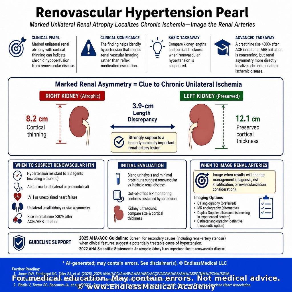
Marked unilateral renal atrophy with cortical thinning can indicate chronic hypoperfusion from renovascular disease.
Clinical significance: The finding helps identify hypertension that merits renal vascular imaging rather than reflex medication escalation alone.
Basic takeaway: Compare kidney lengths and cortical thickness when renovascular hypertension is suspected.
Advanced takeaway: A creatinine rise greater than 30 percent after ACE inhibitor or ARB initiation is concerning, but renal asymmetry more directly localizes chronic unilateral ischemic disease.
🎬 Watch the video explanation: https://t.co/yYiwRhuL7H
🔗 Learn more: https://t.co/2LttR1LdgD
#MedSchool #NursesOfTwitter #Healthcare
@TheLancet @bmaboreddr
Dig deeper on this topic at https://t.co/ofNUUCifgT, where multiple AI models debate, cross-check references, and verify calculations to give you the most updated and accurate medical information with minimal hallucinations and errors.
Further reading:
1. 2025 AHA/ACC/AANP/AAPA/ABC/ACCP/ACPM/AGS/AMA/ASPC/NMA/PCNA/SGIM Guideline for the Prevention, Detection, Evaluation and Management of High Blood Pressure in Adults - (2025) - by Jones DW, Ferdinand ...
Confidence: 100%
Educational value: Supports understanding of the medical concepts in the question.
https://t.co/WSuIJeObOZ
2. Revascularization for Renovascular Disease: A Scientific Statement From the American Heart Association - (2022) - by Bhalla V, Textor SC,...
Confidence: 100%
Educational value: Supports understanding of the medical concepts in the question.
https://t.co/wB8aUPMzSq
Links to sources are provided for optional further reading only.
Verified, approved and authored using AI by Lukasz Kiljanek MD, board-certified in Internal Medicine, Nephrology and Hypertension
*For medical education. May contain errors. Intended for licensed healthcare professionals & educators. Not medical advice. Full disclaimer: https://t.co/d57C8xdFf2
Today we will learn about...
Category: Hypertension > Management of Resistant Hypertension
A 31-year-old woman is evaluated at a rural outpatient clinic for blood pressure that remains above goal despite confirmed adherence to three antihypertensive agents. Pharmacy refill records and pill counts at two prior visits verify daily use of lisinopril 20 mg, amlodipine 10 mg, and chlorthalidone 25 mg. She also takes naproxen 440 mg as needed for osteoarthritis several times per week and methimazole 5 mg daily for recurrent Graves disease. She does not smoke or drink alcohol.
Over the last 4 months she has developed muscle cramps, intermittent headaches, and increased nocturia. Temperature is 36.3 °C, pulse 85/min, respirations 17/min, blood pressure 148/80 mm Hg in both arms while seated, oxygen saturation 97% on room air, and BMI 28 kg/m². Cardiopulmonary examination is normal. No abdominal bruits are heard. There is 1+ bilateral pretibial edema. The thyroid is normal in size without tenderness, and the skin shows no striae or easy bruising.
Laboratory studies show sodium 146 mmol/L, potassium 2.9 mmol/L, chloride 103 mmol/L, bicarbonate 30 mmol/L, blood urea nitrogen 20 mg/dL, creatinine 1.4 mg/dL, glucose 92 mg/dL, and 24-hour urinary potassium excretion 72 mmol. Urinalysis shows specific gravity 1.014 and no protein. Morning cortisol after an overnight 1-mg dexamethasone suppression test is 0.6 μg/dL, and thyroid-stimulating hormone is 1.8 μIU/mL. Abdominal ultrasonography performed locally last month showed normal-sized kidneys without hydronephrosis and no adrenal mass. Computed tomographic angiography was avoided because she develops flushing after iodinated contrast. A 12-lead electrocardiogram and chest radiograph are unremarkable.
Advanced imaging and subspecialty services are not easily accessible in this rural setting. Which single diagnostic test is most appropriate now to identify the specific secondary cause of her resistant hypertension?
🎬 Watch the video explanation: https://t.co/kyCfqysmOU
Visit https://t.co/S0zujDuNBX for the answer and detailed explanation. Access requires a valid US NPI (licensed healthcare providers only).
#Medical #EndlessMedicalAcademy #MedicalEducation #ManagementofResistantHypertension #Hypertension #EndlessMedicalcom #EndlessMedical #MultipleChoiceQuestions #MCQ
Dig deeper on this topic at https://t.co/ofNUUCifgT, where multiple AI models debate, cross-check references, and verify calculations to give you the most updated and accurate medical information with minimal hallucinations and errors.
Further reading:
1. Resistant Hypertension: Detection, Evaluation, and Management: A Scientific Statement From the American Heart Association - (2018) - by Carey RM, Calhoun DA...
Confidence: 100%
Educational value: Supports understanding of the medical concepts in the question.
https://t.co/c6yWCQSyIa
2. Primary Aldosteronism: An Endocrine Society Clinical Practice Guideline - (2025) - by Adler GK, Stowasser ...
Confidence: 100%
Educational value: Supports understanding of the medical concepts in the question.
https://t.co/jCrGo9q6DI
Links to sources are provided for optional further reading only.
Verified, approved and authored using AI by Lukasz Kiljanek MD, board-certified in Internal Medicine, Nephrology and Hypertension
*For medical education. May contain errors. Intended for licensed healthcare professionals & educators. Not medical advice. Full disclaimer: https://t.co/d57C8xdFf2