Cirrhosis, hematemesis and melena for one day. EGD shows this varix.
Which sign is it?
A) Red spots
B) Red wale sign
C) Nipple sign
D) Active bleeding
The case, and why the authors banded quickly, by Rami Musallam, MD and Klaus Mönkemüller, MD, PhD:
From Chapter 30 of The EndoCollab Guide for GI Bleeding: Abdominal Pain, Jaundice and Melena: Think of Hemobilia. A 224-page atlas of real cases by Klaus Mönkemüller and colleagues. Get it on Amazon: https://t.co/CHbVsgJkp3
Abdominal pain, jaundice and melena the day after a liver biopsy? Think hemobilia.
Only 22% of patients show the full triad. It is often missed because the papilla is not closely inspected on EGD.
Tip: use a distal transparent cap or a side-viewing duodenoscope.
Dynamic injection for a submucosal cushion has two parts.
The nurse pulses the syringe.
You move the needle, the wheels, and the shaft, so the blue mix spreads under the whole polyp.
From Klaus Mönkemüller's new 66-minute atlas of the complex colon polyp.
The snare keeps slipping off a flat lesion.
Pre-cutting EMR (hybrid ESD-EMR) in 3 steps:
1. Inject a submucosal cushion
2. Incise around the lesion with the snare tip (usually 1-2 mm exposure)
3. Snare in the groove and resect
Full technique by Klaus Mönkemüller, MD, PhD:
How a cholangiogram corresponds to liver segments.
Numbers indicate drained hepatic segments.
Adapted from Pauli et al. Arch Surg 2012 (Diagram), and Abdominal Key (cholangiogram).
EndoCollab Quiz
This lesion was located in the upper esophagus, just distal to the upper esophageal sphincter, at about 17 cm from the incisors.
What's your diagnosis?
Reply below with your answer.
Closure of complex gastro-colo-cutaneous fistula with two scopes
- Case
- Procedures
- Wire through second oostomy-fistula
- Localization of the orifice of the fistula
- Closed with Clips TTS
- Fistula closed with 12/6 OTSC
Patient with intermittent abdominal pain.
Colonoscopy showed this.
What is the diagnosis?
A. Gastrointestinal stromal tumor (GIST)
B. Neuroendocrine tumor (carcinoid)
C. Inverted diverticulum
D. Large lipoma causing intermittent obstruction
Comment your letter first.
Then check the answer in the first comment.
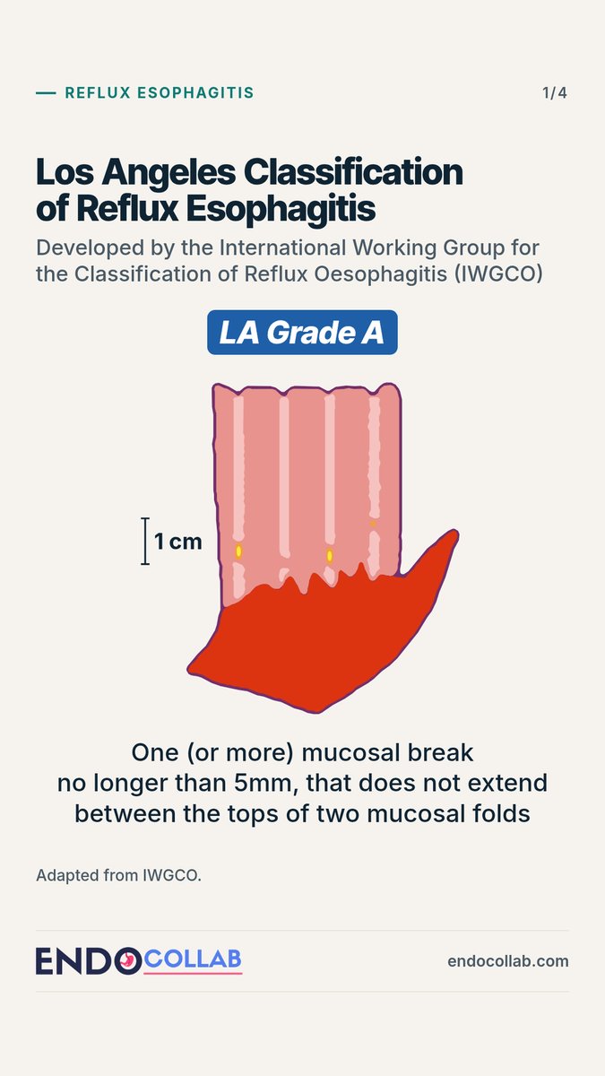
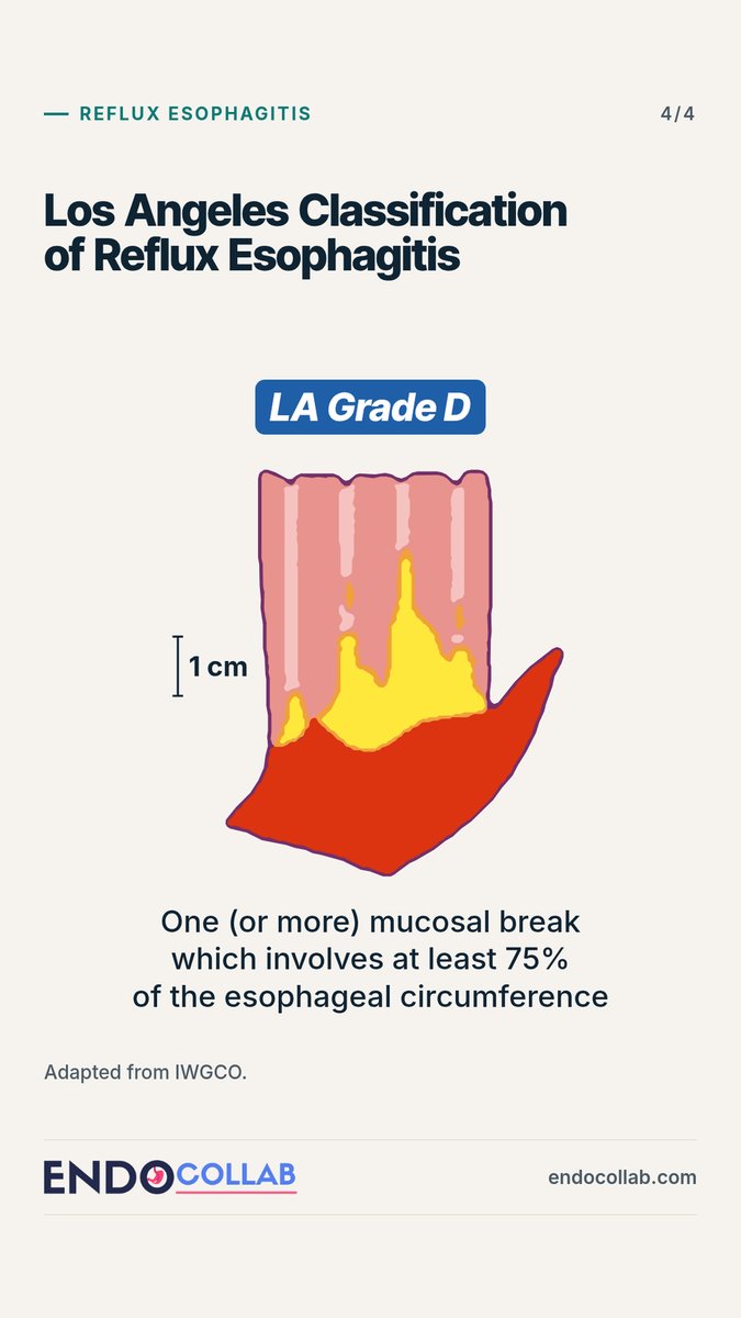
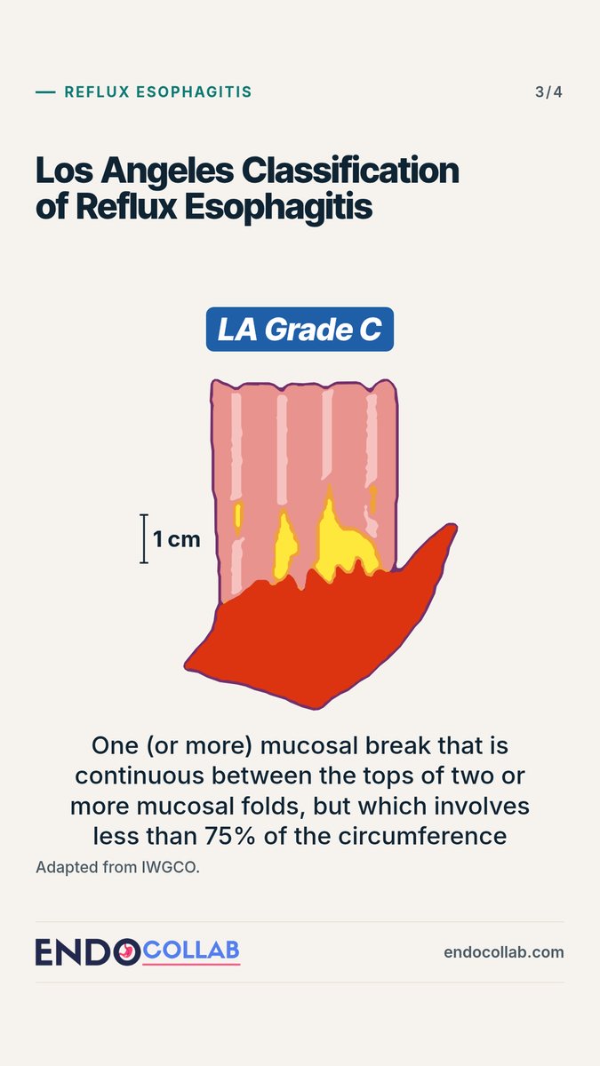
Los Angeles Classification of Reflux Esophagitis.
Grades A to D are defined by:
The length of the mucosal break
Whether it extends between the tops of mucosal folds
Whether it involves at least 75% of the esophageal circumference
Adapted from IWGCO.
"Four-quadrant" injection is a myth.
Two anterior injections can give an adequate submucosal lift. Injecting from behind is not always mandatory.
From Klaus Mönkemüller's Complex Colon Polyps lecture, Panama.
Find the bleeding source.
Duodenum case: recurrent overt GI bleeding, and two previous EGDs without significant findings. On the third look: active bleeding, no ulceration or angiodysplasia. Hemostasis with a through-the-scope clip.
Full case by Klaus Mönkemüller, MD, PhD:
Stuck distal stent? Overtube and cap extraction.
After dilating past a filiform stricture, use an overtube plus distal cap.
Grasp the stent, pull it into the cap and overtube, and extract through that giant working channel to avoid laceration.