Thank you for celebrating #OpenScienceWeek with us!
Continue your #OpenScience exploration with a menu of our most popular atlases, tools, and resources. https://t.co/SV8lNLeUX3
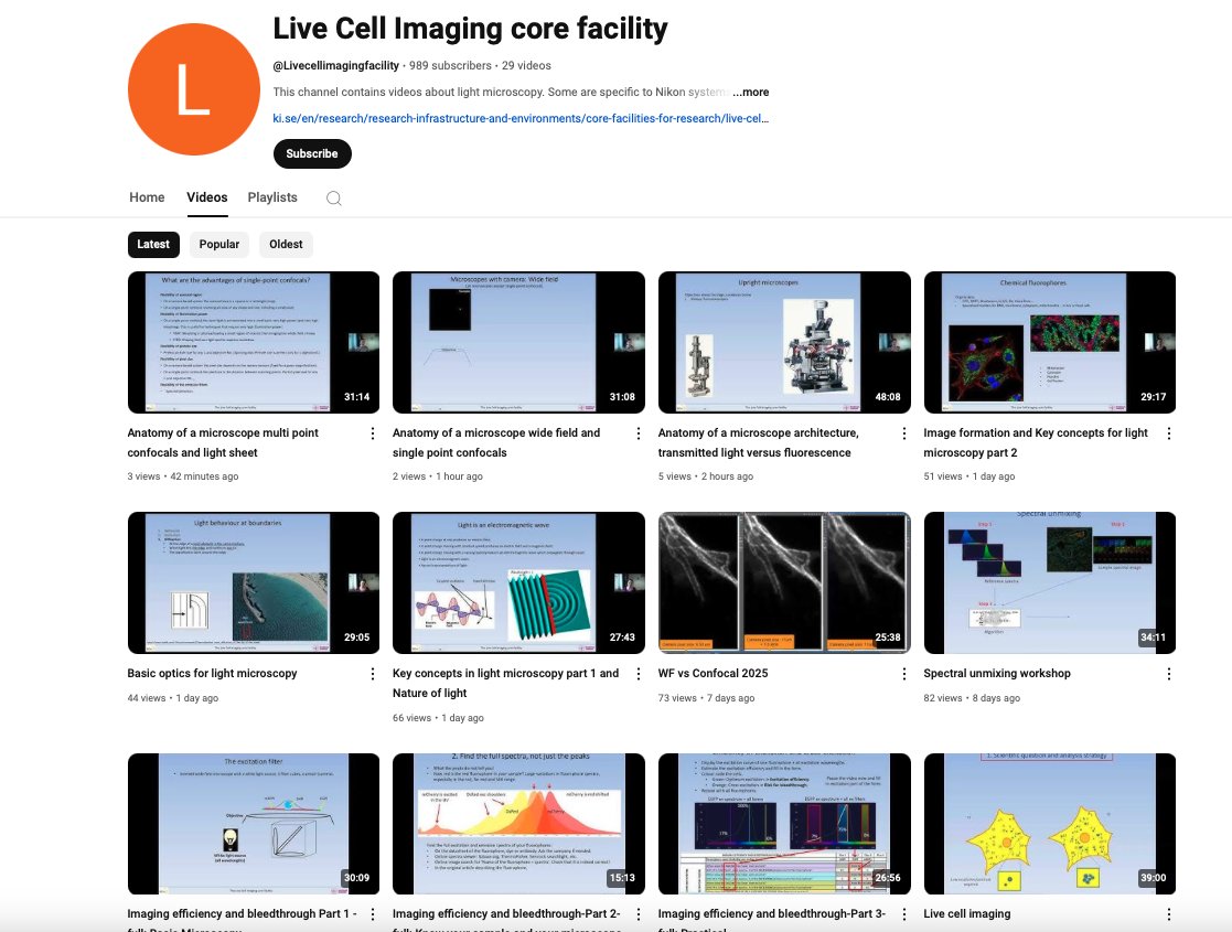
Interested in microscopy? 🔬 Here's a great resource from the Live Cell Imaging Core Facility of the Karolina Institutet, part of our Swedish NMI Node. This YouTube channel includes recordings from their course, “Microscopy: improve your imaging skills - from sample preparation to image analysis”.
Subscribe to get the updated videos 📺: https://t.co/GEVEUW3x3J
Applications are *OPEN* for our @RMS_EarlyCareer Award Competition 2025!
Great opportunity to receive recognition for your work in #microscopy, #ImageAnalysis or #FlowCytometry
Shortlisted entrants will be invited to the final at #mmc2025
Enter now: https://t.co/TJ6aBu6kIq
🔬The February 2025 issue of the @JofMicroscopy is now available to view online!
The issue features 7 original articles and one Methods and Protocols paper, and 6 of the papers are #OpenAccess.
https://t.co/a8bF1XgzNI
@RoyalMicroSoc@RMS_EarlyCareer @michellepeckham
Community is key to scientific discovery. @VolumeEM1 + team are #ImagingTheFuture by expanding the VolumeEM network, connecting experts & novices across the global scientific community
We are delighted to be working with partners in this ambitious programme. to help make transformative scientific advances that will make it possible to develop treatments for previously incurable conditions.
🔬We are pleased to announce the publication of the January 2025 issue of @JofMicroscopy!
The issue features a rapid publication, a Methods and Procotcols paper and 8 original articles.
The issue is available to view here > https://t.co/NkmO6gPG4p
It's been an incredible journey since we first attended the #SEMT at the Natural History Museum 5 years ago. From humble beginnings with just 2 team members and our first katana microtome, we've now expanded to a vibrant team of 6 and have successfully placed nearly 20 katana microtomes around the globe! 🏆📷Join us at our booth to see how far we've come and where we're headed next.
Looking for a funded PhD position? Come and work with @DrPerscitus@paoloactis and myself. Use Nanobiopsy an incredible cool technology to study the interaction between mitochondria and nuclei. Deadline 13th Dec. @nuact_NCL@FMSgradschool Apply here: https://t.co/mio0eMoA3C
Our team in Ghent wants to grow!! If you would be interested in doing electron microscopy with us, please check out the job posted below. Come join a great team @VIBtechnologies! Trust me - it is really a great place to work! https://t.co/Cp876fmaMS
Check out the latest @VolumeEM1 case study from @giulia_casal@EM__Ian@JemimaBurden and Alison Lloyd @LMCB_UCL. They explain how they used array-tomography-SEM to reveal the cellular composition of the Blood Nerve Barrier.
https://t.co/Udqcn88WGq
Congratulations to our Professor of Dermatology and Immunology @Muzz_Haniffa and colleagues, on the publication of their pioneering paper "A prenatal skin atlas reveals immune regulation of human skin morphogenesis". 👏
https://t.co/6XMKrJIDJN