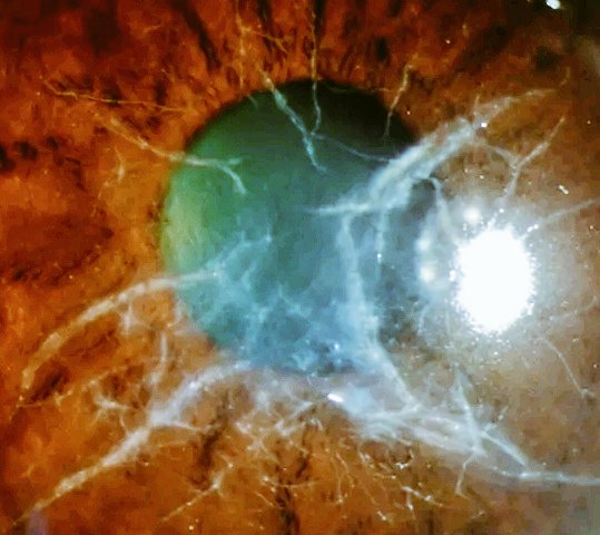
Young patient, bilateral gradual vision loss.
Diagnosis?
A. Granular Corneal Dystrophy
B. Lattice Corneal Dystrophy
C. Reis–Bücklers Dystrophy
#eyes#Awareness#Ophthalmology#medx
What is the most likely diagnosis?A. Retinoblastoma
B. Retinitis pigmentosa
C. Bilateral Diffuse Uveal Melanocytic Proliferation (BDUMP)
D. Choroidal melanoma (primary)
E. Metastatic carcinoma to the choroid
#Awareness#eye#Ophthalmology#eyeshealth
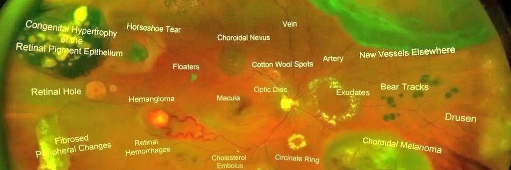
What is the most likely diagnosis based on the fundus image?
A. Age-related macular degeneration (dry type)
B. Moderate to severe non-proliferative diabetic retinopathy
C. CRVO
D. Cytomegalovirus (CMV) retinitis
E. Hypertensive retinopathy (grade 3 only)
F. None
#MedTwitter#eye
Gottsch and colleagues have suggested that this disorder can result from a host response to calgranulin C, a normally hidden antigen expressed by keratinocytes in the corneal stroma. This molecule has also been found in circulating polymorphonuclear leukocytes.
#MedTwitter#eye
Inflammation is not seen in the sclera adjacent to the peripheral ulcers, nor does it affect the underlying Descemet’s membrane. Destruction of the cornea generally affects stromal tissue only, leaving behind an intact endothelium and epithelium.
#XM#MedTwitter#eye_capture
Inflammation is not seen in the sclera adjacent to the peripheral ulcers, nor does it affect the underlying Descemet’s membrane. Destruction of the cornea generally affects stromal tissue only, leaving behind an intact endothelium and epithelium.
#XM#MedTwitter#eye_capture
Ulcers are described as crescent-shaped & can leave behind an opaque & edematous central cornea. Alternatively, they can completely consume the corneal stroma, replacing it with a thin fibrovascular membrane.
#MedTwitter#Awareness#Ophthalmology#eye
Ulcers are described as crescent-shaped & can leave behind an opaque & edematous central cornea. Alternatively, they can completely consume the corneal stroma, replacing it with a thin fibrovascular membrane.
#MedTwitter#Awareness#Ophthalmology#eye
Corneal changes begin within 2-3 mm from limbus, first appearing as grey swellings that rapidly furrow, affecting superficial one-third of cornea & then proceeding circumferentially & centrally over 4-12 months.
#Medx#eye#eyecare#Ophthalmology#optometria
Corneal changes begin within 2-3 mm from limbus, first appearing as grey swellings that rapidly furrow, affecting superficial one-third of cornea & then proceeding circumferentially & centrally over 4-12 months.
#Medx#eye#eyecare#Ophthalmology#optometria
Mooren’s ulcer is characterized by painful peripheral corneal ulceration of unknown etiology. The disease generally begins with intense limbal inflammation & swelling in episclera & conjunctiva.
#medx#Awareness#MedTwitter#Ophthalmology
Mooren’s ulcer is characterized by painful peripheral corneal ulceration of unknown etiology. The disease generally begins with intense limbal inflammation & swelling in episclera & conjunctiva.
#medx#Awareness#MedTwitter#Ophthalmology
Hemorrhagic hypopyon, also termed “candy cane hypopyon,” is a rare presentation that is due to the mixture of hyphema & hypopyon, as both erythrocytes & neoplastic leukocytes accumulate & layer together in the anterior chamber.
#MedTwt#HealthyLiving#Awareness#Eyecare
Hemorrhagic hypopyon is most commonly associated with infectious causes, oftentimes herpetic, but there have also been a few reports of hemorrhagic hypopyon associated with neoplasia.
#MedTwt#MedX#Eyes#Ophthalmology#awareness
Hemorrhagic hypopyon, also termed “candy cane hypopyon,” is a rare presentation that is due to the mixture of hyphema & hypopyon, as both erythrocytes & neoplastic leukocytes accumulate & layer together in the anterior chamber.
#MedTwt#HealthyLiving#Awareness#Eyecare
Hemorrhagic hypopyon, also termed “candy cane hypopyon,” is a rare presentation that is due to the mixture of hyphema & hypopyon, as both erythrocytes & neoplastic leukocytes accumulate & layer together in the anterior chamber.
#MedTwt#HealthyLiving#Awareness#Eyecare
Though it has been observed in both infants and older patients. It is primarily caused by aneurysms and telangiectatic vessels within the temporal retina. These abnormal vessels are leaky and there is exudation in various degrees.
#MedTwitter#eyes
Coats disease is characterized by unilateral (95%), progressive development of abnormal vessels in the retina of the affected individuals. It’s more frequent in males (at least 3:1) than females, and is seen most often in patients aged <8 years.
#MedTwitter#MedClips#Awareness
Coats disease is characterized by unilateral (95%), progressive development of abnormal vessels in the retina of the affected individuals. It’s more frequent in males (at least 3:1) than females, and is seen most often in patients aged <8 years.
#MedTwitter#MedClips#Awareness
He divided the morphology into 3 groups:
Group I: massive subretinal exudate alone
Group II: G 1 + intraretinal and subretinal hemorrhage, and retinal vascular dilatations
Group III: subretinal exudate and retinal arteriovenous malformations & angiomatosis retinae.
#MedTwitter
He divided the morphology into 3 groups:
Group I: massive subretinal exudate alone
Group II: G 1 + intraretinal and subretinal hemorrhage, and retinal vascular dilatations
Group III: subretinal exudate and retinal arteriovenous malformations & angiomatosis retinae.
#MedTwitter
Coats disease is a telangiectatic neovascular disease of the retina of unknown etiology. It usually affects one eye rather than both eyes. In 1908, George Coats described the histopathologic features of enucleated eyes with massive exudations.
#MedTwitter#eyes#Ophthalmology