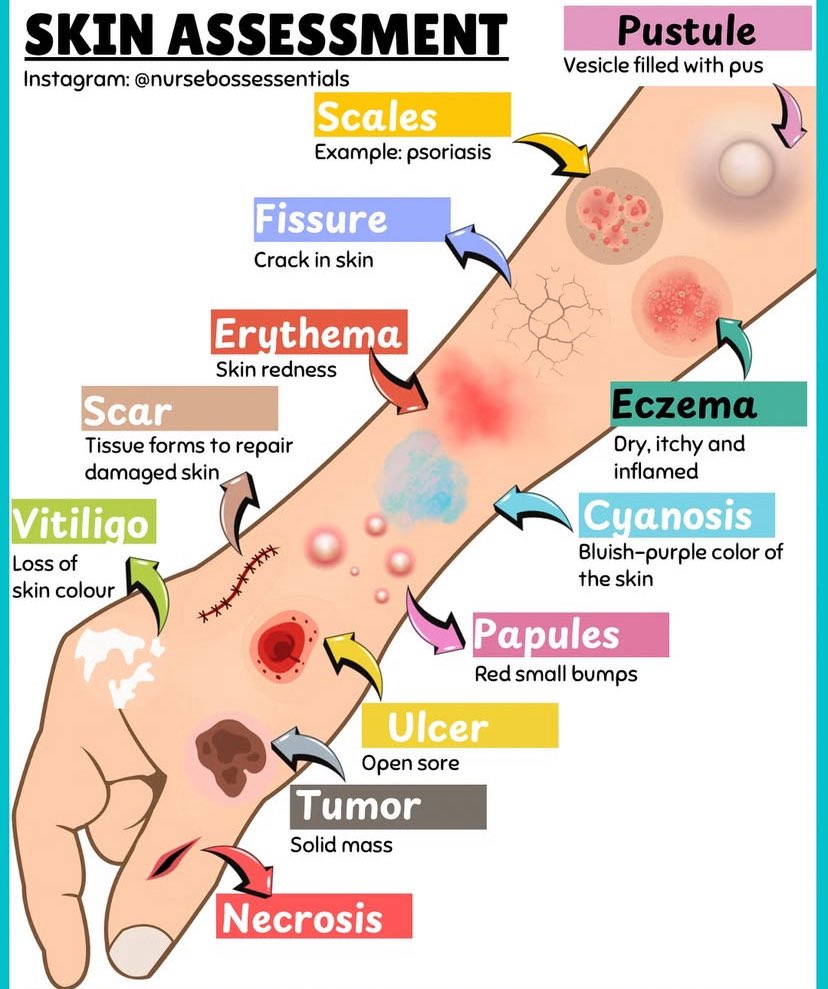
A patient develops ecchymosis around the umbilicus two days after severe acute abdominal pain. Which condition is this sign most associated with?
A. Ruptured spleen
B. Hemorrhagic pancreatitis
C. Liver laceration
D. Renal artery thrombosis
🧵 "What really determines tissue perfusion?"
– and why most explanations get it wrong.
Let’s sort out MAP, CVP, CCP, autoregulation, vasopressors, and the flow that actually reaches your organs.
👇
Watch how the heart’s conduction system controls each heartbeat, starting from the SA node and spreading electrical impulses through the atria and ventricles.
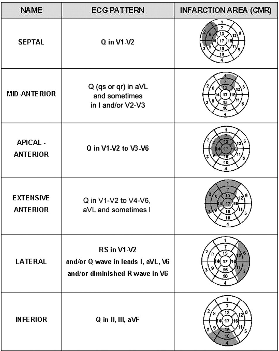
ECG Clues to Localizing Myocardial Infarction (MI): What the Q waves tell us
Different infarct locations produce distinct ECG patterns and CMR helps confirm the myocardial territory involved.
Here’s a breakdown of the 6 major types of Q-wave MI 👇
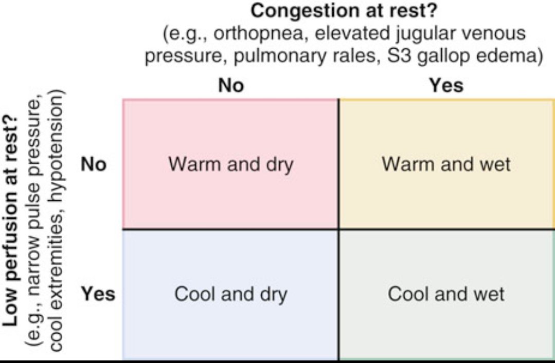
How do we classify heart failure patients at the bedside?
This 2×2 table is a simple clinical tool used to assess patients with heart failure based on two key questions:
1️⃣ Is there congestion at rest?
Look for signs like orthopnea, elevated jugular venous pressure, pulmonary rales, S3 gallop, or leg swelling.
2️⃣ Is there low perfusion at rest?
Check for signs like narrow pulse pressure, cold hands/feet, dizziness, low blood pressure, or fatigue.
Based on yes or no to each, patients fall into one of four profiles:
🔴 Warm and dry
• Good perfusion, no congestion
• This is the ideal, compensated state
🟡 Warm and wet
• Good perfusion, but fluid overload
• Most common in acute decompensated HF
• Needs diuretics and afterload reduction
🔵 Cool and dry
• Poor perfusion, but no fluid overload
• Often seen in advanced HF
• Needs inotropes or optimization of volume
🟢 Cool and wet
• Poor perfusion and congestion
• Worst prognosis
• Requires urgent intervention: diuresis + inotropes ± mechanical support
✅ This simple classification helps guide initial treatment decisions and is widely used in emergency and inpatient settings for managing acute heart failure.
Community leaders from local churches, non-profits, and businesses!
You’re invited to join Mayor Barnes and the Community Development Committee for an important meeting on crime prevention and community development.
Date: June 16 | 6 PM
Location: 324 W. Evans Street
Atrioventricular node (AVN) - Part 1
The atrial extension of the atrioventricular node (AVN) has four branches connected to the atria, resulting in stronger signals in the AN (atrial node) region compared to the N (node) and NH (node-His) regions, with a wider duration and the highest waveform amplitude. This is related to the anatomical structure of the AVN. Another possible reason is associated with the slow pathway region in the atrial extension of the AVN.
The first pathway originates from multiple directions in the atrial extension, so in approximately one-third of the population, the N region signal is not detectable, often masked by fusion with the atrial extension. In the NH (node-His) region, signals are typically detectable, likely due to the fast pathway signals.
The second pathway is related to fast-conducting cells and different conduction routes. The fast and slow pathways are distinct channels that do not interconnect. The slow pathway originates from five directions, while the fast pathway originates from two [see the final figure]. Thus, in the AVN region of a high-resolution ECG (new ECG), if a bimodal or multimodal pattern is observed, independent fast and slow pathway signals—or first and second pathway peaks—can be identified. The signal conduction in the AVN region is characterized by a decremental pattern, typically extending from the peak rightward, downward, and along the X-axis from left to right, regardless of the pattern type. The AVN wave often appears notched or slurred, likely due to the composite nature of the wave formed by multiple cell types.
Another reason for the significant variability in AVN waveform morphology is likely closely tied to AVN conduction delay. The normal physiological delay is approximately 40 ms, but individual variability is substantial, and it varies with each heartbeat. Therefore, AVN delay is likely a critical aspect of studying AVN waveforms and a key focus in arrhythmia research, as delay is one of the essential functions of the AVN.
In new ECG, the AVN potential waveform in the P-wave segment is termed the "N wave," with the following characteristics:
The N wave has a lower frequency response than the H wave.
The N wave begins at the end of the A wave.
The N wave exhibits various forms, including monophasic, multiphasic, or biphasic, with highly variable morphology. It may appear with a steplike notch or slurring.
To be continued
PhysioSign USA
#ECG
#Cardiology
#Electrophysiology
#Medtech
Hi, ER Doc here.
I’ve used this account for over 10 years now to teach people about medical emergencies. It’s my birthday today and I’m hoping to hit 700K followers to be able to reach more people.
Can I get some follows and reposts to help me get there?
We summarized the guideline recs for #anticoagulant& #antiplatelettherapy in ACS & compared them w prior recs in @ACCinTouch@AHAScience & @escardio
guidelines. 🙏🙏 Writing Committee and also a great team who made this summary possible 👇👇:
https://t.co/ZMc0DoXfiN
Wolff-Parkinson-White (WPW) Syndrome: Understanding the Rhythm Patterns
WPW is caused by the presence of an accessory atrioventricular (AV) pathway, which alters the normal conduction pattern of the heart. The rhythm can vary based on the conduction route.
1️⃣ Sinus Rhythm in WPW
During sinus rhythm, the ventricles may be activated by both the AV node and the accessory pathway.
⚪If conduction occurs primarily through the AV node: QRS complex is narrow.
🔵If conduction occurs primarily via the accessory pathway: QRS complex is wide and shows preexcitation.
2️⃣ Orthodromic Tachycardia
In this common form of AV reentrant tachycardia:
⚪Antegrade conduction occurs via the AV node.
🔵Retrograde conduction occurs through the accessory pathway.
➡️The QRS complex is narrow due to use of the normal His-Purkinje system.
3️⃣ Antidromic Tachycardia
This less common reentrant circuit is the reverse:
⚪Antegrade conduction occurs through the accessory pathway.
🔵Retrograde conduction occurs through the AV node.
➡️The QRS complex is wide because the ventricles are activated abnormally.
4️⃣ Atrial Fibrillation in WPW
When atrial fibrillation occurs in a patient with an accessory pathway:
⚪Conduction to the ventricles may occur through both the AV node and accessory pathway.
🔵QRS complexes vary in morphology and timing.
🔴If conduction occurs predominantly over the accessory pathway, it can result in rapid ventricular rates and risk of degeneration into ventricular fibrillation.
Clinical Insight
In WPW, the width and morphology of the QRS complex, along with the conduction direction, help differentiate between sinus rhythm, orthodromic and antidromic tachycardia, and atrial fibrillation with preexcitation.
Reference: The Cardiac Catheterization Handbook
Morton J. Kern
#Cardiology #WPW #ECG #Electrophysiology #Tachycardia #MedEd #ClinicalTips






![physiosign's tweet photo. Atrioventricular node (AVN) - Part 1
The atrial extension of the atrioventricular node (AVN) has four branches connected to the atria, resulting in stronger signals in the AN (atrial node) region compared to the N (node) and NH (node-His) regions, with a wider duration and the highest waveform amplitude. This is related to the anatomical structure of the AVN. Another possible reason is associated with the slow pathway region in the atrial extension of the AVN.
The first pathway originates from multiple directions in the atrial extension, so in approximately one-third of the population, the N region signal is not detectable, often masked by fusion with the atrial extension. In the NH (node-His) region, signals are typically detectable, likely due to the fast pathway signals.
The second pathway is related to fast-conducting cells and different conduction routes. The fast and slow pathways are distinct channels that do not interconnect. The slow pathway originates from five directions, while the fast pathway originates from two [see the final figure]. Thus, in the AVN region of a high-resolution ECG (new ECG), if a bimodal or multimodal pattern is observed, independent fast and slow pathway signals—or first and second pathway peaks—can be identified. The signal conduction in the AVN region is characterized by a decremental pattern, typically extending from the peak rightward, downward, and along the X-axis from left to right, regardless of the pattern type. The AVN wave often appears notched or slurred, likely due to the composite nature of the wave formed by multiple cell types.
Another reason for the significant variability in AVN waveform morphology is likely closely tied to AVN conduction delay. The normal physiological delay is approximately 40 ms, but individual variability is substantial, and it varies with each heartbeat. Therefore, AVN delay is likely a critical aspect of studying AVN waveforms and a key focus in arrhythmia research, as delay is one of the essential functions of the AVN.
In new ECG, the AVN potential waveform in the P-wave segment is termed the "N wave," with the following characteristics:
The N wave has a lower frequency response than the H wave.
The N wave begins at the end of the A wave.
The N wave exhibits various forms, including monophasic, multiphasic, or biphasic, with highly variable morphology. It may appear with a steplike notch or slurring.
To be continued
PhysioSign USA
#ECG
#Cardiology
#Electrophysiology
#Medtech](https://pbs.twimg.com/media/Gslo2qkaMAAYRp7.jpg)