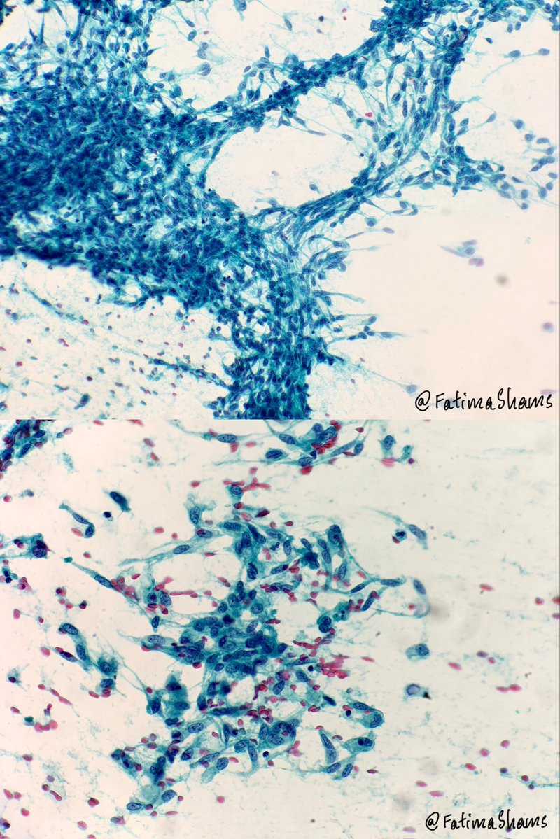
🔬 CYTO–HISTO CORRELATION
👉 A 58-year-old male presented with a small intestinal mass and underwent surgical resection.
Scrapings were taken from fresh cut surface.
❓ Based on the cytomorphological features, what is the most likely diagnosis?
Choose the single best answer:
A. Leiomyoma
B. Gastrointestinal stromal tumour (GIST)
C. Schwannoma
D. Leiomyosarcoma
💬 Share your answer in the comments!
Stay tuned for the histopathological correlation.
#Cytology #Histopathology #CytoHistoCorrelation #PathologyTeaching #DiagnosticCytology #SpindleCellLesion #SurgicalPathology #PathologyCaseOfTheDay #PathologyEducation #MedTwitter
🔬 CYTO–HISTO CORRELATION
👉 A 58-year-old male presented with a small intestinal mass and underwent surgical resection.
Scrapings were taken from fresh cut surface.
❓ Based on the cytomorphological features, what is the most likely diagnosis?
Choose the single best answer:
A. Leiomyoma
B. Gastrointestinal stromal tumour (GIST)
C. Schwannoma
D. Leiomyosarcoma
💬 Share your answer in the comments!
Stay tuned for the histopathological correlation.
#Cytology #Histopathology #CytoHistoCorrelation #PathologyTeaching #DiagnosticCytology #SpindleCellLesion #SurgicalPathology #PathologyCaseOfTheDay #PathologyEducation #MedTwitter
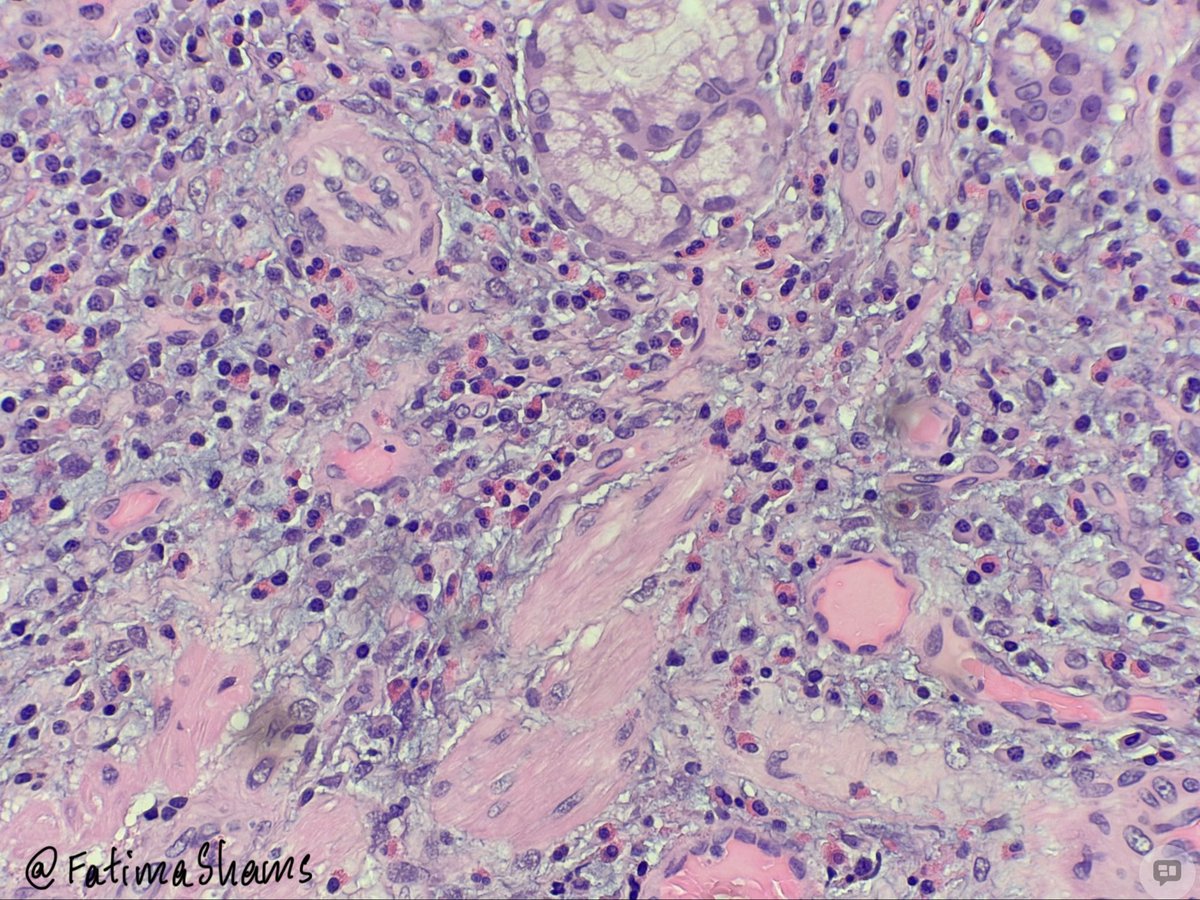
The presence of inflammatory cells, including eosinophils, alone does not qualify a lesion as an inflammatory fibroid polyp; the overall histomorphological features, characteristic stromal architecture and vascular pattern, along with the clinical context, must be carefully considered before arriving at a diagnosis.
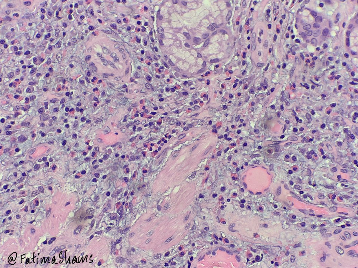
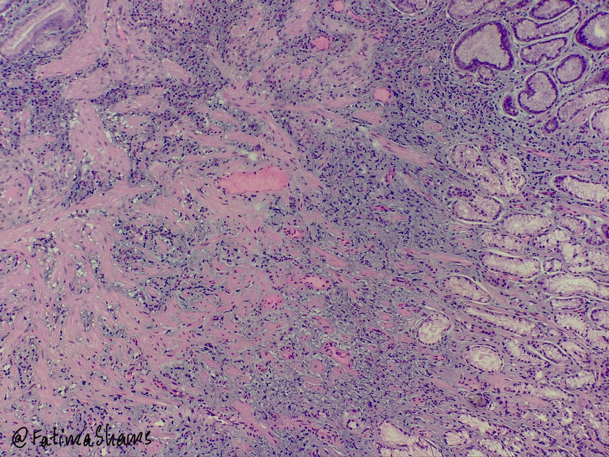
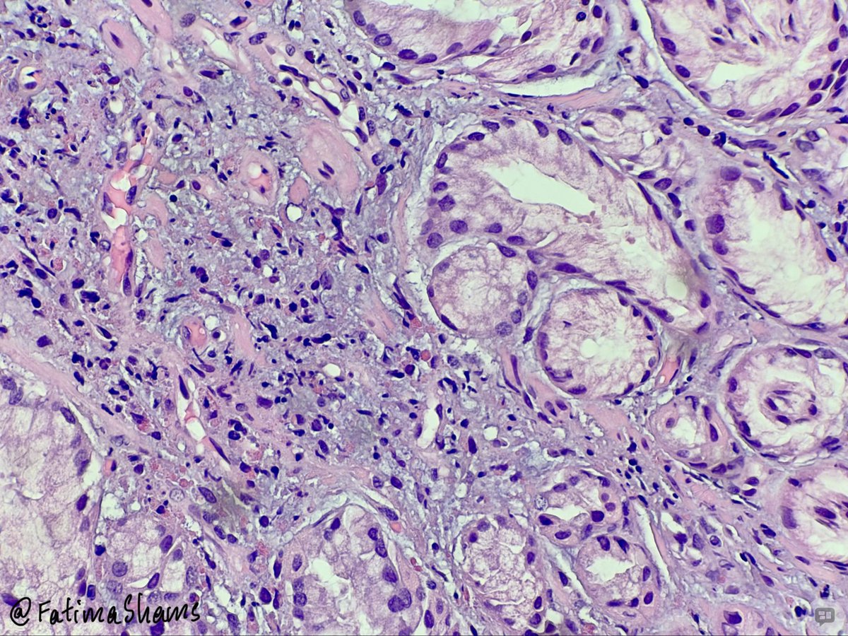
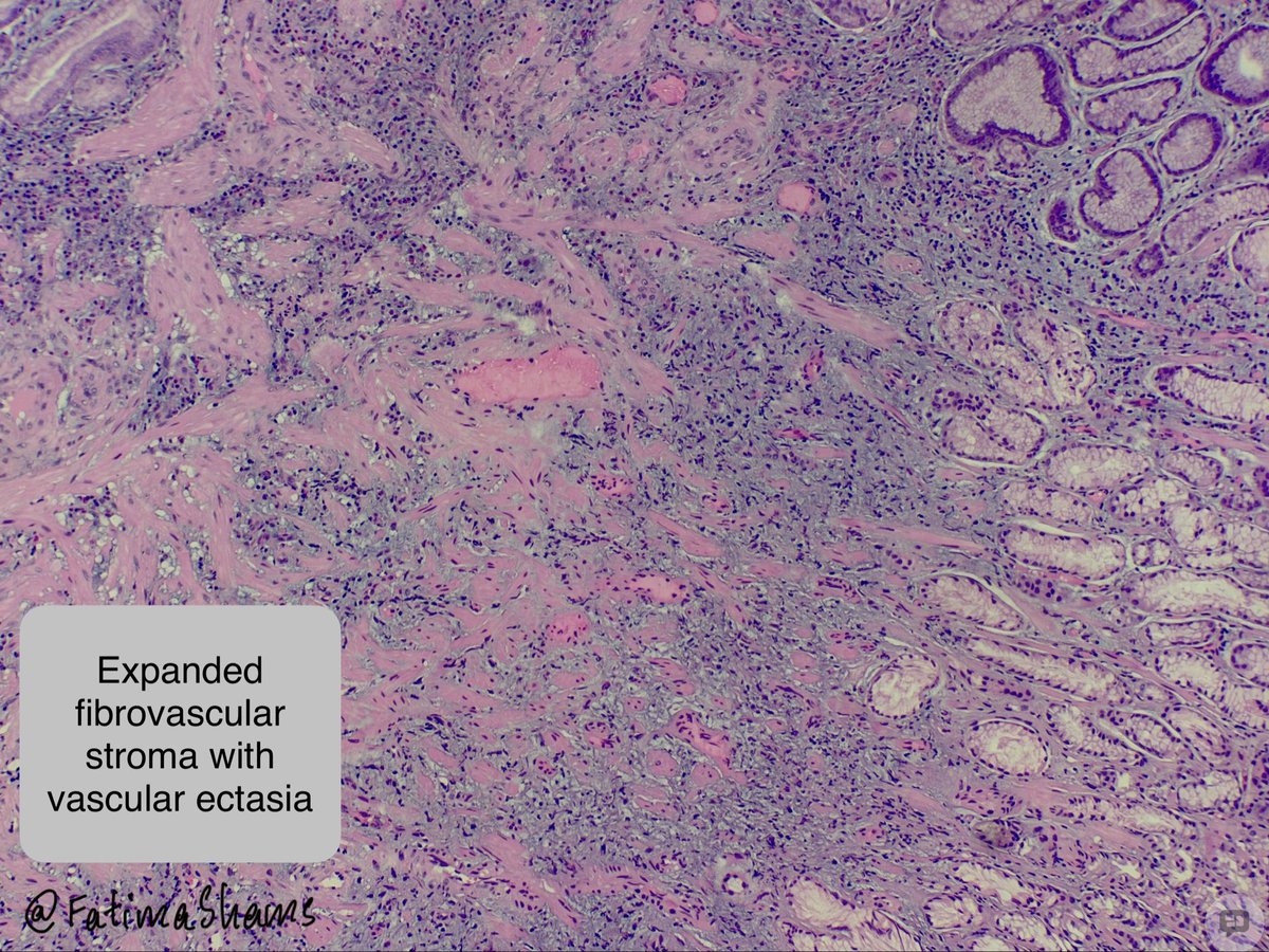
👉 A 70-year-old male with liver cirrhosis undergoes upper GI endoscopy, which reveals a gastric polyp.
🔬 ANSWER: Portal Hypertension-Associated Gastric Polyp
The histological findings include:
• Foveolar hyperplasia with elongated and tortuous gastric glands.
• Prominent proliferation and ectasia of vascular channels within the lamina propria.
• Stromal oedema and vascular congestion.
• Fibromuscular proliferation.
The prominent vascular component, in association with foveolar hyperplasia, is an important histological clue.
💎 Pathological pearl:
- Portal hypertension-associated gastric polyps are an uncommon manifestation of portal hypertension and may mimic conventional gastric hyperplastic polyps.
- The distinction relies on recognising the prominent vascular changes and correlating with the clinical history.
💡An interesting example of how systemic vascular alterations can manifest as distinctive gastrointestinal histopathological changes!
#GIPathology #Histopathology #PortalHypertension #GastricPolyp #PathologyTeaching
👉 A 70-year-old male with liver cirrhosis undergoes upper GI endoscopy, which reveals a gastric polyp.
🔬 ANSWER: Portal Hypertension-Associated Gastric Polyp
The histological findings include:
• Foveolar hyperplasia with elongated and tortuous gastric glands.
• Prominent proliferation and ectasia of vascular channels within the lamina propria.
• Stromal oedema and vascular congestion.
• Fibromuscular proliferation.
The prominent vascular component, in association with foveolar hyperplasia, is an important histological clue.
💎 Pathological pearl:
- Portal hypertension-associated gastric polyps are an uncommon manifestation of portal hypertension and may mimic conventional gastric hyperplastic polyps.
- The distinction relies on recognising the prominent vascular changes and correlating with the clinical history.
💡An interesting example of how systemic vascular alterations can manifest as distinctive gastrointestinal histopathological changes!
#GIPathology #Histopathology #PortalHypertension #GastricPolyp #PathologyTeaching
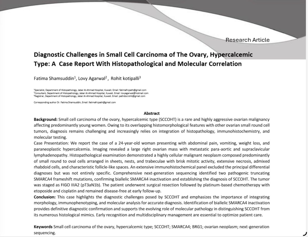
📢 New publication!
Our case report on small cell carcinoma of the ovary, hypercalcemic type (SCCOHT), highlights the diagnostic value of integrating histomorphology, IHC and molecular testing, including SMARCA4 analysis.
Grateful to my co-authors!
Link to article: https://t.co/RfSmt1rBLA
#Pathology #SCCOHT #MolecularPathology
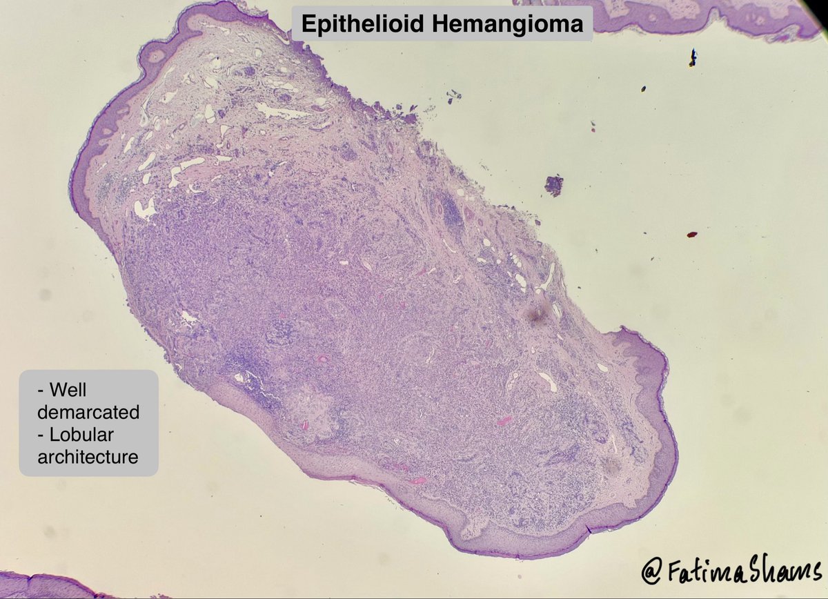
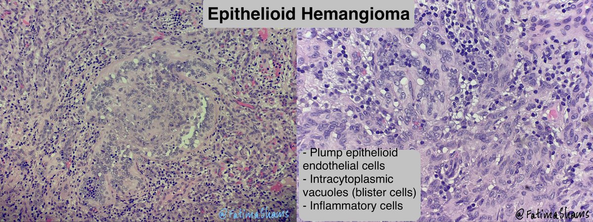
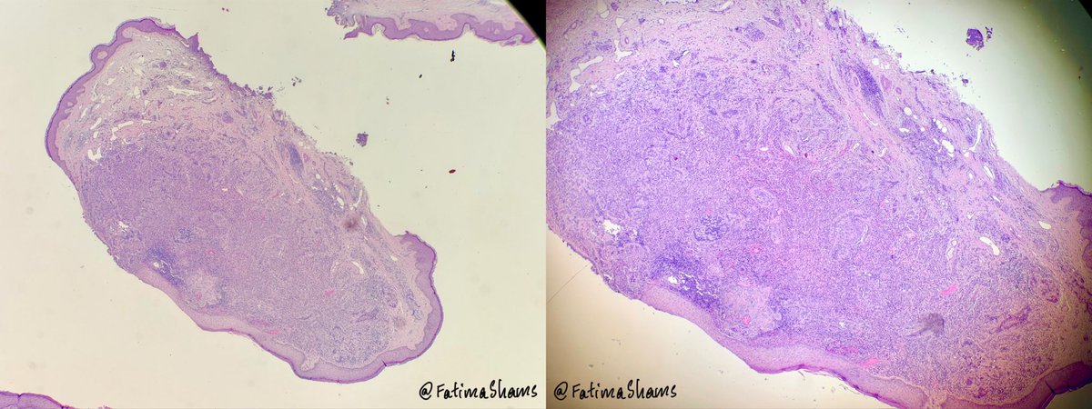
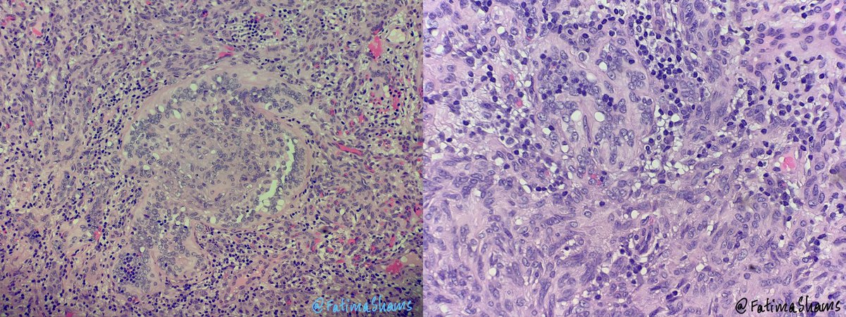
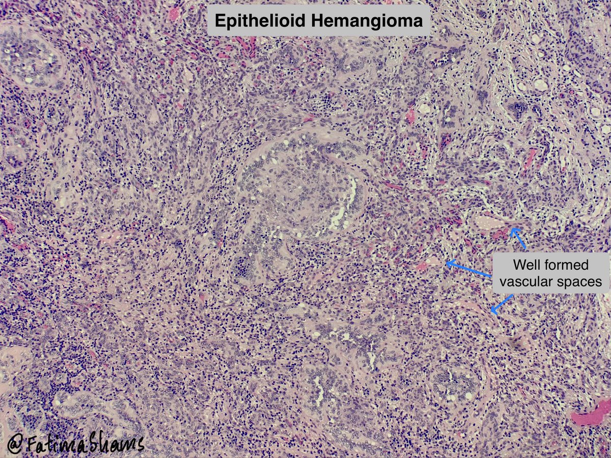
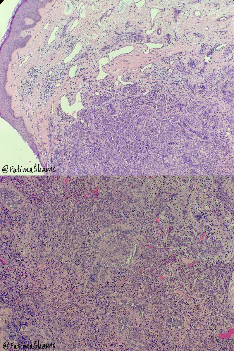
🔬 Epithelioid Hemangioma
•Benign vascular neoplasm composed of well-formed vessels lined by plump epithelioid endothelial cells.
•Typically shows a well-demarcated lobular architecture, with more solid/cellular areas centrally and better-formed vascular lumina peripherally.
•Endothelial cells have abundant eosinophilic cytoplasm, vesicular nuclei, and may show nuclear lobulation/clefting and intracytoplasmic vacuoles (blister cells).
•Cytological atypia is usually mild and mitoses are infrequent.
•A variable lymphocytic/eosinophilic inflammatory infiltrate may be present.
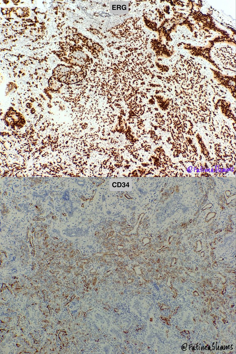
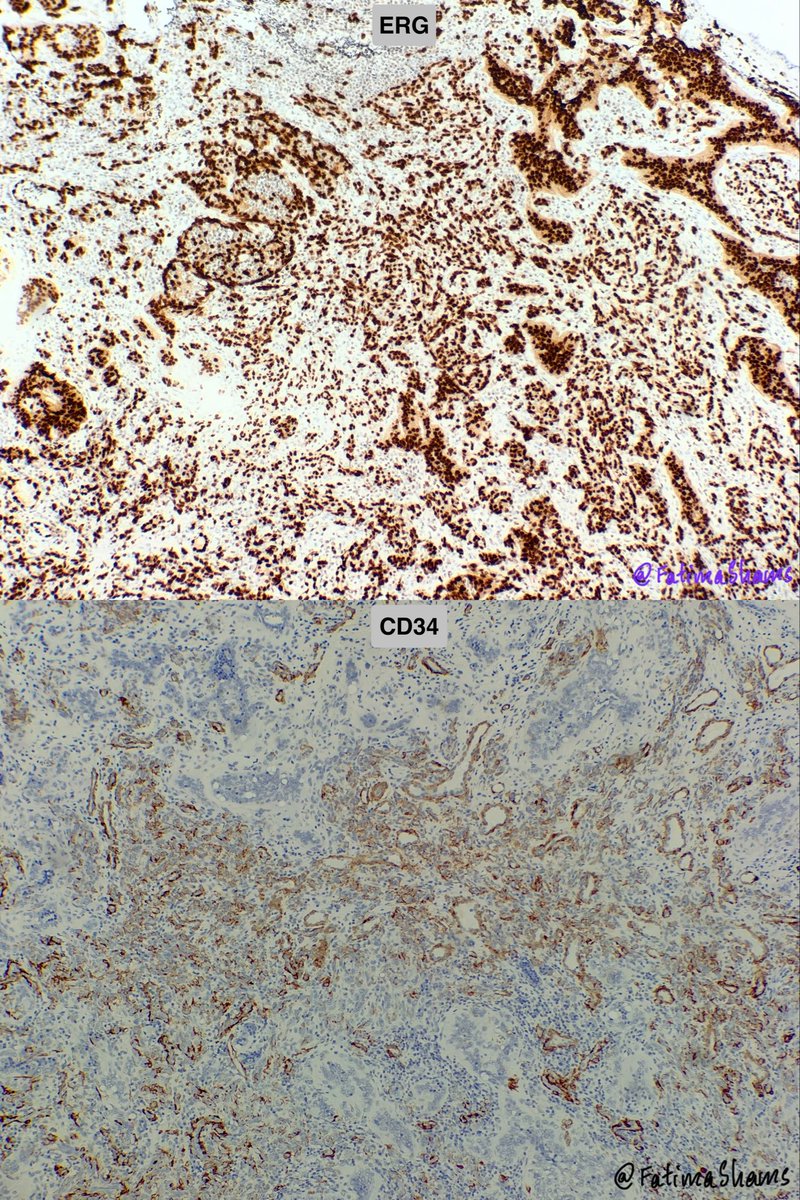
•Endothelial markers: ERG, CD31 and FLI1 positive; CD34 is variable.
•FOS/FOSB rearrangements are identified in approximately half of cases and can support the diagnosis in challenging cases.
•The key is to recognize true vasoformation with epithelioid endothelial lining, rather than diagnosing malignancy based on epithelioid morphology alone.
🌟 Differential diagnosis
•Pyogenic granuloma: Characteristic lobular capillary proliferation, often with surface ulceration; lacks the prominent epithelioid endothelial morphology of EH.
•Epithelioid haemangioendothelioma: Cords/nests of epithelioid cells in myxohyaline stroma, often with intracytoplasmic vacuoles; vasoformation is much less prominent.
•Epithelioid angiosarcoma: Destructive infiltrative growth, marked cytological atypia/pleomorphism, increased mitoses and necrosis, with irregular vascular channels or solid malignant areas.
Take-home message:
👉 Epithelioid morphology does not equal malignancy. In EH, the combination of well-formed vasoarchitecture, characteristic epithelioid endothelial cells, low-grade cytology and appropriate endothelial immunophenotype is the key.
💎 Well-formed vessels + plump epithelioid endothelial cells + inflammatory/eosinophilic background + absence of significant malignant atypia
Case courtesy @annsmiley78
#EpithelioidHemangioma #VascularPathology #Dermatopathology #SurgicalPathology #Histopathology #PathologyEducation #ERG #Immunohistochemistry
🔬 Epithelioid Hemangioma
•Benign vascular neoplasm composed of well-formed vessels lined by plump epithelioid endothelial cells.
•Typically shows a well-demarcated lobular architecture, with more solid/cellular areas centrally and better-formed vascular lumina peripherally.
•Endothelial cells have abundant eosinophilic cytoplasm, vesicular nuclei, and may show nuclear lobulation/clefting and intracytoplasmic vacuoles (blister cells).
•Cytological atypia is usually mild and mitoses are infrequent.
•A variable lymphocytic/eosinophilic inflammatory infiltrate may be present.
•Endothelial markers: ERG, CD31 and FLI1 positive; CD34 is variable.
•FOS/FOSB rearrangements are identified in approximately half of cases and can support the diagnosis in challenging cases.
•The key is to recognize true vasoformation with epithelioid endothelial lining, rather than diagnosing malignancy based on epithelioid morphology alone.
🌟 Differential diagnosis
•Pyogenic granuloma: Characteristic lobular capillary proliferation, often with surface ulceration; lacks the prominent epithelioid endothelial morphology of EH.
•Epithelioid haemangioendothelioma: Cords/nests of epithelioid cells in myxohyaline stroma, often with intracytoplasmic vacuoles; vasoformation is much less prominent.
•Epithelioid angiosarcoma: Destructive infiltrative growth, marked cytological atypia/pleomorphism, increased mitoses and necrosis, with irregular vascular channels or solid malignant areas.
Take-home message:
👉 Epithelioid morphology does not equal malignancy. In EH, the combination of well-formed vasoarchitecture, characteristic epithelioid endothelial cells, low-grade cytology and appropriate endothelial immunophenotype is the key.
💎 Well-formed vessels + plump epithelioid endothelial cells + inflammatory/eosinophilic background + absence of significant malignant atypia
Case courtesy @annsmiley78
#EpithelioidHemangioma #VascularPathology #Dermatopathology #SurgicalPathology #Histopathology #PathologyEducation #ERG #Immunohistochemistry
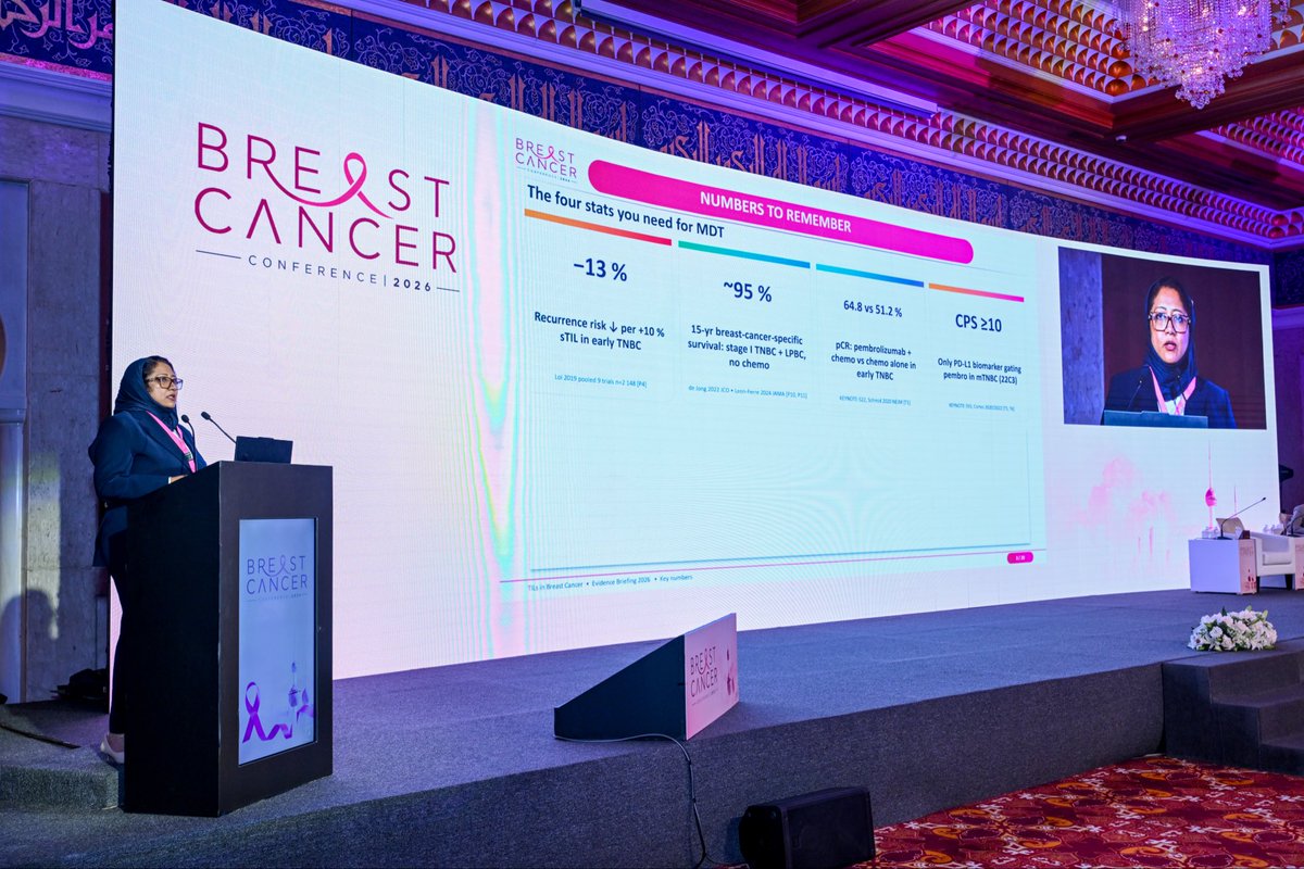
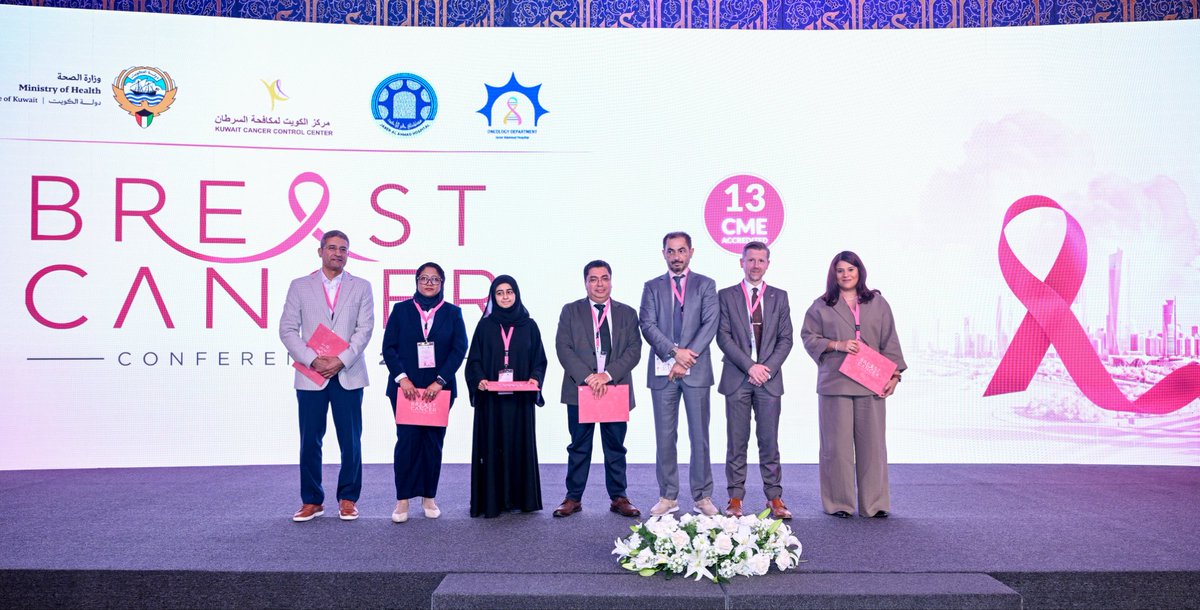
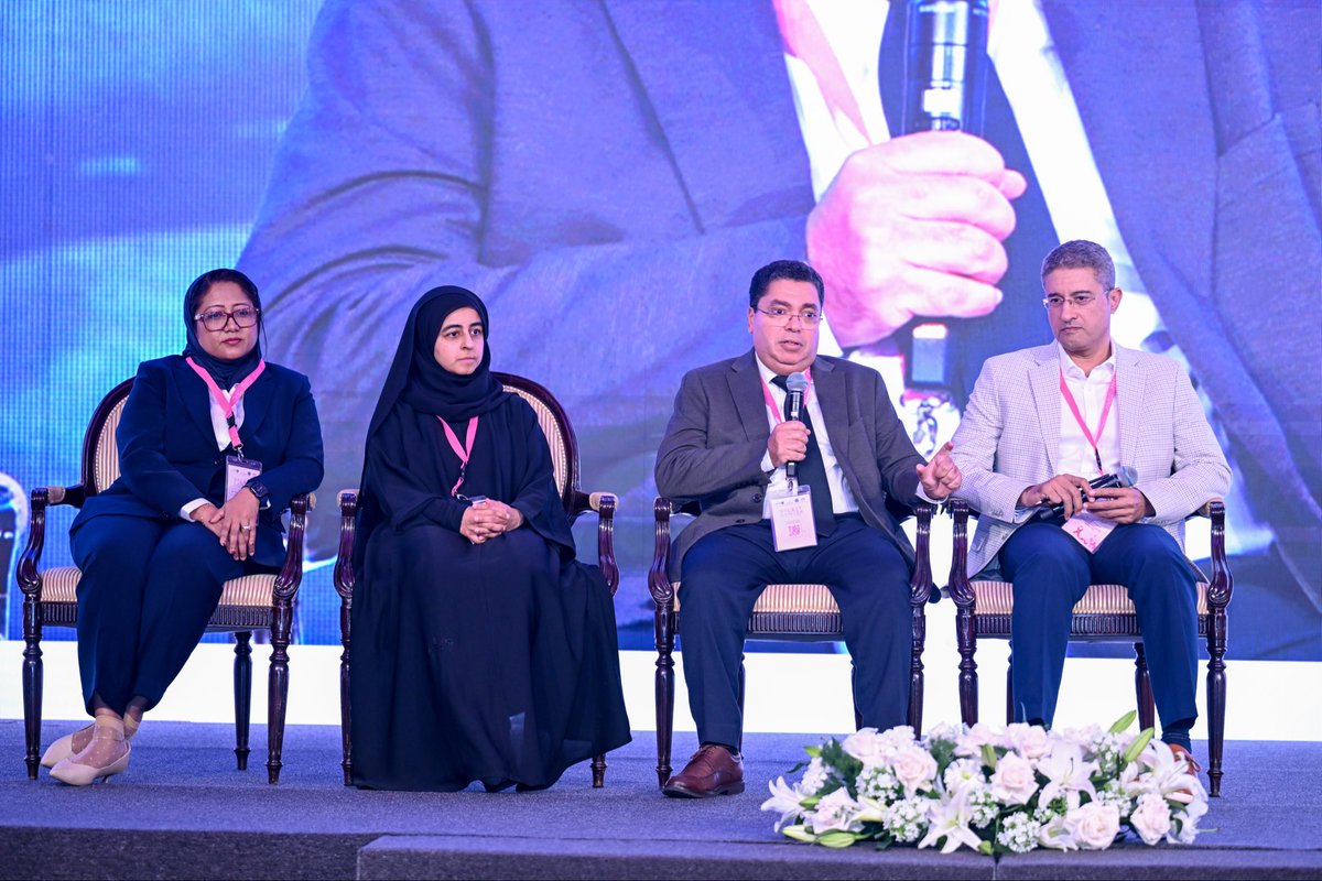
Honoured to be invited as a Guest Speaker at the Breast Cancer Conference 2026, delivering two lectures on TILs in breast cancer—scoring, prognosis & treatment.
Grateful to the Scientific Committee, Breast MDT team, my Department Head, colleagues, friends & family for their support and encouragement. 🌸
#BreastCancer #TILs #BreastPathology #Pathology
🫀 Giant-cell-rich left atrial myxoma — a rare histological variant!
•A 24-year-old man, with no significant past medical history, was found to have a large left atrial mass during evaluation of nonspecific ECG changes detected during a pre-employment medical examination.
•Echocardiography showed a large mass attached to the interatrial septum, partially prolapsing through the mitral valve, with trace mitral regurgitation.
🔬 Histopathology
•Myxoid to focally collagenized stroma with scattered bland stellate and spindle-shaped myxoma cells, seen singly and in cords within the myxoid matrix.
•Striking presence of numerous multinucleated giant cells, some containing multiple relatively uniform nuclei.
•Focal hemorrhage, fibrosis, Gamna-Gandy bodies and degenerative changes.
•Calretinin: positive in the lesional myxoma cells, supporting the diagnosis.
💡 What makes this case interesting?
•Giant-cell-rich cardiac myxoma is rare, with only a small number of reported cases.
•The multinucleated giant cells can be a striking microscopic feature and may raise concern for malignancy.
•However, giant or bizarre cells do not necessarily indicate malignant transformation in a cardiac myxoma.
•Ultrastructural observations in a reported case suggested that the giant cells may result from fusion of individual myxoma cells.
•Cardiac myxomas most commonly arise in the left atrium, particularly around the interatrial septum.
•Calretinin positivity is a useful diagnostic feature of cardiac myxoma and can help distinguish it from myxoid thrombus.
📚 References
1.Hemachandran M, Kakkar N, Khandelwal N. Giant-cell-rich myxoma of right atrium: an ultrastructural analysis. Cardiovasc Pathol. 2003;12:287–289. PubMed
2. Tariq H, Mamoon N. Giant-cell rich atrial myxoma: report of two cases and review of literature. J Ayub Med Coll Abbottabad. 2016;28:197–198. PubMed
#CardiacMyxoma #AtrialMyxoma #GiantCellRichMyxoma #CardiacPathology #Histopathology #SurgicalPathology #Pathology #Calretinin
💡When Keratin Misleads: Keratotic BCC Mimicking PTT!
A 51-year-old man with a longstanding sun-exposed scalp lesion developed recent discharge.
Histology showed a prominent nodular, basaloid to squamous proliferation with keratin-filled cystic structures, creating a challenging differential:
🔹 Keratotic BCC
🔹 SCC with follicular differentiation
🔹 Proliferating trichilemmal tumour (PTT) – benign/malignant
Interestingly, SCC with follicular differentiation received the maximum votes, followed by malignant PTT. Keratotic BCC was not the leading choice — and I had the same dilemma initially!
🧩 The diagnostic clue: IHC
BerEP4: diffuse, strong positivity → strongly supports BCC and argues against conventional SCC and PTT.
BCL2: diffuse positivity → supportive of BCC.
EMA: negative → favours BCC over SCC.
CD34: Negative in tumor cells - favors BCC over PTT
🎯 The take-home point
Don’t be fooled by the keratin!
Keratotic BCC can show prominent squamous/follicular differentiation and closely mimic SCC or PTT.
👉 Look carefully for the underlying BCC architecture and use BerEP4 as a valuable adjunct when morphology is equivocal.
Final diagnosis: KERATOTIC BASAL CELL CARCINOMA.
Sometimes the most difficult cases are those where the morphology makes you believe the wrong diagnosis first! 😉
#Dermatopathology #KeratoticBCC #BasalCellCarcinoma #ProliferatingTrichilemmalTumor #SCC #Histopathology #Pathtwitter
Published! 🔬📚
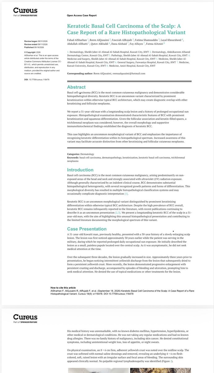
A case I previously shared here has now been published in Cureus: Keratotic Basal Cell Carcinoma of the Scalp — A Rare Histopathological Variant.
An interesting diagnostic pitfall where prominent keratinization and follicular association can mimic other keratinizing/follicular neoplasms.
Grateful to the Dermatology team at Sheikh Jaber Al-Ahmad Al-Sabah Hospital and all co-authors for the collaboration. ❤️
Cureus 2026;18(9):e116478 | DOI: 10.7759/cureus.116478