65 yo M present with severe aquagenic pruritus (hot water more of a trigger than cold). Otherwise well. Physical exam normal. CBC, differential and peripheral smear normal.
Do you test for Jak2 mutation?
رُوي عن الإمام الحسن المجتبى عليه السلام :
الحمد لله الذي من تكلم سمع كلامه ، ومن سكت علم ما في نفسه ، ومن عاش فعليه رزقه ، ومن مات فإليه معاده .
📒 بحار الأنوار
Episode III: Return of the Elastic State
Venous Return Wars
It is a period of haemodynamic uncertainty. The gradient has been challenged, the numerator has struck back, and the old equation can no longer explain itself. From the compliant depths of the venous circulation, a forgotten state is about to return…
From pressure and flow to volume
Levy had exposed the weakness in treating the venous return equation as a one-way causal statement. When flow was imposed, the pressure difference changed with it. Right atrial pressure and flow varied together; neither was altered independently of the other. The experiments therefore did not reveal one variable commanding the other. Guyton himself had acknowledged the deeper problem: in the intact circulation, pressure and flow were both dependent variables.
But that left an obvious question unanswered. Dependent on what?
The equation related venous return to mean systemic filling pressure, right atrial pressure and resistance, but said little about the vascular properties from which those pressures arose. Blood did not occupy a rigid system of pipes. It was distributed through vessels that expanded, recoiled and changed shape. The same total blood volume could therefore produce very different pressures, depending on the vessels containing it.
This was the territory explored most systematically by Carl Rothe. Rather than beginning with the pressure difference said to drive venous return, he examined how the circulation contained blood: where that blood resided, how easily the vessels expanded, and how smooth-muscle activity altered the available vascular space.
The veins were central to this account. Their large resting volume and high compliance mean that they contain most of the systemic blood volume and provide most of the circulation’s ability to accommodate changes in volume. Their importance was therefore not simply that blood passed through them on the way to the heart. Venous elastic and contractile properties helped determine how the blood volume was distributed and what pressures appeared throughout the circulation.
Read on for free on SubStack. Link in reply.
دعاء مولاتي فاطمة الزهراء عليها السلام يوم الجمعة:
اللهمّ اجعلنا من أقرب من تقرّب إليك، وأوجه من توجّه إليك، وأنجح من سألك وتضرّع إليك، اللهمّ اجعلنا ممّن كأنّه يراك إلى يوم القيامة الذي فيه يلقاك، ولا تمتنا إلاّ على رضاك، اللهمّ واجعلنا ممّن أخلص لك بعمله، وأحبّك في جميع خلقك.
اللهمّ صلّ على محمّد وآل محمّد واغفر لنا مغفرة جزماً حتماً لا نقترف بعدها ذنباً ولا نكتسب خطيئة ولا إثماً، اللهمّ صلّ على محمّد وآل محمّد، صلاة نامية دائمة زاكية متتابعة متواصلة مترادفة برحمتك يا أرحم الراحمين.
الشيعه
لهم طريقتهم
لهم طقوسهم
قالوا لك تعال تشيع
طقوا باب بيتكم
اعزموك
حلفوا عليك
قالوا لك عطنا رايك
تحرشوا فيك
في طوائف ثانيه و أديان ما سمعت نقد
و انتشار فيديوهات و تعليقات عليهم
المهم
البعض مثلاً كان يقول شجرة الكريسماس تسامح اديان🤨زين ليش مع الشيعه ما تنطم و تسكت و تفعل ما تقول
انت بتحاسبهم لع👊
مفتاح الجنه بمخباتك لع👊
تبي تحاججهم حط حجتك بكل احترام
كفانا تفرقه
I used to teach that venous congestion reduces organ blood flow because it reduces the pressure gradient across the capillary bed.
Higher venous pressure.
Lower driving pressure.
Less flow.
Simple.
But I don’t think that explanation survives first principles.
The next article on my Substack will be: venous congestion, back-pressure, and why MAP − CVP does not explain organ perfusion. Stay tuned!
https://t.co/54f9IPVCXf
@ThinkingCC@NephroP@ArgaizR
عن أميرالمؤمنين علي بن ابي طالب عليه السلام :
إِنَّ الدُّنْيَا سَرِيعَةُ التَّحَوُّلِ، كَثِيرَةُ التَّنَقُلِ، شَدِيدَةُ الْغَدْرِ، دَائِمَةُ الْمَكْرِ، فَأَحْوَالُهَا
تَتَزَلْزَلُ، وَنَعِيمُهَا يَتَبَدَّلُ، وَرَخَاؤُهَا
يَتَنَقَصُ، وَلَذَّاتُهَا تَتَنَغَصُ،
💊 Perioperative antiplatelet management is not “stop aspirin 7 days before surgery.”
It is a balance between surgical bleeding and catastrophic arterial thrombosis.
Recent PubMed indexed guidance is clear: in non cardiac surgery, the highest risk patient is not the one taking aspirin. It is the patient with a recent coronary stent, recent ACS, recent stroke, or high thrombotic burden in whom interruption of antiplatelet therapy may trigger myocardial infarction, stent thrombosis, or stroke (Thompson et al., 2024; Douketis & Spyropoulos, 2023).
For elective surgery, timing matters. After PCI, elective non cardiac surgery should ideally be delayed until the minimum recommended DAPT period is completed. If surgery cannot wait, aspirin should usually be continued when bleeding risk is acceptable, especially in patients with coronary stents. P2Y12 interruption, when necessary, should be as short as possible: clopidogrel usually 5 days, ticagrelor 3 to 5 days, and prasugrel 7 days before surgery (Thompson et al., 2024; Swan et al., 2024).
Emergency surgery is different. The decision becomes procedural urgency, bleeding site compressibility, last dose, platelet function recovery, and whether the antiplatelet effect can be tolerated. Platelet transfusion may partially reverse irreversible agents such as aspirin and clopidogrel, but it is much less reliable for ticagrelor because circulating drug can inhibit transfused platelets (Swan et al., 2024).
Recent evidence also challenges dogma. In stable patients with previous drug eluting stents undergoing low to intermediate risk non cardiac surgery, aspirin continuation did not clearly reduce ischemic events compared with temporary interruption, although minor bleeding increased (Kang et al., 2024). This does not mean “stop aspirin in everyone.” It means individualize.
The practical question is not:
“Should antiplatelets be stopped?”
It is:
What is more dangerous for this patient: bleeding today, or thrombosis tomorrow?
#Anesthesiology #PerioperativeMedicine #Cardiology #AntiplateletTherapy #Aspirin #Clopidogrel #Ticagrelor #Prasugrel #NonCardiacSurgery #PatientSafety
References 📚
Douketis, J. D. NEJM Evidence, 2(6). https://t.co/zms5Bz8MAE
Kang, D. Y. Journal of the American College of Cardiology, 84(24), 2380–2389. https://t.co/p9iMZBqx7M
Swan, D., Research and Practice in Thrombosis and Haemostasis, 8(6), 102548. https://t.co/61A5TyzWbO
Thompson, A., Journal of the American College of Cardiology, 84(19), 1869–1969. https://t.co/ExOBujTyfY
😴Consciousness in the ICU should not be reduced to “GCS 8.”🤔
The Glasgow Coma Scale remains useful because it is simple, familiar, reproducible, and embedded in emergency medicine, trauma, neurology, and ICU communication.But in the ICU, the classic GCS has important flaws.
The verbal component becomes untestable in intubated patients. Sedation, neuromuscular blockade, aphasia, delirium, language barriers, facial trauma, hearing impairment, and metabolic encephalopathy can all distort the score. Most importantly, GCS does not directly assess brainstem reflexes or respiratory pattern, which are central in deep coma and neurocritical care.
Recent evidence supports a more nuanced approach.
The FOUR score appears particularly useful in ICU and emergency patients because it removes the verbal component and adds brainstem reflexes, breathing pattern, eye response, and motor response. A 2025 systematic review found that both GCS and FOUR have strong reliability and validity, but FOUR showed slightly higher overall performance and may be especially suitable for ICU assessment (Brun et al., 2025). Another systematic review found FOUR may be superior to GCS for ICU mortality prediction, with better responsiveness in deeply impaired patients (Schey et al., 2025).
Sedated ICU patients require a different logic. RASS and SAS are sedation scales, not coma scales. They should be used to titrate sedation depth, while CAM ICU or ICDSC should be used when delirium is assessable. A 2025 PADIS focused update reinforces structured assessment of pain, agitation, sedation, delirium, immobility, and sleep in adult ICU patients (Lewis et al., 2025).
The 2025 French expert consensus on severe acute encephalopathy recommends using delirium scores such as CAM ICU or ICDSC, coma scores such as GCS or FOUR, and specifically suggests FOUR over GCS in coma because it better captures brainstem responses and respiratory patterns (Sonneville et al., 2025).
The practical ICU message:
Use RASS to define sedation level.
Use CAM ICU or ICDSC to detect delirium when the patient is arousable.
Use FOUR, not only GCS, in coma or intubated patients.
Use CRS R when prolonged impaired consciousness requires recovery tracking.
Do not interpret a low score without knowing sedation, analgesia, paralysis, metabolic state, and airway status.
The question is not only:
“What is the GCS?”
It is:
What is the patient’s brain doing, and what is the drug doing to the brain?
#CriticalCare #ICU #NeurocriticalCare #Consciousness #Sedation #Delirium #GCS #FOURScore #RASS #CAMICU #IntensiveCare
References
Brun, F. K.. Australian Critical Care, 38(1), 101057. https://t.co/DhIlQ6isvp
Lewis, K., Critical Care Medicine, 53(3), e711–e727. https://t.co/TiOVn3apvR
Schey, J. E., et al. (2025). Neurocritical Care. https://t.co/hdpso6BynF
Sonneville, R. Annals of Intensive Care, 15, 37. https://t.co/JKzksc2e3S
🫀🦠 In the ICU, elevated troponin during sepsis is often automatically labeled as “ACS.”...The reality is far more complex.🤔
Recent evidence suggests that septic patients may present with at least 4 distinct cardiac phenotypes:
• Type 1 myocardial infarction from plaque rupture/thrombosis
• Type 2 myocardial infarction from oxygen supply demand imbalance
• Sepsis induced myocardial injury without infarction
• Sepsis induced cardiomyopathy with reversible ventricular dysfunction
This distinction matters because management changes completely.
A troponin elevation alone is NOT enough to diagnose ACS in sepsis. Dynamic ECG changes, ischemic symptoms, regional wall motion abnormalities, hemodynamic profile, lactate kinetics, vasopressor burden, and echocardiographic findings must all be integrated into the evaluation.
In septic shock, myocardial injury is frequently driven by:
• Cytokine mediated toxicity
• Microcirculatory dysfunction
• Mitochondrial injury
• Catecholamine excess
• Tachycardia
• Hypoxemia
• Anemia
• Increased myocardial oxygen demand
One of the major clinical mistakes is treating all septic troponin elevations with a “routine ACS package” despite absence of evidence for type 1 MI.
The opposite error is equally dangerous: Missing a true STEMI or high risk NSTE ACS because the patient “only has sepsis.”
The modern approach should therefore focus on:
• Phenotype identification
• Serial ECG and hs troponin trends
• Early echocardiography
• Hemodynamic profiling
• Identification of mixed septic cardiogenic shock
• Careful selection of invasive coronary evaluation
Perhaps the most important concept from recent literature:
In many septic patients, the primary treatment of myocardial injury is not antithrombotic escalation, but correction of the underlying physiologic stress: Source control, oxygenation, perfusion optimization, vasopressor strategy, anemia correction, and reduction of excessive adrenergic stimulation.
Critical care cardiology is increasingly becoming a field of physiology rather than isolated biomarkers.
References📚
Byrne, R. A., et al. (2023). European Heart Journal, 44(38), 3720–3826. https://t.co/yQ3AQtTa72
Rao, S. V., et al. (2025). Circulation, 151(13), e771–e862. https://t.co/ikEUfp1CCJ
Gajardo, A. I. J., et al. (2025). Critical Care, 29, 76. https://t.co/kqTkHDqScW
Zheng, P., et al. (2023). Immunity, Inflammation and Disease, 11(9), e1014. https://t.co/L2dkqtJQ8A
#ICU #CriticalCare #Cardiology #Sepsis #SepticShock #ACS #Hemodynamics #Echocardiography #FOAMed #IntensiveCare #AcuteCardiovascularCare
🫁One of the most overlooked variables in assisted ventilation may not be compliance.
It may be resistance.
This ATS viewpoint argues that elevated inspiratory airway resistance can amplify lung injury during assisted ventilation by increasing:
• respiratory effort
• negative pleural pressure swings
• pendelluft
• hydrostatic edema
• regional stress and strain.
The implication is important:
A patient can have “acceptable” plateau pressures and tidal volumes while still generating injurious mechanics through excessive inspiratory effort against high resistance.
Especially relevant during:
🫁 pressure support ventilation
🫁 spontaneous breathing phases
🫁 weaning
🫁 ARDS with high respiratory drive
The paper also highlights that:
ETT narrowing, secretions, turbulent flow , obstructive physiology may substantially worsen these mechanisms.
Perhaps future lung protection will require not only monitoring pressure and volume, but also understanding how much effort is being spent simply overcoming resistance.
📖 Castellvi Font A, Goligher EC. Am J Respir Crit Care Med. 2026.
📖 Castellvi Font A, Am J Respir Crit Care Med. 2026;212(5):893-896. https://t.co/9IwnhbsNv9
👵🏼Older adults already account for the majority of ICU days in many healthcare systems.
Yet modern critical care is still largely designed around:
⚙️ disease centered models
⚙️ organ centered endpoints
⚙️ short term survival metrics
rather than:
🧠 cognition
🚶 mobility
🏠 independence
👥 quality of recovery
❤️ what actually matters to older patients.
The document repeatedly emphasizes that:
• frailty
• baseline functional status
• cognition
• disability
• mobility
• social vulnerability
often predict outcomes better than age itself.
Notably, the guideline highlights principles that are often overlooked in traditional ICU culture: 📌 minimizing restraints
📌 reducing unnecessary catheters
📌 deprescribing anticholinergics
📌 hearing optimization
📌 mobility preservation
📌 occupational therapy integration
📌 delirium prevention strategies
This reflects an important evolution:
Modern critical care is no longer only about survival. It is increasingly about survivorship.
One particularly important concept is the Age Friendly Health System “4Ms” framework:
🧠 Mentation
💊 Medications
🚶 Mobility
❤️ What Matters Most
In practice, this means ICU decisions should not focus exclusively on:
• vasopressor dose
• ventilator settings
• creatinine trends
but also:
• whether the patient may walk again
• preserve cognition
• return home independently
• maintain social identity and autonomy
The delirium recommendations are also notable.
The guideline suggests:
⚠️ avoid prophylactic antipsychotics for delirium prevention in older ICU adults.
This is important because antipsychotics are often perceived as “low risk ICU routine medications,” while older adults are particularly vulnerable to:
• oversedation
• falls
• prolonged cognitive dysfunction
• inappropriate continuation after discharge
Another fascinating area: 🩸 permissive hypotension in older vasodilatory shock patients.
The panel ultimately made no formal recommendation regarding MAP 60-65 mmHg versus traditional targets >65 mmHg because evidence remains uncertain.
However, the discussion reflects a broader critical care transition:
⚖️ balancing organ perfusion against vasopressor burden and iatrogenic harm.
Perhaps the most clinically meaningful line in the entire document:
Older adults often prioritize maintaining independence over survival itself.
That single concept should fundamentally change: • goals of care discussions
• ICU communication
• rehabilitation planning
• discharge decisions
• outcome measurement in research
This guideline is less about “geriatric ICU protocols” and more about redefining what successful critical care truly means.
📖 Society of Critical Care Medicine Guidelines on Caring for Older Adults in the ICU. Critical Care Medicine, 2026.
DOI: 10.1097/CCM.0000000000007085
🧠 Coma of unknown origin: from chaos to structured diagnosis
One of the most challenging scenarios in critical care is not the unstable patient
It is the unexplained coma
This new review proposes something we have all needed for years:
👉 A stepwise, physiology-driven diagnostic strategy
⚠️ Key shift: from “order tests” → to “follow a structured pathway”
The approach is simple in concept but powerful in execution:
1. Stabilize first
2. Identify reversible causes immediately
3. Localize dysfunction clinically
4. Guide diagnostics, not shotgun them
5. Reassess continuously
👉 Coma is not static
👉 It is a dynamic process requiring iterative decisions
🧠 1. Clinical examination is still king
Despite all technology:
✔️ Brainstem reflexes
✔️ Motor patterns
✔️ Respiratory patterns
→ These define localization and urgency
👉 Imaging confirms
👉 Exam directs
🧬 2. Think in two axes: structural vs non-structural
This is a critical diagnostic mindset:
🟥 Structural
• Hemorrhage
• Stroke
• Mass lesions
🟦 Non-structural
• Metabolic
• Toxic
• Infectious
• Endocrine
👉 First rule
Exclude what you can treat immediately
⚡ 3. EEG is not optional anymore
A major modern message:
✔️ Detect non-convulsive seizures
✔️ Assess thalamocortical integrity
✔️ Stratify prognosis
👉 Up to 18% of coma patients have subclinical seizures
Without EEG, you miss them
🧠 4. CT is first, but MRI is truth
✔️ CT → fast, rules out catastrophic lesions
✔️ MRI → detects subtle and diffuse injury
👉 Normal CT ≠ normal brain
🧪 5. Labs are not routine, they are lifesaving
Must not miss:
• Hypoglycemia
• Electrolyte disorders
• Hyperammonemia
• Toxic exposures
• Endocrine crises
👉 Many “coma” cases are reversible within minutes if recognized early
💉 6. CSF: timing is critical
✔️ Early lumbar puncture when infection suspected
✔️ PCR panels accelerate diagnosis
👉 Delay = neurological damage
🔁 7. Reassessment is the real algorithm
This is the most important concept:
✔️ Every intervention changes the diagnosis
✔️ Every hour changes the patient
👉 Recommended early reassessment
Every 15-30 minutes initially
🚀 8. The future is already here
Emerging tools:
• Advanced MRI
• Continuous EEG analytics
• Automated pupillometry
• Multimodal AI
👉 Moving from:
Phenotype → Endotype
Protocol → Precision neurocritical care
⚠️ Final thought
Coma management is not about ordering more tests
It is about asking the right question, in the right order, at the right time
📚 Silva S et al. Intensive Care Medicine 2026
DOI: 10.1007/s00134-026-08418-1
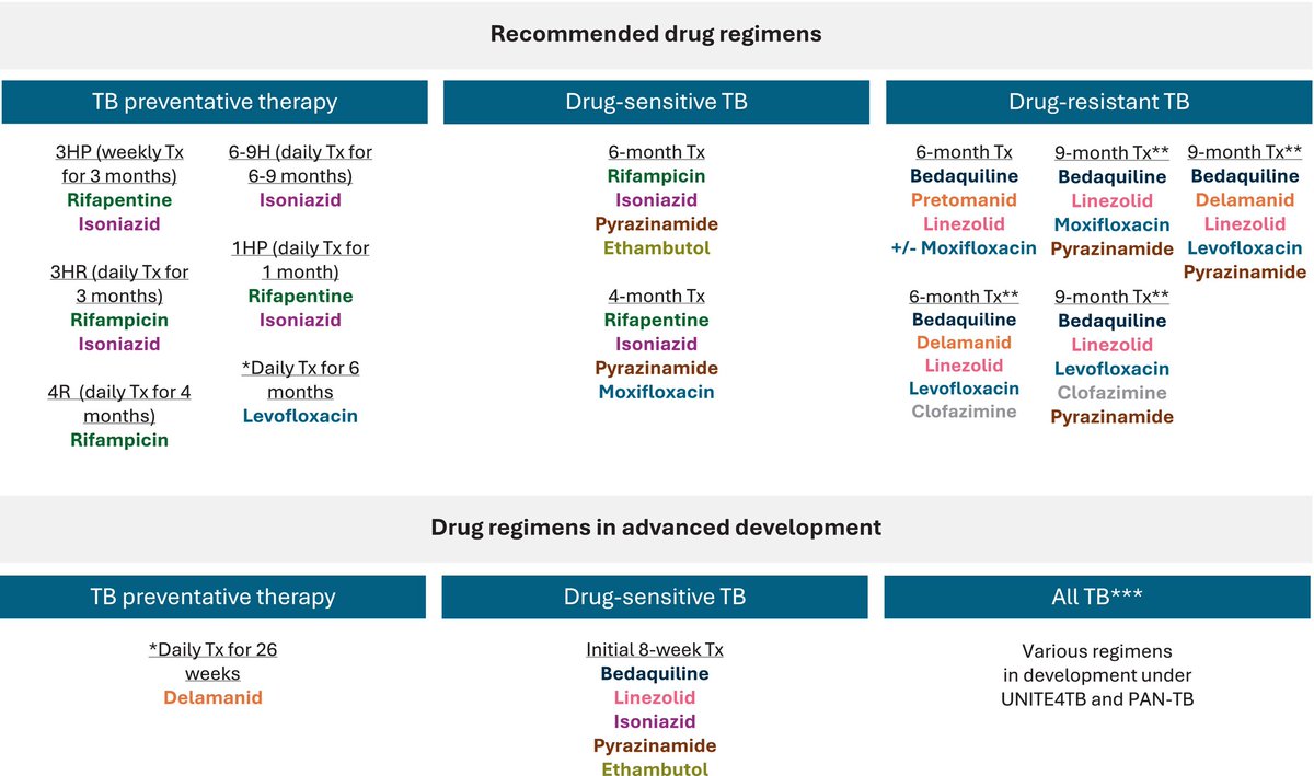
Today's Paper of the Day is:
Tuberculosis: An Update for the Clinician
https://t.co/JKgcYjlUQ5
Join us to read 1 paper per day and stay up-to-date as we cover the spectrum of critical care across 2026
🫀 Septic shock is not just about pressure. It’s about coupling.
We often treat septic shock with a familiar mindset:
➡️ Fluids
➡️ Vasopressors
➡️ Maybe inotropes
But we rarely ask the deeper question:
👉 Is the heart actually working efficiently with the arterial system?
⚙️ Enter: Ventriculo-arterial coupling (LVAC)
LVAC = Ea / Ees
• Ea → arterial load
• Ees → ventricular contractility
➡️ This ratio reflects how efficiently energy is transferred from the heart to the circulation
🧠 Why this matters
The cardiovascular system is not just a pump.
It is an energy transmission system.
From the pressure-volume perspective:
• Stroke work = useful energy
• Potential energy = wasted energy
• Total energy (PVA) ∝ myocardial O₂ consumption
👉 Efficiency depends on coupling, not just output.
📊 Key physiological insights
✔️ Optimal mechanical efficiency
→ LVAC ≈ 0.5
✔️ Maximal stroke work
→ LVAC ≈ 1
✔️ Septic shock
→ Often LVAC > 1 (uncoupling)
🚨 What happens in septic shock?
A complex mix:
• Vasodilation → ↓ Ea (sometimes)
• Myocardial dysfunction → ↓ Ees
• Microcirculatory chaos
• Variable preload
➡️ Result:
👉 Frequent ventriculo-arterial uncoupling
And here’s the key:
❗ Normal MAP ≠ optimal coupling
❗ Improved BP ≠ improved flow
💉 Therapeutic implications
Same MAP, different physiology:
🔵 Patient A
→ NE increases Ees > Ea
→ ↓ LVAC → ↑ SV (responder)
🔴 Patient B
→ NE increases Ea > Ees
→ ↑ LVAC → no SV improvement (non-responder)
🧬 Clinical reality check
Even more provocative:
• LVAC-guided resuscitation → faster lactate clearance
• BUT → no mortality benefit yet
And importantly:
👉 Optimizing coupling does NOT guarantee microcirculatory perfusion
🔥 Take-home message
We should move from:
❌ Pressure-driven resuscitation
To:
✅ Efficiency-driven hemodynamics
Because:
👉 The goal is not just to push blood
👉 The goal is to transfer energy effectively to tissues
📚 Caicedo Ruiz JD. et al. (2026)
Journal of Critical Care
https://t.co/1ZNMHqwBl4
🫀⚠️ Post-resuscitation vasoplegic shock: are we targeting the right physiology?
After ROSC, we often focus on blood pressure.
But the problem is much more complex.
🧠 What is really happening?
Post-resuscitation shock is not a single entity:
👉 Myocardial dysfunction
👉 Vasoplegia
👉 Relative hypovolemia
➡️ And their contribution changes over time in the same patient
📌 Current standard
✔️ MAP target: ≥ 60-65 mmHg
❌ No strong evidence supporting higher targets (>70 mmHg)
👉 Higher MAP may improve myocardial injury and cerebral oxygenation
👉 But no clear outcome benefit
💉 Vasopressor reality
• Norepinephrine = first-line
• Epinephrine → ↑ mortality, arrhythmias (observational data)
• Dopamine → ↑ arrhythmias
➡️ Catecholamines are necessary… but not harmless
🔥 The hidden problem: vasoplegia
Driven by:
• Ischemia-reperfusion injury
• Cytokine surge
• Nitric oxide overproduction
• Vasopressin deficiency
➡️ Leading to loss of vascular tone and responsiveness
⚖️ Are we overusing catecholamines?
High adrenergic load may cause:
• Myocardial injury
• Arrhythmias
• Immune dysregulation
• Receptor desensitization
➡️ This is why catecholamine-sparing strategies are emerging
🧩 What about vasopressin?
✔️ Physiological rationale exists
✔️ May reduce catecholamine exposure
❌ No strong evidence for routine use post-ROSC
❌ Possible ischemic risks
📊 Key concept: hemodynamic phenotype
Not all patients are the same:
• Early phase → low CO + high SVR
• Later phase → high CO + low SVR
➡️ Same MAP ≠ same perfusion
👉 This is where multimodal monitoring becomes essential
🚨 Critical insight
Optimizing MAP alone:
❌ Does NOT guarantee microcirculatory perfusion
❌ Does NOT ensure organ protection
🔥 Take-home message
Post-resuscitation shock is:
👉 Dynamic
👉 Heterogeneous
👉 Frequently oversimplified
And:
➡️ Vasopressor therapy should be individualized, not protocolized
📚 Jendoubi A. et al. (2026)
Critical Care
https://t.co/AdBRWWeHHP
🔥 Cytokine storm: when the immune system becomes the disease
We often say “hyperinflammation”…
But that doesn’t capture the reality.
👉 This is not just inflammation.
👉 This is a self-amplifying biological cascade.
📌 What actually happens
Cytokine storm is driven by:
• Excess cytokine release (IL-1, IL-6, TNF, IFNγ)
• Innate immune overactivation
• A positive feedback loop between cytokines and cell death (PANoptosis)
Once triggered:
➡️ Cytokines → cell death
➡️ Cell death → more cytokines
➡️ Loop continues → systemic collapse
⚠️ Why patients crash fast
It’s not linear. It’s exponential.
From the temporal model:
🟢 Hours–Days 1–2 (Onset)
Fever, early cytokine release
🟡 Days 2–5 (Amplification)
Immune recruitment, vascular leak
🔴 Days 5–10 (Crisis)
ARDS, coagulopathy, multiorgan failure
⚫ After Day 10
Either recovery… or immune exhaustion and death
🧠 Key clinical insight
👉 The damage is not only from cytokines
👉 It is from cytokine-driven cell death + endothelial injury
This explains:
• Capillary leak
• Microthrombosis
• Organ failure
• Shock physiology
🧪 Triggers are diverse
• Sepsis
• COVID-19
• Autoimmune diseases (SLE, MAS)
• Malignancy
• CAR-T therapy
Different triggers ➡️ Same final pathway
💡 The mistake
Treating it as “just inflammation”
Because:
❌ Not all cytokines are equal
❌ Not all phases respond to the same therapy
❌ Timing is everything
🚨 Bottom line
Cytokine storm is:
👉 A runaway immune feedback system
👉 Driven by PANoptosis + cytokine loops
👉 Leading to multisystem failure if not interrupted early
📚 Karki R. et al. (2026). Cytokine storm. Nature Reviews Disease Primers. https://t.co/5vXjDnlgI4
🩻 Mechanical ventilation in obesity: the physiology changes everything
We often apply “standard” ICU ventilation strategies…
But in obesity, the rules are different.
📌 The core problem
Obesity is not just weight.
It is a respiratory mechanics disease.
👉 ↑ Pleural pressure
👉 ↓ Functional residual capacity (FRC)
👉 ↑ Chest wall elastance
👉 Early airway closure + atelectasis
➡️ Result: hypoxemia, hypercapnia, and high VILI risk
🧠 Critical insight most people miss
👉 The lung is not bigger in obesity
So:
❌ Tidal volume based on actual body weight = dangerous
✅ Tidal volume based on predicted body weight (PBW)
➡️ 6-8 mL/kg PBW remains the cornerstone
⚠️ Airway pressures can mislead you
In obesity:
• Plateau pressure ↑
• Driving pressure ↑
But:
👉 This may reflect chest wall stiffness, NOT lung overdistension
➡️ Airway pressure ≠ lung stress
🔥 PEEP is not optional here
Because:
• High pleural pressure collapses alveoli
• FRC is critically low
👉 Many patients require higher and individualized PEEP
But:
❗ Optimal titration is still unclear
❗ One-size ARDS tables are insufficient
🧪 Advanced monitoring matters
To truly understand physiology:
• Esophageal pressure → transpulmonary pressure
• EIT → regional ventilation
👉 Move from “numbers” to mechanics-based ventilation
🚨 Airway management is high risk
• Rapid desaturation
• Difficult intubation
Best strategy:
✔️ Head-up / ramped position
✔️ Positive-pressure preoxygenation (NIV/HFNC)
✔️ Video laryngoscopy first-line
🔄 Extubation is not the end
👉 High work of breathing post-extubation
👉 Increased risk of failure
✔️ Consider prophylactic NIV
✔️ Assess carefully before liberation
💡 Bottom line
Ventilating obese ICU patients is not “standard ventilation + adjustments”
It is:
👉 A different physiological model
👉 Where pleural pressure dominates
👉 And interpretation matters more than numbers
📚 Kitisin N. et al. (2026)
Intensive Care Medicine
https://t.co/jf7fUxCIMf