@GIPathJC Since pathologists do not categorize dysplasia as conventional or non-conventional dysplasia, it is difficult to answer this question at this time.
@GIPathJC In our experience, non-conventional dysplasia was often reported as indefinite for dysplasia or had a descriptive diagnosis (such as ‘glandular atypia,’ ‘crypt atypia’, ‘atypia,’ ‘favor reactive atypia,’ etc) without a definite diagnosis of dysplasia.
@GIPathJC However, in our analysis of 207 consecutive total colectomy or proctocolectomy specimens of IBD patients, we found a total of 49 missed dysplastic lesions in 27 patients (13%).
@GIPathJC So, I would say that “false negative” cases are not uncommon, as many non-conventional dysplasia cases were ignored or missed as negative for dysplasia.
@GIPathJC In addition, it is reported that SATB2 loss could be potentially useful in identifying IBD-associated dysplasia; however, in our experience, SATB2 is often patchy and weak (even in normal colon) and difficult to interpret, so we don’t recommend this stain at this time.
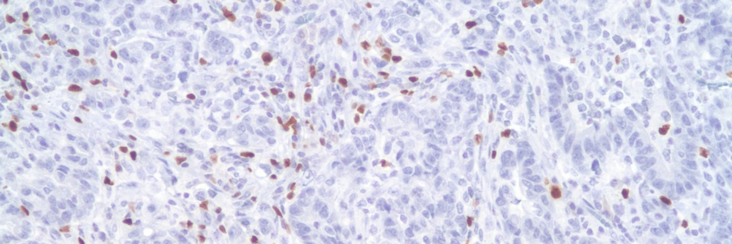
@GIPathJC We do not regularly perform p53 immunostaining in IBD-associated dysplasia cases. A diagnosis of dysplasia should be based on morphologic findings.
@GIPathJC However, wild-type staining pattern does not exclude a diagnosis of dysplasia, and pathologists should not be deterred from making a diagnosis of dysplasia even if p53 is weak or patchy (wild-type)
@GIPathJC If p53 staining is weak or patchy in a potential crypt cell dysplasia case, we often use a descriptive diagnosis (e.g., “crypt cell atypia”) or a diagnosis of indefinite for dysplasia.
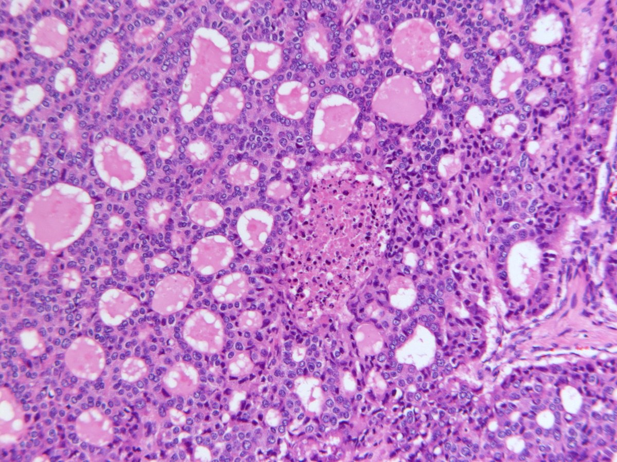
@GIPathJC However, in challenging situations like crypt cell dysplasia (mild enlargement and hyperchromasia of round/mildly irregular, non-stratified nuclei limited to the crypt base, no surface involvement), strong and diffuse p53 staining can be potentially helpful.
#GIPathJC
- Undetected dysplastic lesions were often associated with non-conventional dysplasia, flat/invisible gross appearance, and a smaller number of biopsies per colonoscopy
- Study recommends increased random biopsy sampling
#GIPathJC
Greater proportion of the undetected (19%) or previously detected (23%) dysplasia group had concurrent PSC compared with only 3% in the group without dysplasia
#GIPathJC
- Three (11%) patients in the undetected dysplasia group also had undetected CRC, of which two (67%) were found in the same colonic segment as nonconventional dysplasia