🌍Live from IPNTN Season VII, Session 1🌍
Head and Neck Pathologies, Take 5! with @amyfjuliano
💡Think carefully about the wording of reports! Mastoiditis is a clinical diagnosis, coalescent mastoiditis and other complications of mastoiditis should be assessed by imaging. And remember, not all mastoid opacification is infectious.
Hoy, en "Cosillas de cabeza y cuello"... (1/2)
Paciente con bultoma entre nariz y labio superior desde hace años, crecimiento lento. Al elevar el labio se observa un abultamiento de la mucosa alveolar, anterior al maxilar superior. Imagen de TC:
🎙️ Join the ESHNR Webinar!
📅 Jan 27 | ⏰ 18:00 CET
Imaging Approach to Dysphonia
👨⚕️ Speaker: @Alok_A_Bhatt
🎓 Moderator: @SalmanQureshiDr
🔗 Register: https://t.co/9kVZA8qj42
📖 Related reading: https://t.co/6W1QTr4iLW Insights into Imaging
👀 A sneak preview of the talk
Early diagnosis prompts IV antibiotics and ENT consult (consider I&D of subperiosteal abscess, myringotomy, +/-cortical mastoidectomy).
Complications include intracranial infxn (epidural abscess, meningitis, or temporal lobe brain abscess like here https://t.co/3NAqy0d4e0).
In the roetgenogram era, radiologists did not know about the squamous air cells, as they’re small and harder to see.
But even now with CT/MRI, suppurative infection in pneumatized squamous temporal bone is still rarely called as such.
🩻This mass originates in the right maxillary sinus and extends into the nasal cavity via the maxillary ostium. Can you appreciate the alternating lines of hyperintensity and hypointensity on the T2-weighted and post-contrast images? This is the "convoluted cerebriform pattern" typical of an inverted papilloma.
🔬These account for 3 - 5% of sinonasal tumours. While benign, there is a significant risk of malignant transformation (5 - 15%), usually to squamous cell carcinoma, and a propensity for recurrence.
💡Focal loss of the cerebriform pattern may indicate a site of malignant transformation.
💡On CT, an area of hyperostosis suggests the site of tumour origin; this will need to be removed after stripping the overlying mucosa.
💡The cerebriform pattern is not unique to inverted papillomas and can also be seen, rarely, in other sinonasal tumours.
MRI is recommended for evaluation of sudden sensorineural hearing loss. Do you know the yield and most common pathologies?
See this podcast based on the @TheAJNR paper by @klreinshagen
https://t.co/BGY1R7il0X
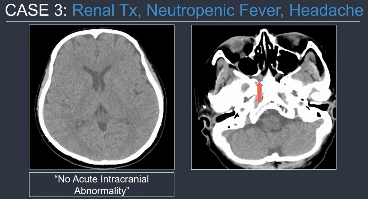
Immunocompromised patient, headache, head or face CT. You see sphenoid mucosal thickening.
Scrutinize the fat spaces. 👀
This was a case of invasive fungal sinusitis. The right pterygopalatine fossa is infiltrated. 🚩
I've seen this same miss!
@tabby_kennedy at #RSNA25
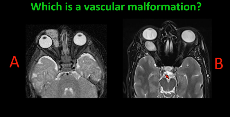
Hemangiomas are moderately T2 bright and may have flow voids.
Venous malformations (formerly/incorrectly known as cavernous hemangiomas) are very T2 bright and don't have flow voids.
@DShatzkes at #RSNA25
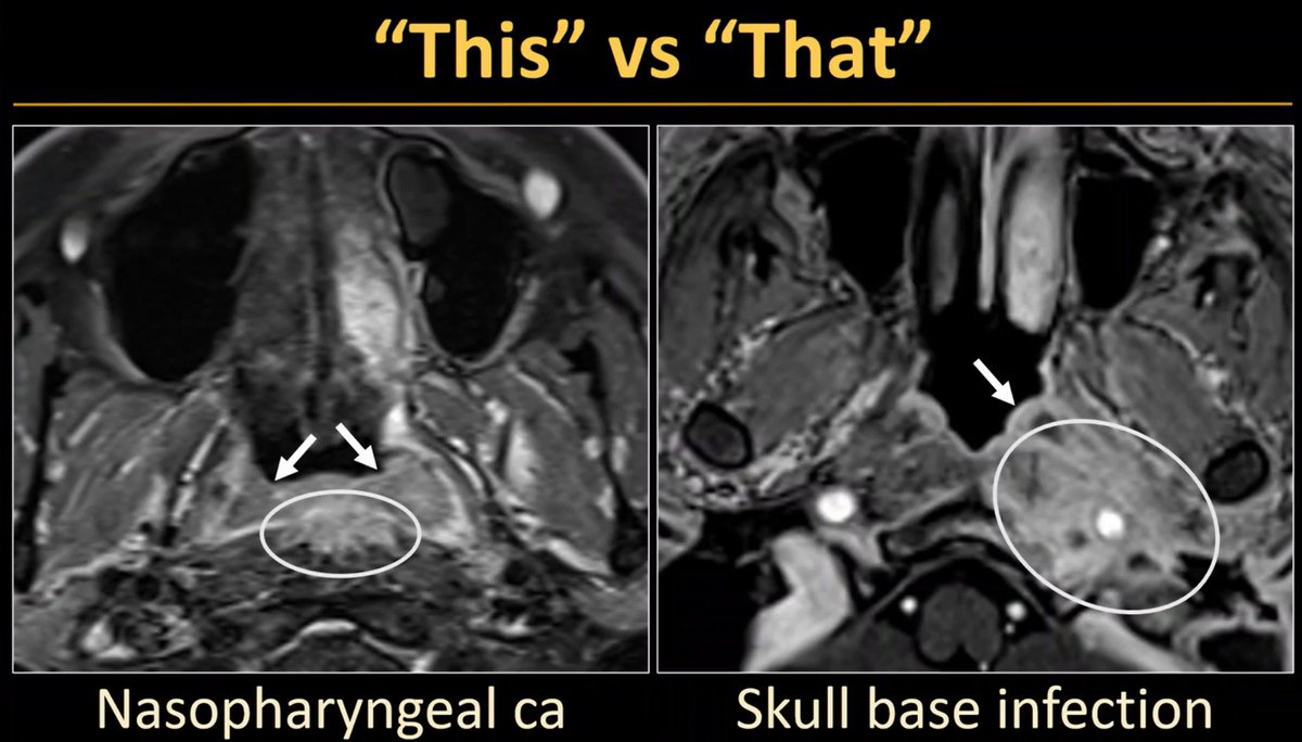
Even though we're not mucosa doctors, we can still look at the mucosa to guide our differential and certainty. Here, carcinoma involves mucosa, whereas skull base osteomyelitis related inflammation spares mucosa.
@pbunchmd at #RSNA25