Since 1939, Hitachi has developed and manufactured various Electron Microscopes including unique tabletop and ultrahigh voltage as well as SEM, TEM and FIB.
Happy Oral Health Month ! 😬🦷🪥👄
(June)
Cross-sectional SEM of a permanent tooth (wisdom tooth)
Cross-sections of a permanent tooth (wisdom tooth) were prepared by BIB (Broad Ion Beam) milling and observed using a Variable Pressure SEM. The two elongated holes in the whole image are root canals containing blood vessels and nerves. The tooth root surface is covered with a cementum layer showing a banded structure like trees thickening with age to form an annual-ring structure. This property has been used to determine the age of mammals. The cross-sectional image of the tooth neck shows the boundary between dentin and enamel. Though distinction between these two has been difficult with a torn surface, ion milled surface allows the identification. Four holes in the enlarged image are cross-sections of dentinal tubules regarded as to transmit pain and temperature.
To know more about BIB (Broad Ion Beam) milling, check out the following post !
Happy National Egg Day ! 🥚🍳🐔
(June 3)
Low-voltage & Low-vacuum SEM of food samples
Various food samples were observed with SEM without coating : fat crystals of chocolate, powder of corn potage, boiled egg yolk, and cross section of buckwheat. Low accelerating voltage SEM and low vacuum SEM revealed their fine crystal structures and surface topography.
To know more about the latest Low-voltage & Low-vacuum SEM, check out the following post !
After 35 years at #UMichPath, Yinru Sieracki is retiring and leaving behind a legacy of excellence and dedication to patient care. From work in electron microscopy to mentoring generations, her impact will be felt for years to come. Wishing Yinru a wonderful retirement filled with travel, art, and new adventures!
Read her story: https://t.co/IzXtK0GIGq
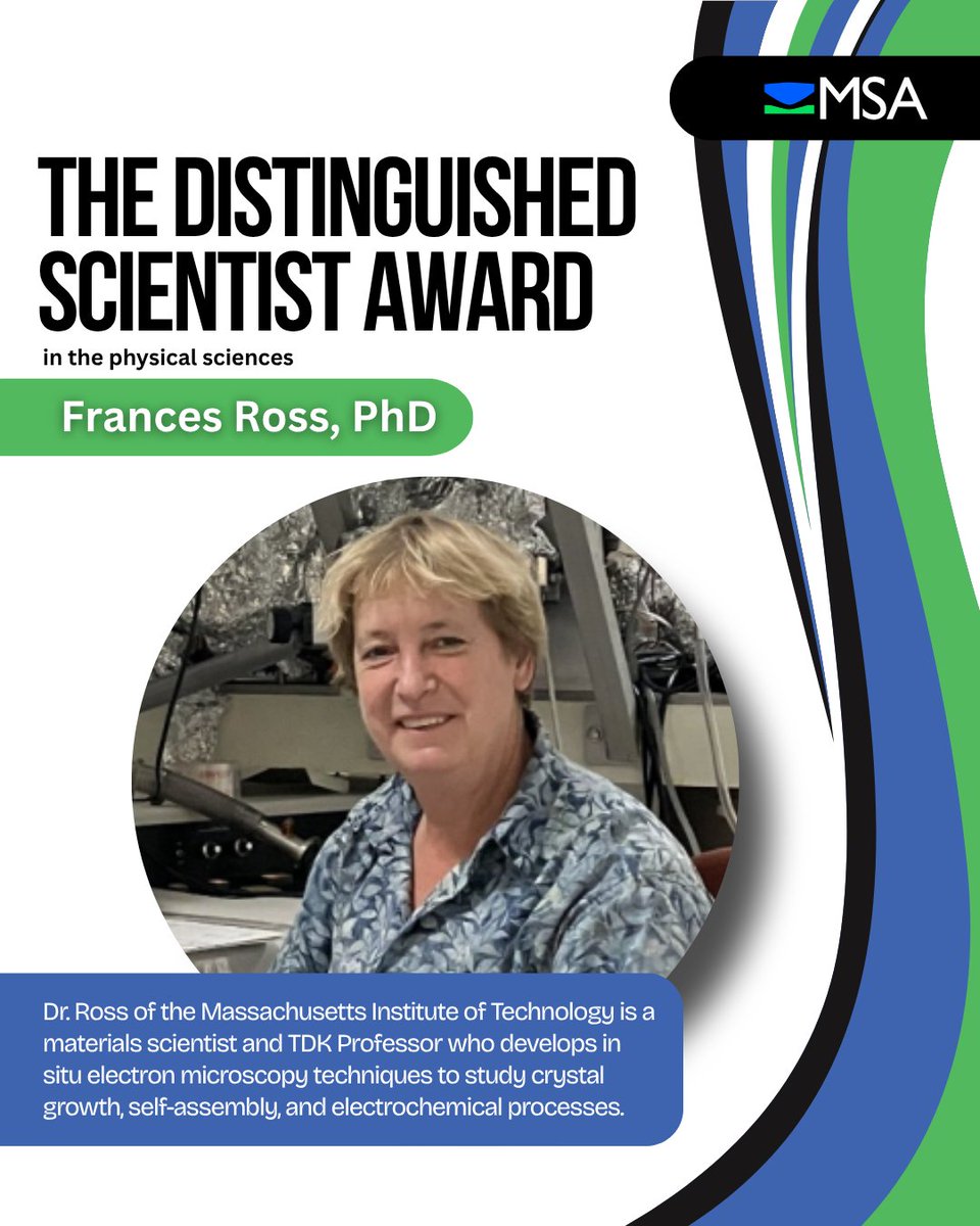
Frances Ross, PhD, of Massachusetts Institute of Technology, receives the Distinguished Scientist Award for her pioneering work in in situ electron microscopy, advancing our understanding of crystal growth, self-assembly, and liquid-phase processes.
https://t.co/1PkafdA5Om
🤖 Meet our "Aeye-1" — the world's first AI-driven transmission electron microscope system.
⚡ 300× faster image analysis
📸 4,000+ images/day
🧪 2 weeks of operation ≈ 1 year of conventional TEM workload
From materials to life sciences, AI is transforming how we explore the atomic world.
#CAS #AI #Microscopy #ElectronMicroscopy #TEM #AIForScience #MaterialsScience
The paper we use every day looks simple —
but under a microscope, its structure is completely different.
We compared these two:
Toilet paper
• Lots of gaps between fibres
• Breaks down easily in water
Paper wipes
• Thicker, more tightly tangled fibres
• Less likely to shed lint
Even though they are both paper,
their microscopic structures change depending on their purpose.
The closer we look at everyday things,
the more hidden design we discover.
If you found this interesting, let us know with a like!
Happy World Milk Day ! 🥛🐄🐮
(June 1)
SEM of liquid dairy products
Dairy products were observed in a liquid using newly-developed “Vitro” detector. The new detector utilizes the difference of dielectric constant for image formation to allow high-contrast imaging of unfixed or unstained light-element samples while suppressing sample damage. Milk-fat globules with a diameter of 150 nm to 3 μm were observed in the milk and the infant formula mixed in water. Many casein-micellar-like particles were recognized in the milk. A few particles found in the infant formula may be casein-micellar particles.
To know more about the Vitro Detector, check out the following post !
Happy National Smile Day ! 😄😸😊😎😆
(May 31)
“Smiling Pico-plankton”
This is a small organism of plankton collected in the northern Atlantic Ocean. The population of this plankton is high in cold water regions such as the Antarctic Ocean and the Arctic Ocean. People have very little knowledge about this plankton. Biological and ecological studies as well as other scientific investigation have just begun. Do you think that this plankton is smiling at you in the picture? It may appear in your dream tonight.
This work was presented at the "photo contest" hosted by the Japanese Society of Microscopy.
To know more about the image, check out the following post !
Happy Mint Julep Day ! 🍃🍸🍹
(May 30)
Cryo SEM of oil glands on a plant leaf (Apple mint)
To know more about plant imaging, check out the following post !
China developed the world's first AI-driven transmission electron microscope system, named "Aeye-1", marking a leap from manual operation to AI-driven fully autonomous operation.
The system can analyze 168 samples per day on average and acquire 4,000+ images, then even auto-generate professional analytical reports.
#TechChina #SciTech #AI
Join us at the Microscopy Society of Canada/Société de Microscopie du Canada (MSC-SMC) Summer School & Annual Meeting in Montréal! Come learn about our latest lineup of microscopy and microanalysis products and solutions.
🔴 2026 MSC·SMC Summer School & Annual Meeting
🗓️ May 25-29, 2026
📍 McGill University, Montreal, QC, Canada
📩 Contact us at [email protected] to make an appointment
#microscopysocietyofCanada #electronmicroscopy #microanalysis #mcgilluniversity
Available tools for this measurement are :
Field Emission Transmission Electron Microscope
https://t.co/Gh9tSD9iYr
Further details and previously posted applications can be found on our YouTube channel. Looking forward to your visit.
https://t.co/sL2jHVgsyG
Au nanoparticles were observed by TEM in liquid using K-kit. Observation in the liquid allows confirmation of the dispersion state of the Au nanoparticles. Crystal lattice fringes of the Au nanoparticles were clearly observed at higher-magnification even in the liquid.
To know more about the imaging, check out the following post !