It's that time again! The students here @IL_Wesleyan have submitted another set of #microscopy images for our annual competition. The top 10 images are in the thread below-- Feel free to tell us your favorite 🔬🦠🔬🦠🔬🦠🔬🦠🔬🦠🔬🦠🔬🦠
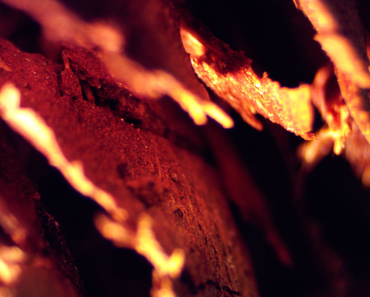
This is a picture of a snakeskin's eye area I captured using the AMScope camera. I used the dissecting microscope with reflective light at a magnification of 10x.
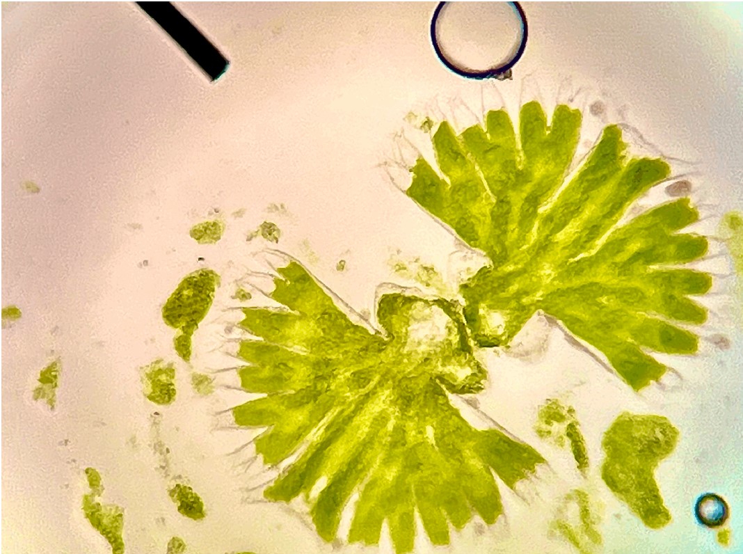
This is a image taken of the Micrasterias Organism at 300x magnification and a compound microscope. In this image the lobes of the organism are visibly present (clear ends), the chloroplasts (green areas of organism), as well as one Micrasterias proceeding through cell division!
A 15x magnification with a dissecting microscope and reflected light. The object shown in the image is a leaf changing colors with differing shades. You can see the blades and the lamina of the leaf in this photo!
Hello #ScienceTwitter! The students in #GenBio lab @ILWesleyan have been learning a bit about microscopy 🔬🔬🔬 and have some images to share with you. We have a little friendly competition going. Scroll through the thread below and like your favorite #microscopy image! 1/11
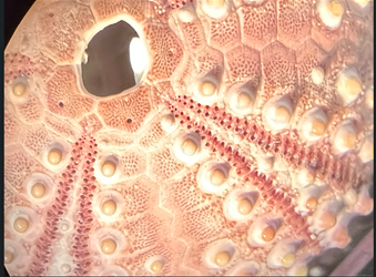
This is an image of a purple/pink sea urchin test at a total magnification of 45x. The test is a hard shell with creme colored bumps on it. You can see the hole where the anus of the sea urchin would have been, the pores for the tube feet, and the primary tubercles. 11/11
Hello #ScienceTwitter! The students in #GenBio lab @ILWesleyan have been learning a bit about microscopy 🔬🔬🔬 and have some images to share with you. We have a little friendly competition going. Scroll through the thread below and like your favorite #microscopy image! 1/11
Sea urchin test at a magnification of 10x. The surface of the dried sea urchin test is covered with white salt crystals. You can also see many bumps where the sea urchin spines were attached. 10/11