This evidence-based guide breaks down the Surviving Sepsis 2026 bundle: from fluids and vasopressors in the first hours to antibiotics, source control, and adjunctive thresholds every clinician needs to know.
We’ve all spent hours debating volume status at the bedside and it’s still one of the hardest things to assess in medicine. A quick visual summary of how physical exam findings, POCUS, and BNP perform for identifying (and ruling out) volume overload #MedTwitter
ATLS has come out with some new key takeaways to help with management of massive hemorrhage. A key difference is the x-ABCDE focus on stabilization that emphasizes stopping the “eX-sanguinating bleed” first before ensuring an airway.
The Renove Trial was completed to assist in understanding what clinicians should consider for patients with high VTE recurrence risk. It showed low dose DOACs were noninferior to full dose DOACs in preventing future VTEs. 1/2
Delighted to present our clinical case report at the National Kidney Foundation Spring Clinical Meeting 2026! Inspired by the ground breaking research transforming kidney care!
#NKFSCM2026@nkf
Autoimmune hepatitis can masquerade as many things! Recognizing the clues early matters. A tweetorial on diagnosis, pitfalls, and management pearls. 🧵 #MedTwitter#LiverTwitter#Hepatology
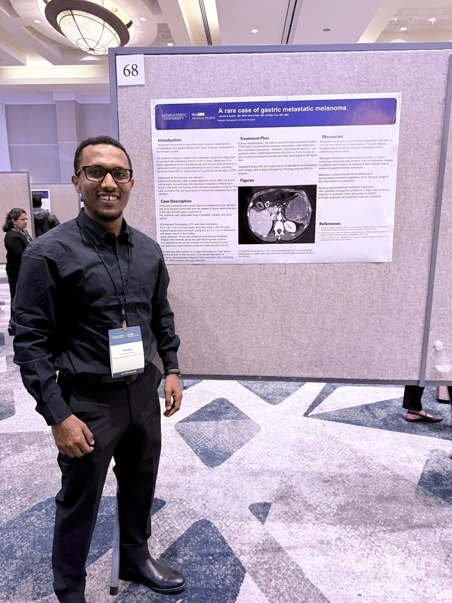
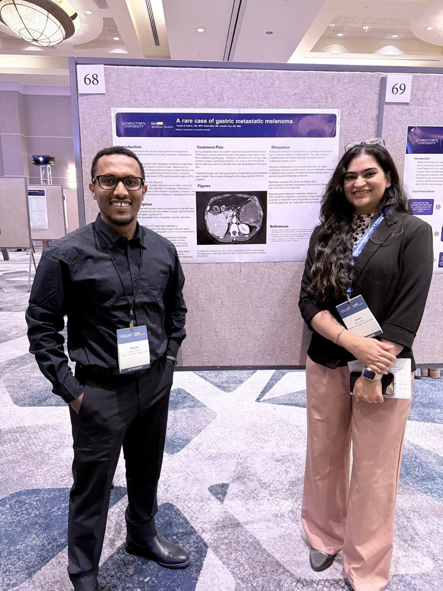
Today, I presented a case of pyogenic liver abscess with transdiaphragmatic extension and concurrent lung abscess due to Streptococcus intermedius. Was nervous but excited to talk through such a cool case!
🏠 Take-home:
1. Echo gives a PASP estimate, not a diagnosis
2. Use ESC probability (Low/Intermediate/High)
3. Normal echo does NOT rule out PH if suspicion is high
4. RHC is required to confirm dx, measure PVR, and classify hemodynamic profile
🫁 Your patient has dyspnea on exertion. The echo report says "PASP 52 mmHg - consider pulmonary hypertension."
But does that mean they have PH?
Not necessarily. Here's what echo can (and cannot) tell you. 🧵 #MedEd#Cardiology#Pulmonary
PH is classified into 5 WHO Groups:
1️⃣ PAH (idiopathic, CTD, drugs)
2️⃣ Left heart disease ← common
3️⃣ Chronic lung disease/hypoxia
4️⃣ CTEPH
5️⃣ Multifactorial
Why does this matter? Because Group 2 & 3 do NOT get PAH therapy. Getting the group right changes everything
So, what do you do with an abnormal echo?
Clinical suspicion → 🔬 TTE → Low prob: look for alternatives → Intermediate/High: V/Q scan → ➡️ Right Heart Catheterization
Echo probability guides who goes to cath - not whether PH exists.
The overdiagnosis problem is real.
When borderline elevation on echo is compared to invasive mPAP?
~50% correlation.
High PASP on echo alone ≠ PH diagnosis. It's an indication to look further - not a diagnosis to hang your hat on.
Fisher MR https://t.co/zwZcEdlglK PMID 19164700
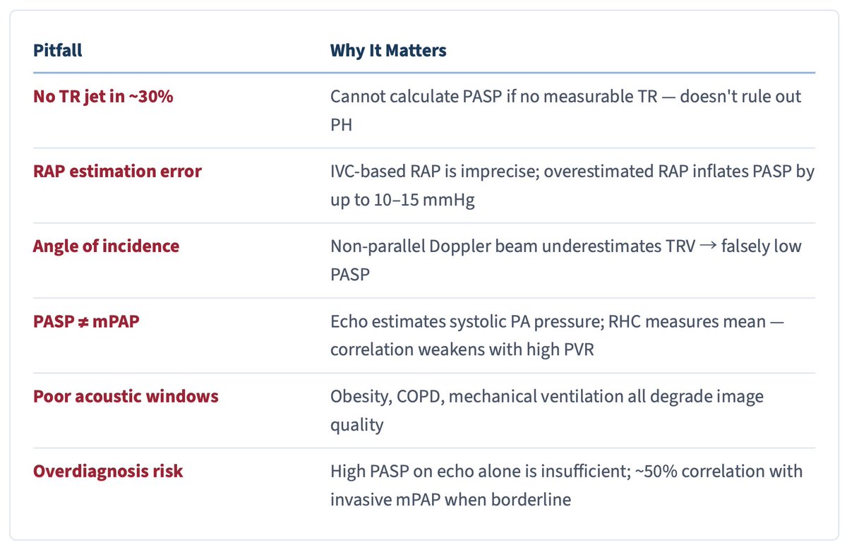
However, Echo lies. Here are some of the ways how:
❌ No TR jet in ~30% of patients → can't calculate PASP (absence ≠ no PH)
❌ IVC-based RAP overestimates by up to 10–15 mmHg
❌ Non-parallel Doppler beam → underestimates TRV
❌ PASP ≠ mPAP - they diverge as PVR rises
The 2022 ESC/ERS Guidelines moved away from a single TRV cutoff.
Now we use echo probability categories (TRV + signs):
✅ TRV ≤2.8 + no signs → Low
🟡 TRV ≤2.8 + signs OR 2.9–3.4 → Intermediate
🔴 TRV ≥3.4 → High
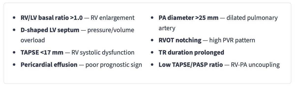
"Signs" = RV dilation, D-septum, TAPSE <17mm, PA dilation