A lesion that looked like #sarcoma.
MRI was concerning.
Biopsy showed only necrosis.
Grossly, it even resembled #strangulated#bowel.
The diagnosis?
A chronic expanding #hematoma related to prior #liposuction
Sometimes the most important finding isn’t on the slide—it’s history
12 consecutive outpatient Family Medicine comparisons of #creatinine-based vs #cystatinC based #eGFR
📊 33% good agreement
⚠️ 25% mild discordance
🚨 42% major discordance
Even in routine outpatient practice, kidney function assessment may not be as straightforward as it seems
#BRCA management is evolving:
• Breast: Annual MRI surveillance an option for selected patients. • Ovary: Risk-reducing salpingo-oophorectomy remains the standard.
Why?
• MRI highly sensitive for breast screening. • Ovarian cancer lacks an equivalent early detection tool.
BRCA-positive no longer automatically means bilateral prophylactic mastectomy.
With modern guidelines supporting annual MRI surveillance for selected patients, now more personalised
Q
Should #BRCA1 & 2 carriers be managed differently, given distinct biology and risk profiles?
Most lab errors occur before analysis.
How do you monitor specimens that are:
✅ Collected
❌ Not Received
Dashboards? RFID? Tube integration? Daily reconciliation?
Curious to learn what works in your organization.
#HealthcareIT#LabMedicine
Not every “cancer” is cancer.
IgG4-related disease can present as a hard mass in the salivary gland, pancreas, orbit, kidney, lung, and many other organs—often mimicking malignancy on imaging and clinically.
Before calling it a tumor: sometimes the immune system is the culprit.
Congratulations to May’s #PathArt winner, Ruchi M. Patel, MD, FCAP, as voted by the CAP’s Digital Content Committee. Check out Dr. Patel’s piece, titled “The Vascular Butterfly.”
CAP members, submit your PathArt for next month’s contest here: https://t.co/vS0RswPlyn
Every patient remembers two things:
How we treated their illness.
And how we treated them as a person.
The second often lasts longer.
❤️ #PatientExperience#Healthcare
Great healthcare is not built by individual expertise alone—it is created when medical practitioners unite around a shared purpose. Through collaboration, clear communication, & innovation, transform knowledge into action, and patient care into lasting impact.
“Diagnose by behavior, not by shape.”
Polypoid melanocytic lesions remind us that large size, dark pigmentation, and a pedunculated appearance can be misleading. Histology—not clinical drama—determines the diagnosis. 🔬 #Melanoma#DermPath#PathologyCases
Can you make the diagnosis from this pic? Only one tumor looks like this (ok, maybe 2).
WSI digital slide 🔬 https://t.co/WBuuWU9cA9.
Answer & DDX ✅ https://t.co/ZGPtZVODD4.
Video ⏯️ https://t.co/xXjLu0RpAO
#BSTpath#pathologists#pathology#pathTwitter
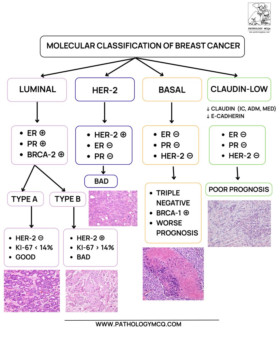
🔬✨ Molecular Classification of Breast Cancer — Made Visual & Easy
🧬 From hormone-driven luminal tumors to aggressive triple-negative and claudin-low variants, this chart connects IHC profile + morphology + prognosis in one glance.
#Pathology#BreastCancer#MolecularPathology
Bread and butter post: Inflammatory cloacogenic polyp is a mucosal prolapse polyp at the junction of rectal glandular mucosa and anal squamous mucosa. Note that the the lamina propria is filled with smooth muscle rather than the usual complement of inflammatory cells.
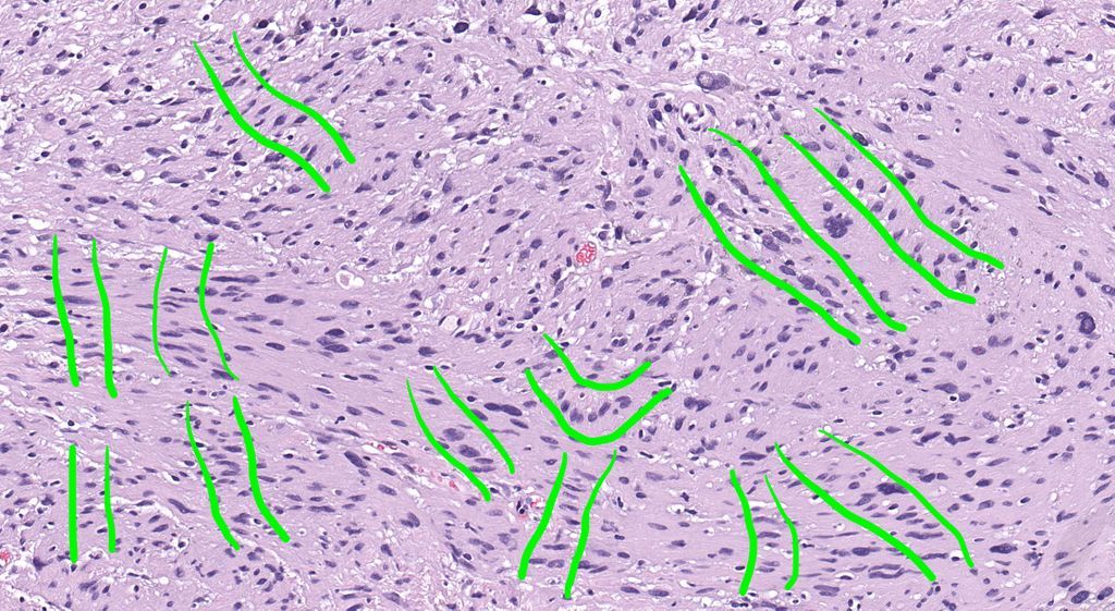
Soft tissue mass. S100+. Diagnosis?
Answer ✅ https://t.co/RLP2My4DDK
What am I highlighting with these green lines?
Explanation & more pics https://t.co/bj7B2HJ7cD
WSI digital slide 🔬https://t.co/rxy2be8cLV
#pathology #pathologists #pathTwitter #dermpath #dermatology #dermtwitter #BSTpath
Case of the Week: radiation-associated epithelial changes in duodenal mucosa — aberrant p53 expression can reflect upregulation after chemoradiation, not mutation. Read: https://t.co/kyDpE6jqht #Pathology#GIPathology#Radiation