A collaboration between Institut Fresnel at Aix Marseille Univ and Centre de Biologie Intégrative at Toulouse Univ has made EDF-RIM possible. Special congratulations to the co-first authors #LorryMazzella@TMangeat
https://t.co/EcegqLaANJ
@CNRS@InstitutFresnel@CNRS_DR12@CBI
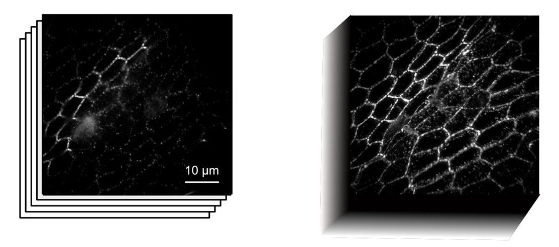
Published in Light: science & Application. Mazzella et al Introduce EDF-RIM, a technique to capture 3D objects combining super-resolved structured illumination and extended depth imaging.
https://t.co/81YuD2vlbY…
#Microscopy#Biology#fluorescence@LightSciAppl
This image compares EDF-RIM with a standard extended depth wide-field. Demonstrating how EDF-RIM allows improved resolution and decreased background.
#imaging
🧐Une nouvelle imagerie projective pour l’étude des tissus biologiques ou comment voir vite, profond et super-résolu avec des illuminations aléatoires ?
L'article vient de paraitre dans Light: Science & Applications
Abstract➡️https://t.co/wb6XDG1wAq
Full📰https://t.co/7xXxPoBM8q
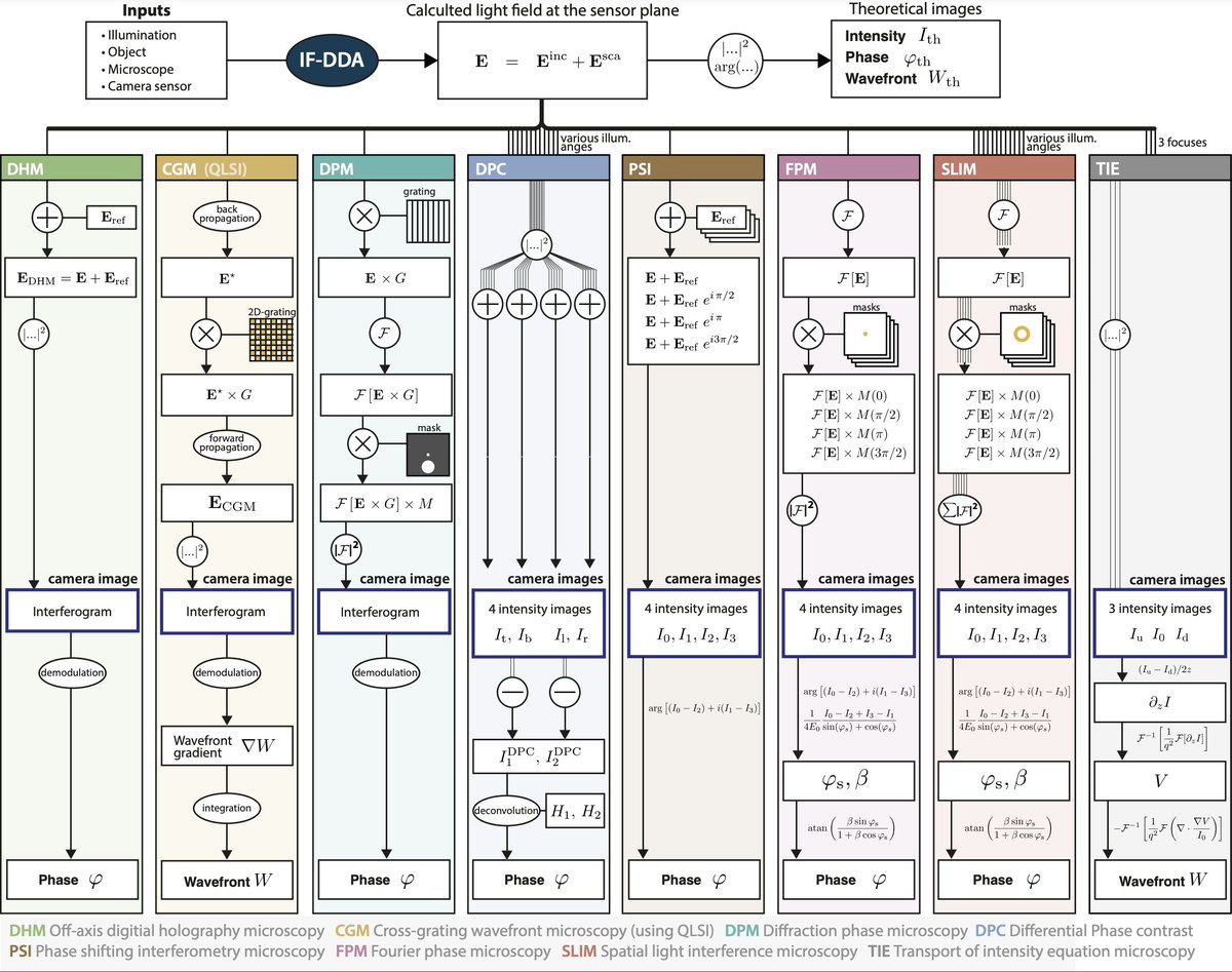
Results highlight key differences in artifact and noise handling. Techniques like DHM and CGM stand out for their robustness, while SLIM and FPM show limitations with large objects. Full insights in the @LightSciAppl article!
4/4
@CNRS@InstitutFresnel@CNRS_DR12
A recent work from the imaging group supervised by @gbaffou at Fresnel was published in @LightSciAppl.
Find below the link:
https://t.co/Z4kBKAxcE5
1/4
The authors compare 8 major #QPM techniques like DHM, CGM, DPC, and SLIM among others. They explore their precision, accuracy, and limitations, using a custom-built numerical toolbox (IF-DDA) aimed to model any microscopy technique.
3/4
Congratulations to all contributors, especially to first author Annafrancesca Rigato. This work is a collaboration between Loic Le Goff from mosaic group at Institut Fresnel and the @RichardSmithLab at the John Innes Centre, Norwich. Read more here: https://t.co/OLEryrMcdK
4/4
In a publication in PLos Biology, researchers at Institut Fresnel uncovered a new morphogenetic process in the developing Drosophila epidermis. https://t.co/OLEryrMcdK #Drosophila#DevelopmentalBiology
1/4
The study demonstrates that this shape change is due to a buckling mechanism, using longitudinal imaging in larvae, genetic perturbations, laser dissections, optical tweezers, and simulations.
#buckling
3/4
Check out our joint tutorial with Ivo Leite, Robert Prevedel (@Prevedel_Lab@EMBLHeidelberg), & Thomas Chaigne (@TomChaigne@InstitutFresnel) on optical wavefront shaping with photoacoustic feedback. Thank @cziscience for supporting open science research. https://t.co/eEWWsW4Gq4
Congratulations to all contributors, especially to first author Barbara Sarri. This work is a collaboration between mosaic group @InstitutFresnel, Lightcore Technologies, and the Cancer Research Center o Marseille @crcm_marseille.
Read more here: https://t.co/Lk9jNXICsU
4/4
In a recent publication in npj Imaging, researchers at Institut Fresnel developed a non-destructive imaging technique (Stimulated Raman Histology) to study patient-derived tumor organoids, enhancing preclinical drug screening.
#Organoids
https://t.co/Lk9jNXICsU
1/4
Using Stimulated Raman Histology (SRH), this technique targets molecular vibrational signatures for virtual staining, preserving organoid viability. "SRH allows in-depth, specific screening of each model in minutes," explains Hervé Rigneault from CNRS. #SRH#Microscopy
3 /4