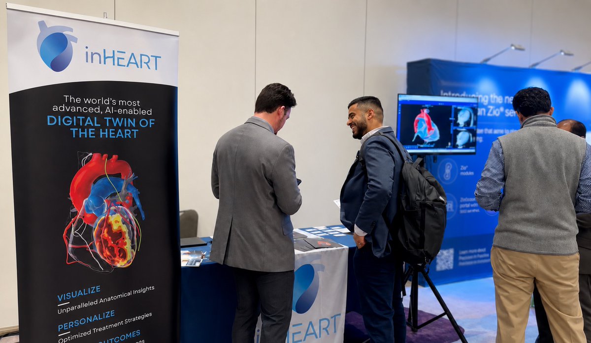
Faster, clearer and more precise LBB area visualization on fluoroscopy—is it possible?
With the integration of inHEART’s 3D anatomical imaging into your #fluoroscopy system, you can bring added clarity and streamline your workflow. See it in action below.
LBB Area Pacing with Dr. Nicolas Combes from @cliniquepasteur.
Want to know how this could integrate into your practice? Visit us at booth E400 at EHRA, April 12–14.
@GEHealthCare@Medtronic #epeeps #fluoroscopy #LBB
What can CT and MRI imaging reveal about the arrhythmogenic substrate?
In this case of Post Myocardial infarction VT, CT and LGE-MRI were combined in a patient-specific inHEART 3D model to characterize scar, wall thickness, fatty infiltration, calcifications, and potential conduction channels - supporting VT ablation planning.
Read the full case discussed with Vincent Pannetier, Imaging Analyst Manager at #inHEART, to discover how multimodality imaging can be translated into actionable 3D insights.
🔗 https://t.co/0CEDP6fIF0
#epeeps #ischemic #VTablation #cardiacimaging
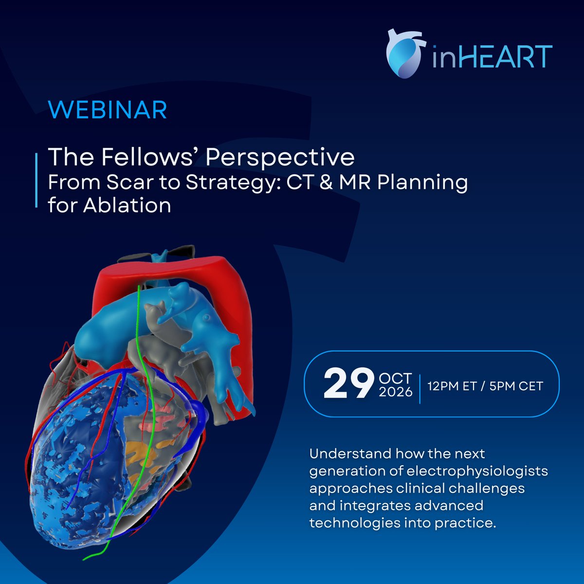
CT and MR imaging can offer valuable insights when preparing for complex ablation procedures. But how do these insights translate into procedural planning?
Join us for The Fellows Perspective, an #inHEART webinar bringing together clinical perspectives on imaging-based planning for ablation.
🗓️ October 29, 2026
⏰ 12 PM ET | 5 PM CET
📍 Online via Zoom
Can’t attend live? Register anyway, and we’ll send you the recording after the webinar!
👉🏻 Register now: https://t.co/IGMQ7jaMa6
Dr. Francesco Notaristefano, MD, Dr. @DrSouvikKDas, MD, Dr. @GZingarini, MD, Dr. @Davilandre, MD, PhD #epeeps #cardiacimaging #ablationVT #ablationplanning
Great to see imaging and mapping come together so clearly in this recent case with Dr. @EsseimS and the @UHhospitals Cleveland team 💪🏻
An initial ILAM map demonstrated deceleration zones along the border of fatty infiltration in the RV. The #VT was easily inducible and hemodynamically tolerated, allowing mapping in VT as well. The circuit location correlated nicely with the imaging and was confirmed by entrainment.
The VT terminated after only a few lesions and, after additional ablation in the area of interest, was rendered non-inducible. The entire procedure was completed in under three hours.
Thank you Mitchell Kershner for the excellent mapping.
#epeeps #cardiacimaging #VTablation #inHEART
Shout-out to our amazing team who took on the #BreastCancerAwarenessMonth challenge by running 5.5 km together! 🩷
Thank you for your commitment to raising awareness about breast cancer and the importance of prevention and early detection.
#BreastCancer#PinkOctober #RubanRoseChallenge #inHEART
On October 29, join #inHEART Fellows Webinar to hear how the next generation approaches CT & MR planning and the challenges that come with it.
👉🏻 Register now: https://t.co/IGMQ7jaMa6
On the agenda: complex real-world cases, practical insights into VT ablation planning, and an open discussion on the fellows' experiences and perspectives.
🗓️ October 29, 2026
⏰ 12 PM ET | 5 PM CET
📍 Online via Zoom
Presenters
Dr. Francesco Notaristefano, MD - Cardiologist & Electrophysiologist, Azienda Ospedaliera di Perugia, Italy
@DrSouvikKDas, MD - Cardiologist & Electrophysiology Fellow, Beth Israel Deaconess Medical Center, Boston, USA
Panelists
Dr. @GZingarini, MD - Cardiologist & Electrophysiologist, Azienda Ospedaliera di Perugia, Italy
Dr. @Davilandre, MD, PhD - Director, Cardiac Arrhythmia Service, The Harvard Thorndike EP Institute, Beth Israel Deaconess Medical Center, Harvard Medical School, Boston, USA
Can’t make it live? Register anyway and we’ll send the recording straight to your inbox!
#webinar #cardiacimaging #VTablation #electrophysiology
Curious about how image integration can support ablation procedures in patients with congenital heart disease? Join us virtually today at #EPAC conference for Prof. Francis Bessière’s presentation: “Navigating Complex Anatomy: How Image Integration Is Changing Ablation in Congenital Heart Diseases”. 🗓️Wednesday, October 7th ⏰ 2pm CET ��� https://t.co/Som5wWNnzd @DrBessiere #EPACEgypt #congenital #cardiacimaging #epeeps #electrophysiology
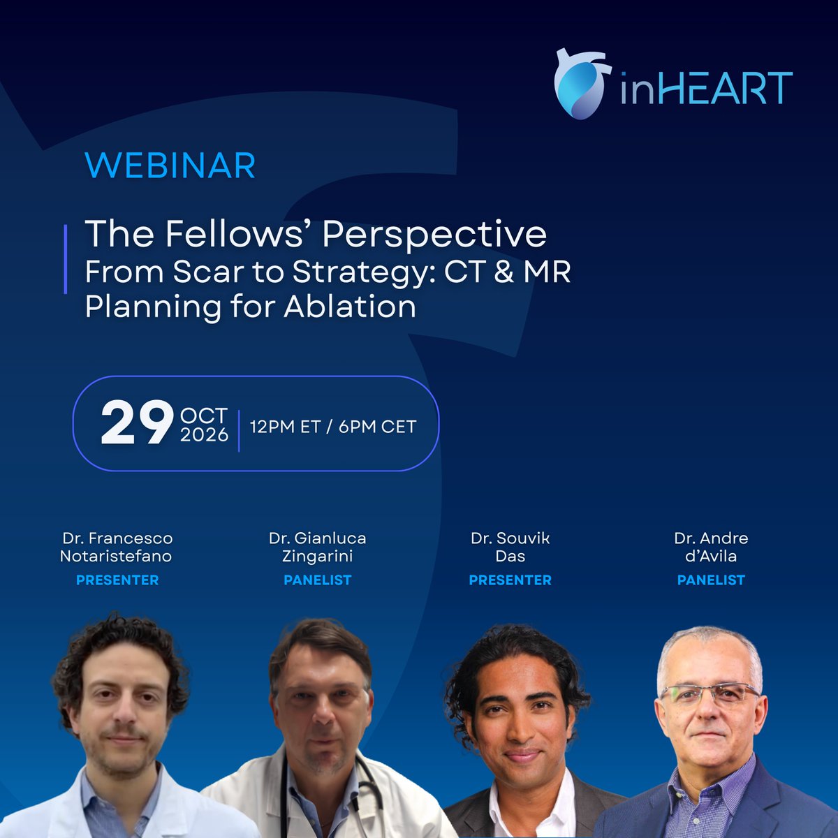
🎙️ Join us on October 29, 2026 for #inHEART next webinar, “The Fellows Perspective | From Scar to Strategy: CT & MR Planning for Ablation.”
Presenters
Dr. Francesco Notaristefano, MD - Cardiologist & Electrophysiologist, Azienda Ospedaliera di Perugia, Italy
Dr. Souvik Das, MD - Cardiologist & Electrophysiology Fellow, Beth Israel Deaconess Medical Center, Boston, USA
Panelists
Dr. @GZingarini, MD - Cardiologist & Electrophysiologist, Azienda Ospedaliera di Perugia, Italy
Dr. @Davilandre, MD, PhD - Director, Cardiac Arrhythmia Service, The Harvard Thorndike EP Institute, Beth Israel Deaconess Medical Center, @harvardmed, Boston, USA
Can’t make it live? Register anyway and we’ll send the recording straight to your inbox!
👉🏻 Register now: https://t.co/IGMQ7jbjZE
@bethisraellahey #cardiacimaging #VTablation #electrophysiology
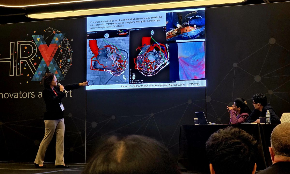
We still can’t get over how great Dr. @utedrow presentation was last weekend at the #HRX Innovation Summit! 👏🏻
Thank you for showcasing how #inHEART’s advanced imaging technology can extract key information from MRI and CT to support the planning and guidance of complex EP ablation procedures.
#HRX #epeeps #cardiacimaging #VTablation
What an incredible first day at the #AIAC Congress in Bologna, Italy! 🇮🇹
It feels great to connect with the Italian #EP community and discuss the latest advancements in the treatment of cardiac arrhythmias.
Stop by booth 17 to discover how #inHEART can support ablation planning and help reduce procedure time with patient-specific 3D cardiac models.
#epeeps #AIAC #ablationVT #AFib #cardiacimaging
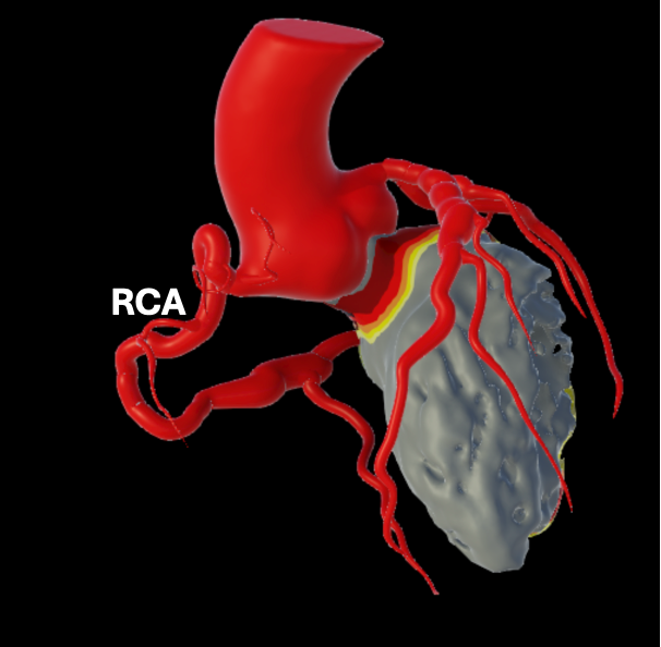
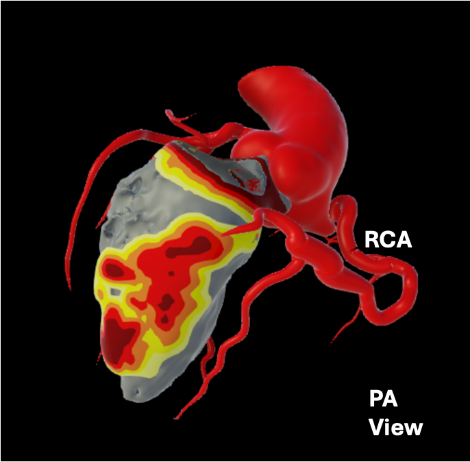
Congratulations, Dr. @EsseimS, on this excellent case highlighting the value of combined detailed anatomical and electrical mapping in a patient with coronary aneurysms. Great to hear that the patient is now VT free.
#epeeps#VTablation#aunerysm#inHEART
Young pt w/ Kawasaki's and recurrent VT despite amio. Great ex. of coronary aneurysms on 3D inHeart model. Discrete inferior scar arising from dominant RCA. Wall thinning matched up perfectly w/ scar and WADL mapping. VT free >6 mo & counting. #AblateVT@inheartmedical
Great to join Dr. @slatercharles1 and the team at Hospital Samaritano for the first inHEART case in Rio de Janeiro, Brazil. Obrigado! 🇧🇷
Patient had a large ischemic scar following a circumflex infarction, with multiple potential channels throughout the inferior wall. Combining pre-procedural #inHEART imaging with Deep VT Mapping and ILAM helped correlate the scar architecture with the electrical substrate and identify areas most relevant to the VT. Strategic linear ablation and selective substrate homogenization ultimately rendered the VT non-inducible.
Always rewarding to see imaging and mapping come together in a complex VT case.
#Epeeps #VTAblation #CardiacImaging #EnSiteX #ILAM
The inHEART team has just arrived at ESC Congress 2026!
This year’s spotlight on #AI strongly resonates with what we do every day at inHEART: creating AI-powered digital twins of the heart to support physicians in pre-procedural planning and enable faster and safer ablation procedures.
Looking forward to connecting with the cardiology community in Munich!
@escardio #ESCCongress #epeeps #cardiacimaging
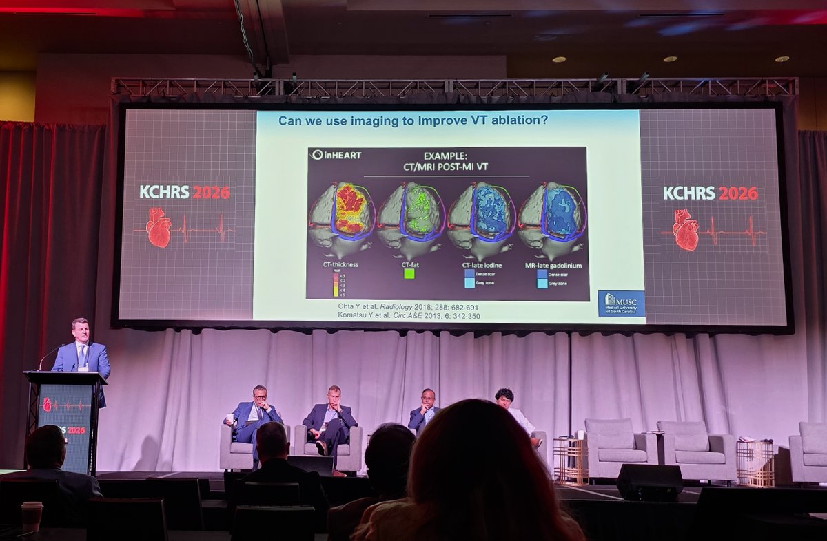
Another great #KCHRS in Kansas City!
Thrilled to see inHEART featured in presentations from Dr. @JRWinterfield on imaging in VT ablation and Dr. @jczerpa highlighting epicardial substrate in a Chagas case.
A standout moment was seeing Dr. William Stevenson honored for his extraordinary contributions to electrophysiology and VT. A truly well-deserved recognition! 👏🏻
It's always a pleasure to reconnect with friends and colleagues across the EP community. Congratulations to the KCHRS team on another outstanding program!
#KCHRS2026 #Electrophysiology #VT #CardiacImaging #inHEART
Congratulations to Dr. Esseim Sharma on this complex VT procedure and excellent outcome👏🏻
A great example of how pre-procedural cardiac imaging can provide valuable insights in complex VT cases. Thank you for sharing.
#epeeps#VTablation#cardiacimaging#electrophysiology
5/ VT morphology didn't look particularly epicardial, but was clearly LV apical.
Could pre-op imaging predict whether epi-only can succeed? @inHeartmedical showed extensive wall thinning + potential VT channels right in the predicted area of interest. Promising!
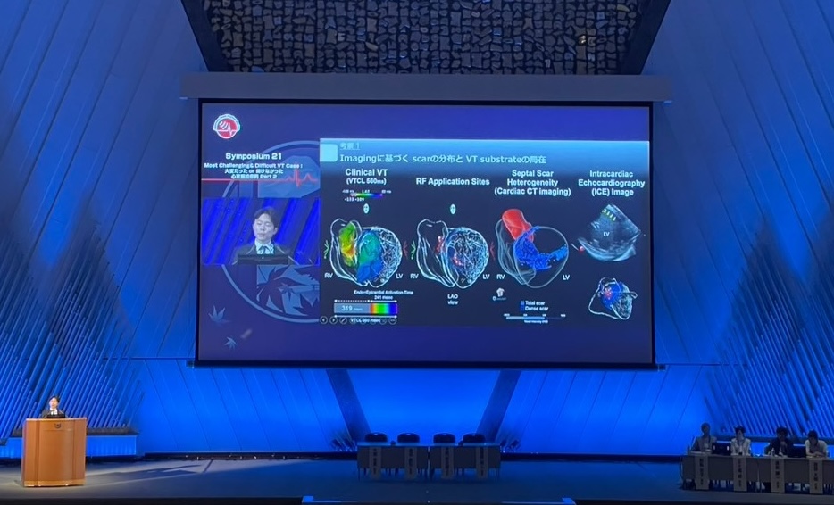
We are proud to see Dr. Masato Hachisuka presenting once again at the VT Symposium during #JHRS2026! 👏🏻
This year, he showcased how #inHEART imaging analysis, combined with pathological findings and micro-CT analysis, can provide deeper insights into ventricular tachycardia substrates.
#JHRS26 #epeeps #electrophysiology #cardicimaging
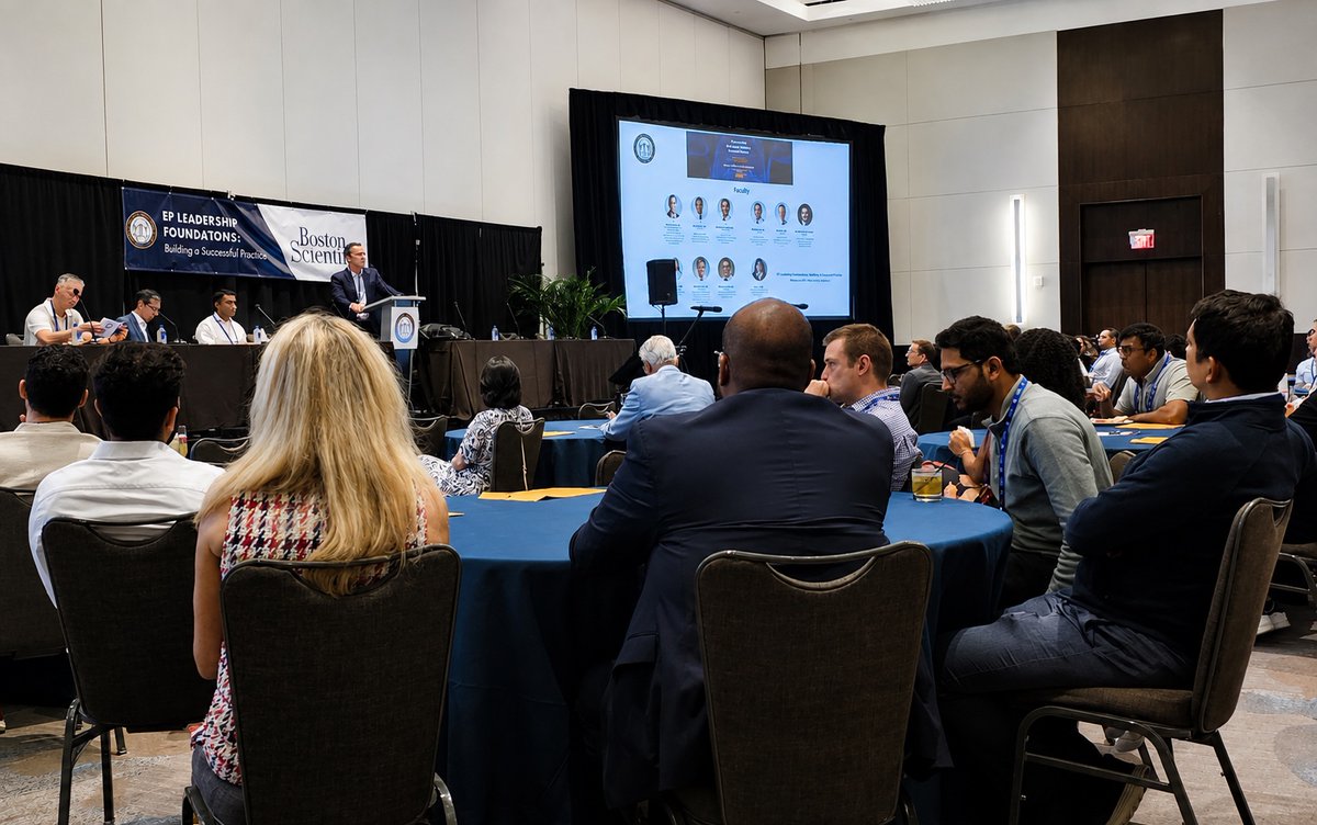
Just wrapped up a fantastic weekend at the Electrophysiology IEP Fellows & Early Attendings' Conference in Atlanta! Great conversations with the next generation of EPs on the future of #electrophysiology and the growing role of patient-specific imaging in procedural planning.
#epeeps #EPFellows #felloweducation #inHEART #cardiacimaging