When the Filter Becomes the Source: POCUS Detection of Inferior Vena Cava Filter Thrombosis Extending Into the Right Atrium
Journal of Clinical Ultrasound
https://t.co/AWJjIj0fmH
Shareable-link:
https://t.co/oJQKn6cyAw
#POCUS#IVC#pulmonaryembolism
When the Filter Becomes the Source: POCUS Detection of Inferior Vena Cava Filter Thrombosis Extending Into the Right Atrium
Journal of Clinical Ultrasound
https://t.co/AWJjIiZHx9
Shareable-link:
https://t.co/oJQKn6c0KY
#POCUS#IVC#pulmonaryembolism
Distinguishing infectious from ischemic colitis at the bedside: the role of ultrasound in a case of Clostridium difficile colitis
World Journal of Emergency Medicine
DOI: 10.5847/wjem.j.1920-8642.2026.099
#POCUS#Intestine#Colitis#Clostridium
Distinguishing infectious from ischemic colitis at the bedside: the role of ultrasound in a case of Clostridium difficile colitis
World Journal of Emergency Medicine
DOI: 10.5847/wjem.j.1920-8642.2026.099
#POCUS#Intestine#Colitis#Clostridium
Lung Consolidation as an Acoustic Window for Pulmonary Artery Visualization on Point‐of‐Care Ultrasound
Sonography
https://t.co/Iml811Q8cb
Shareable link: https://t.co/ad7eMCuEAy
#POCUS#Lungultrasound#Pulmonaryartery#Consolidation
A patient with poor-grade aneurysmal subarachnoid hemorrhage in whom B-mode transcranial ultrasound demonstrates third ventricular dilatation with a mobile hematoma within the ventricle.
#TranscranialUltrasound#NeurocriticalCare#POCUS
Right pleural ultrasound showing a complex septated pleural effusion with heterogeneous echogenic material, highly suggestive of empyema in a patient with pneumonia.
#POCUS#Empyema#Lungultrasound#pneumonia#Sonography
#POCUS#Nephpearls#MedEd 📑
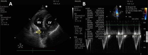
Correlation Between RVOT VTI Measured via a Modified Subcostal View and LVOT VTI in Critically Ill Patients.
“An RVOT VTI > 14.6 cm predicted LVOT VTI ≥ 17 cm with 80.7% sensitivity (95% CI 62.5%–92.5%) and 100% specificity (95% CI 73.5%–100%).”
- by @Issac80 and team
🔗 https://t.co/a6kinNW9sN
Color Doppler imaging of the aorta and pulmonary artery in the upper esophageal view by transesophageal echocardiography.
#TEE#transesophageal#Echocardiography
Journal of Clinical Ultrasound
Correlation Between RVOT VTI Measured via a Modified Subcostal View and LVOT VTI in Critically Ill Patients
DOI: 10.1002/jcu.70280
Shareable-link: https://t.co/KsKGfeETtj
#POCUS#VTI#Strokevolume#cardiacoutput#Echo
Parasternal long-axis view in a critically ill patient with left-sided pneumonia admitted to the ICU, demonstrating visualization of the left pulmonary vein as it enters the left atrium.
#Echocardiography#POCUS#CriticalCareUltrasound#ICU
TEE mid-esophageal long-axis view at 131° in a mechanically ventilated ICU patient, demonstrating left ventricular systolic dysfunction with segmental septal wall motion abnormality, mild mitral regurgitation, and significant aortic regurgitation.
#POCUS#TEE
Beyond the heart: transesophageal echocardiography can reveal much more than cardiac structures.
In this video, the bifurcation of the left pulmonary vein is visualized through left lung consolidation, which acts as an acoustic window and enables extra-cardiac imaging.
#POCUS