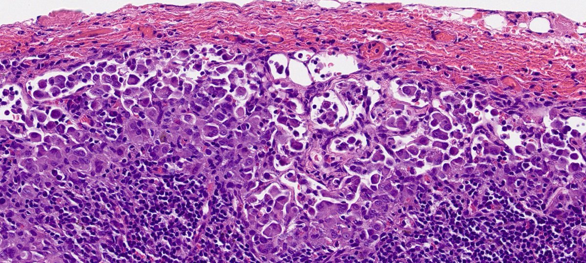
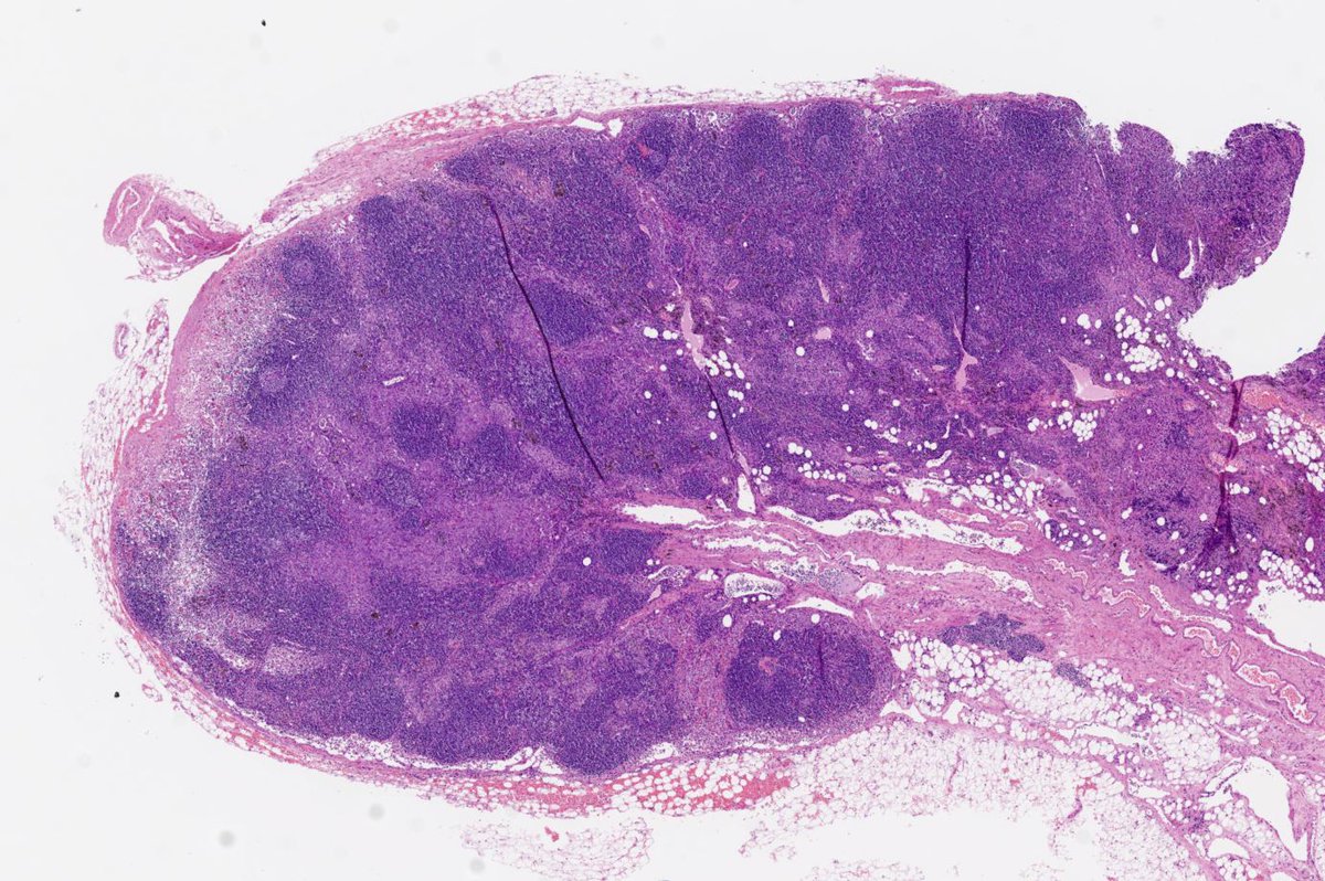
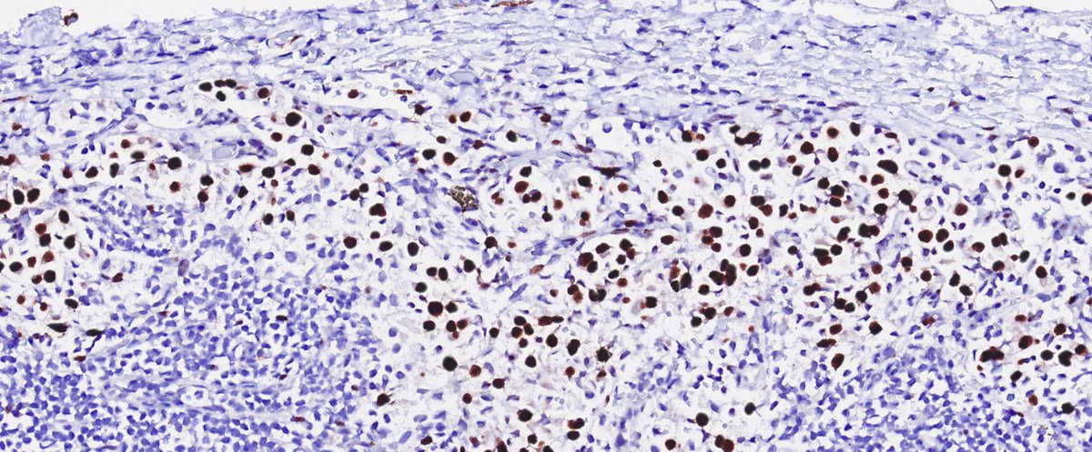
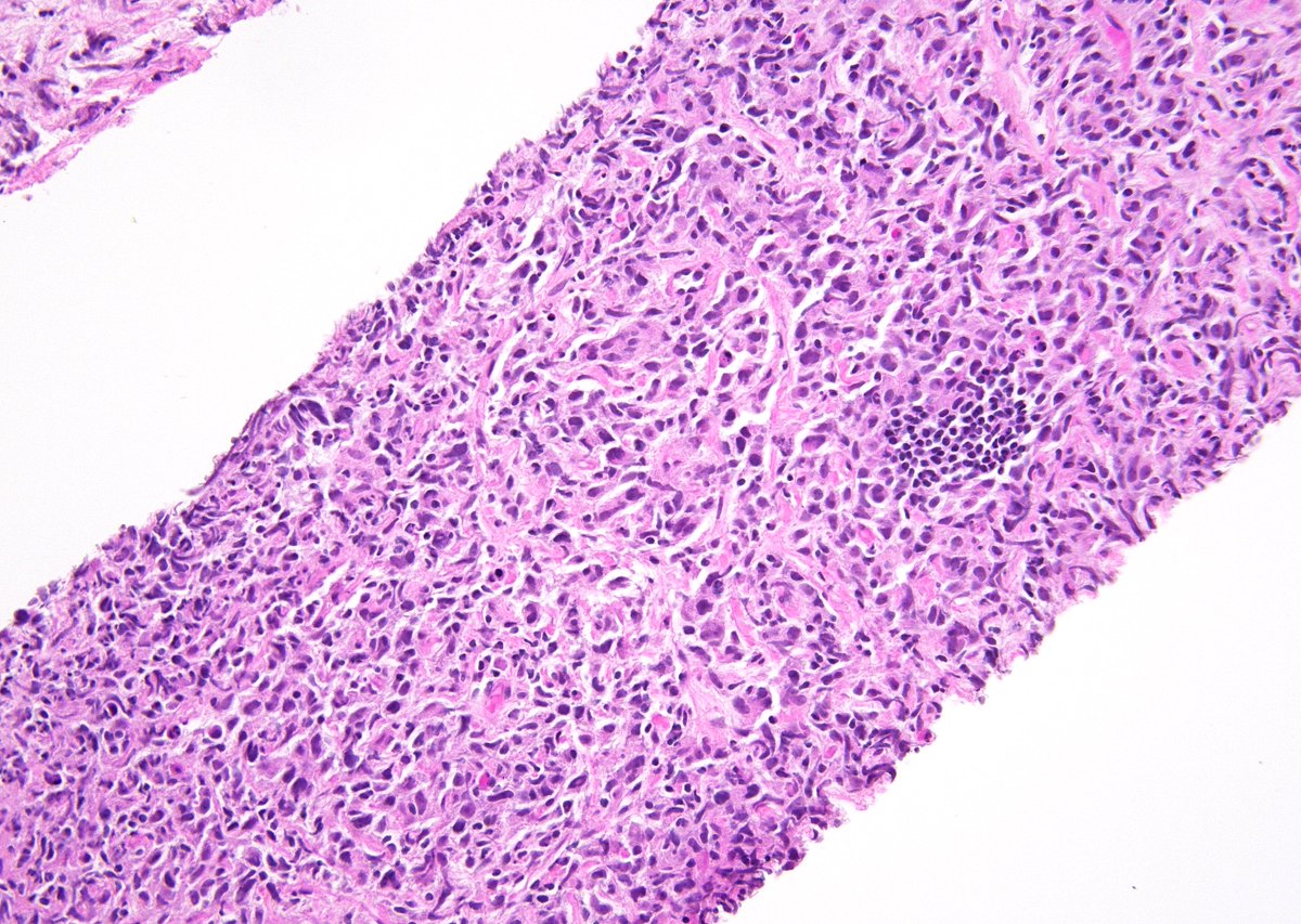
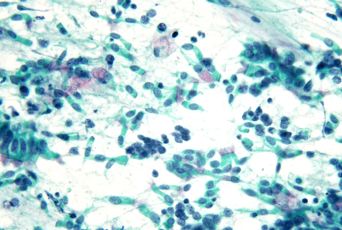
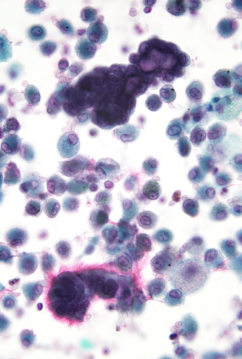
Man, 60s, non-enlarged mediastinal LN resected during cardiac surgery. No known neoplasm. Post op chest CT to r/o pulmonary embolism shows mostly post-surgical changes. CKAE1/AE3 and WT1 shown. Calretinin+. BerEP4-, MOC31-. How to interpret these cells?
Vol 53 (3). López-Ríos F y col. Actualización de las recomendaciones para la determinación de biomarcadores predictivos en el carcinoma de pulmón de célula no pequeña avanzado. Consenso Nacional de la SEAP y de la SEOM. https://t.co/MEDyPxJflJ
I put together a "Basic Cytology" guide geared for junior residents. It's posted on my teaching website, "Kurt's Notes" (https://t.co/OKmaPvoHbB)
"Basic Cytology" Guide: https://t.co/nTCjxPpzPb
@SteveLongMD@shyamraghavan
What to do if the #FNA material is stuck in the needle hub? Safely recap the needle using the plastic cover, a rubber top tube, a rubber safety recapping device or simply an eraser. Then perform the needle flick technique as demonstrated here by @SaeedAsiry@MyCytopathology
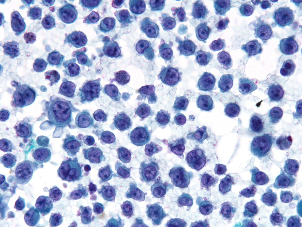
A highly UNUSUAL cytomorphology of a pleural effusion in a 54/M. Without the history of an AML-M6, this would have been a real tough case. The cytoplasmic "snouts" are so unusually prominent and could likely be an artifact of cytospinning.
You may not see such AMAZING morphology again - Cell block of a pleural effusion with "Micropapillary Adenocarcinoma" of the lung. Prognosis of this subtype is significantly poor.
An exciting case to start the day - 68/F, axillary node, h/o lung Sq Cell CA and Mesothelioma. IHCs excluded mets from the two known primaries and confirmed an "Anaplastic Large Cell Lymphoma" (ALCL). NEVER forget to add ALCL to the short list of "Great Mimickers" in Cytology.
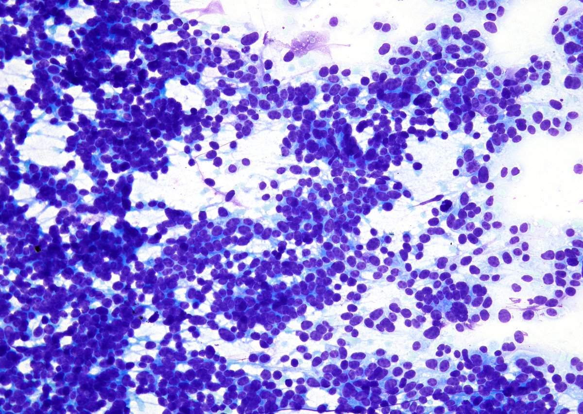
Cellularity alone never helps distinguish a Carcinoid Tumor (CT) from Small Cell Carcinoma (SmCC) of the lung (but lack of mitoses, karyorrhexis and necrosis will). This CT is as hypercellular as any SmCC. Remember, lack of stroma = Hypercellularity.
This is NOT a Papillary Thyroid Carcinoma (PTC). Lung FNA; Well-differentiated adenocarcinomas of the lung (lepidic-predominant on resection), often display overlapping nuclear features with PTC (with INCIs and rarely, psammoma bodies as well).
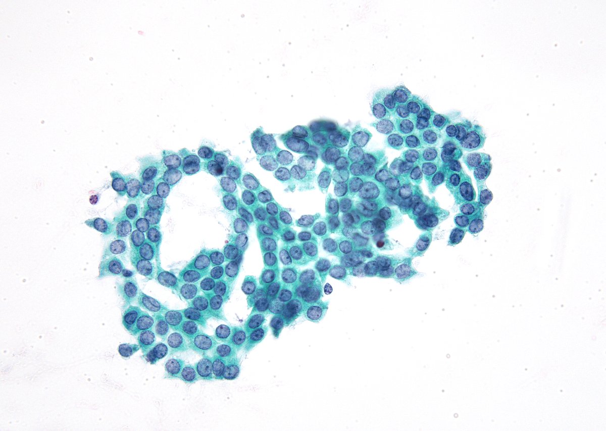
The "AMAZING" Pap stain; bronchial brush of a "Carcinoid Tumor" (CT). The difference between the chromatin texture is extremely subtle but is definitely there. Most cells around the top middle are CT nuclei.