Clinical Observation — Gingival Recession
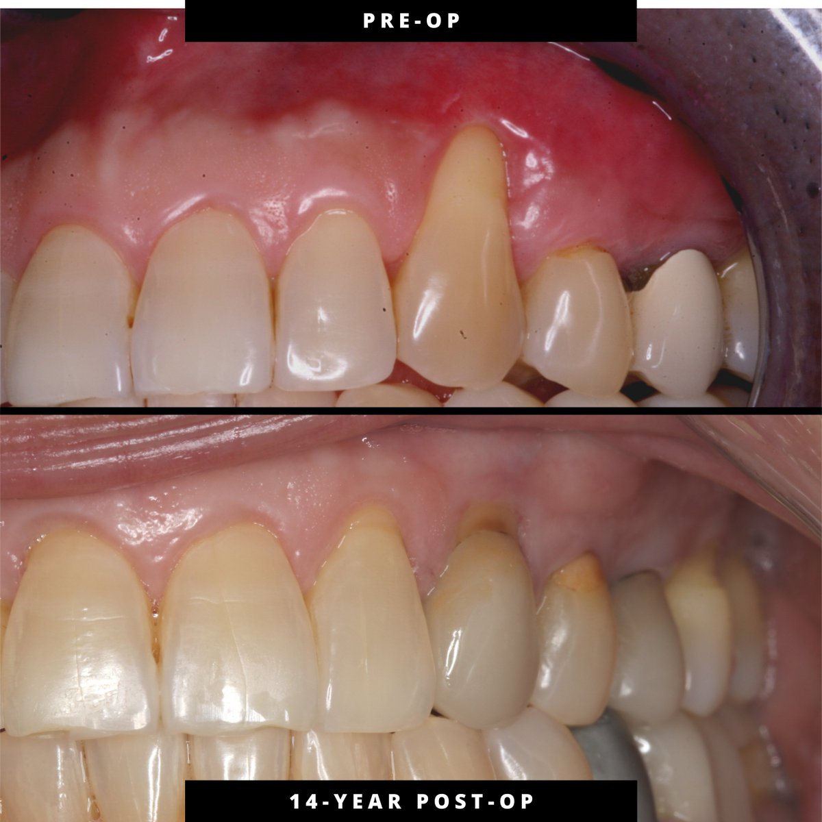
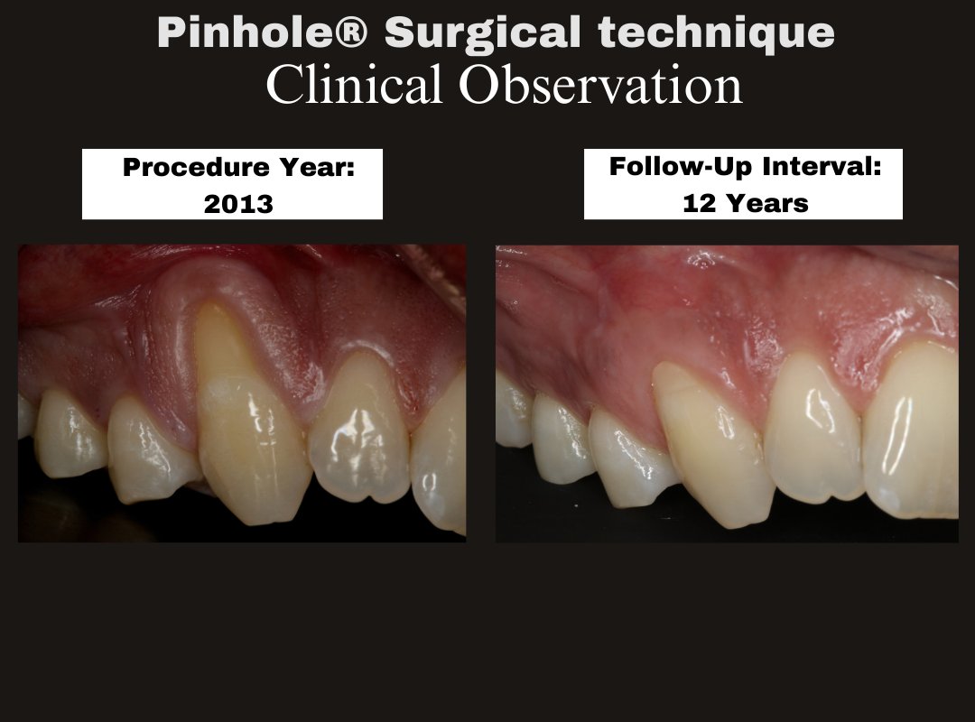
Pinhole® Surgical Technique | Teeth #3–5 | Procedure year: 2013 | Follow-up: ≈13 years (2026 observation)
At long-term intervals, interpretation centers on tissue health and stability of the soft-tissue environment.
Dr. Stephen Kelleher (Periodontist, New York) shares his thoughts after watching Dr. John Chao’s live Pinhole® Surgical Technique surgery at Pinhole® Academy.Hands-on training. Real cases. Clinical decision-making in real time.
Dr. Nikola Mitrović (Oral Surgeon, Serbia) shares his perspective after attending Pinhole® Academy in-person training with Dr. John Chao.
This approach to treating gum recession is unique—and seeing it taught hands-on makes all the difference.
Clinical Observation — Post-op Day 1 at #20–21 following Pinhole® Surgical Technique performed 02/09/26 (11:41–1:01, 80 min). Expected acute edema/erythema with maintained early tissue positioning. Final margin position has not yet declared.
Grateful to have had Dr. Nikola Mitrović join us for the Pinhole® Surgical Technique course — all the way from Serbia 🇷🇸
Thank you, Dr. Mitrović, for traveling so far, bringing great energy, and engaging so thoughtfully throughout the training.
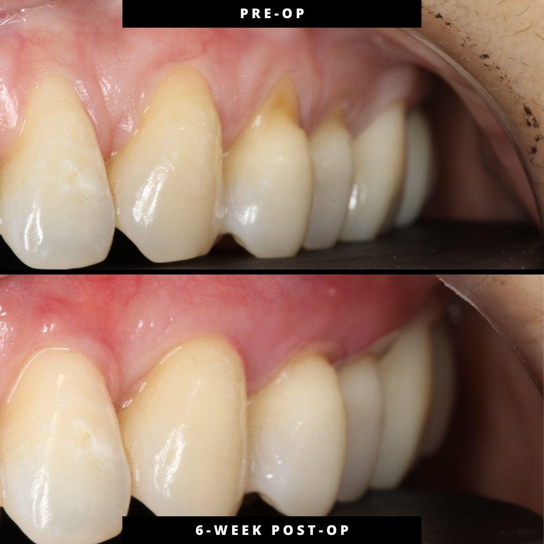
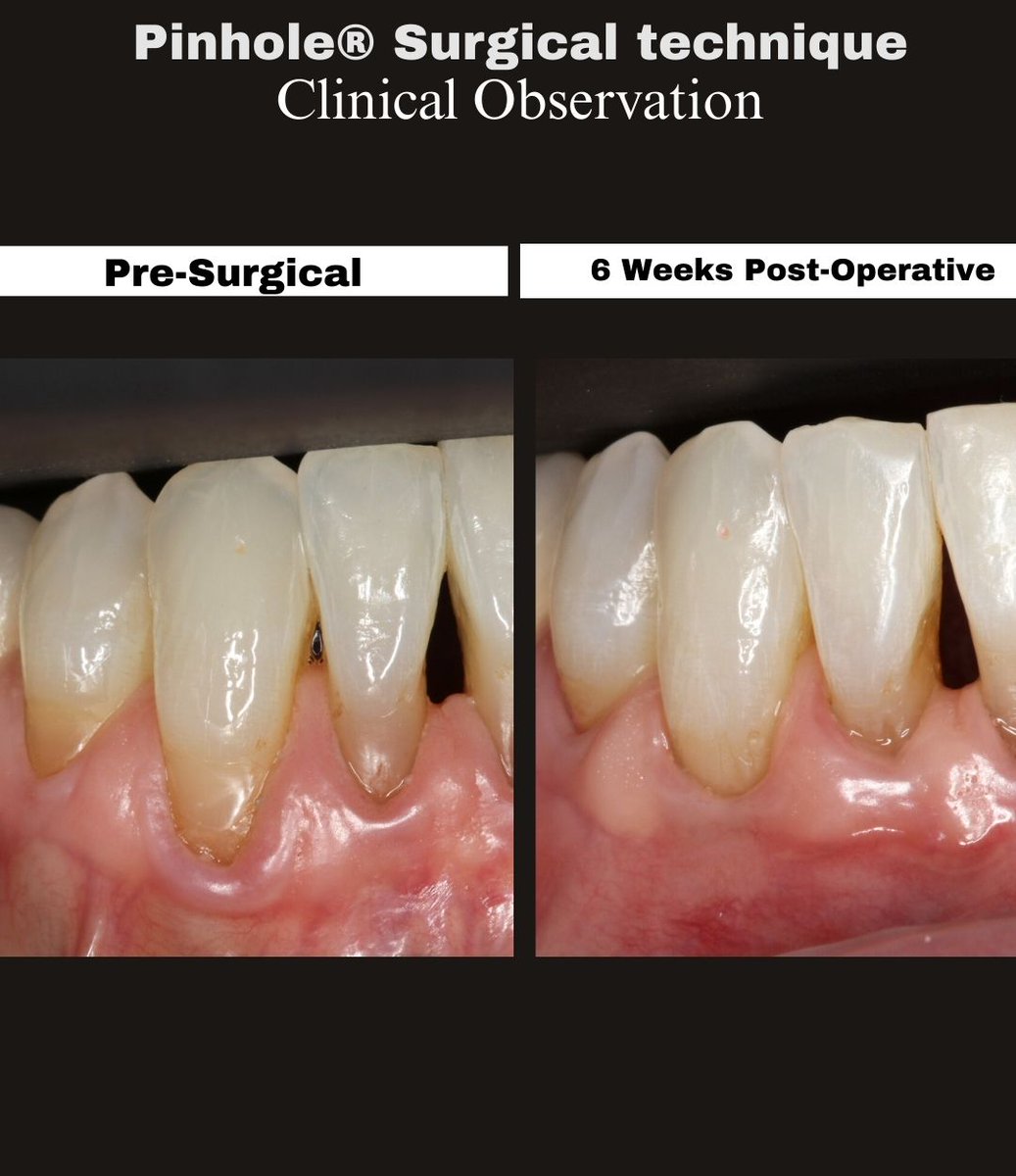
6-week PST follow-up (#21–28,31): improved gingival margin position and tissue adaptation in the mandibular arch.
Early remodeling—more maturation ahead. #pinholesurgicaltechnique#gumrecession#dental
Pinhole® Surgical Technique — Clinical Observation
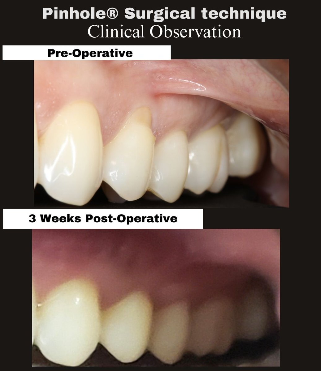
3-week post-op following treatment of gingival recession at #3–5 and #12–15.
Early healing demonstrates tissue continuity and expected erythema. Final margin position remains biologically undetermined.
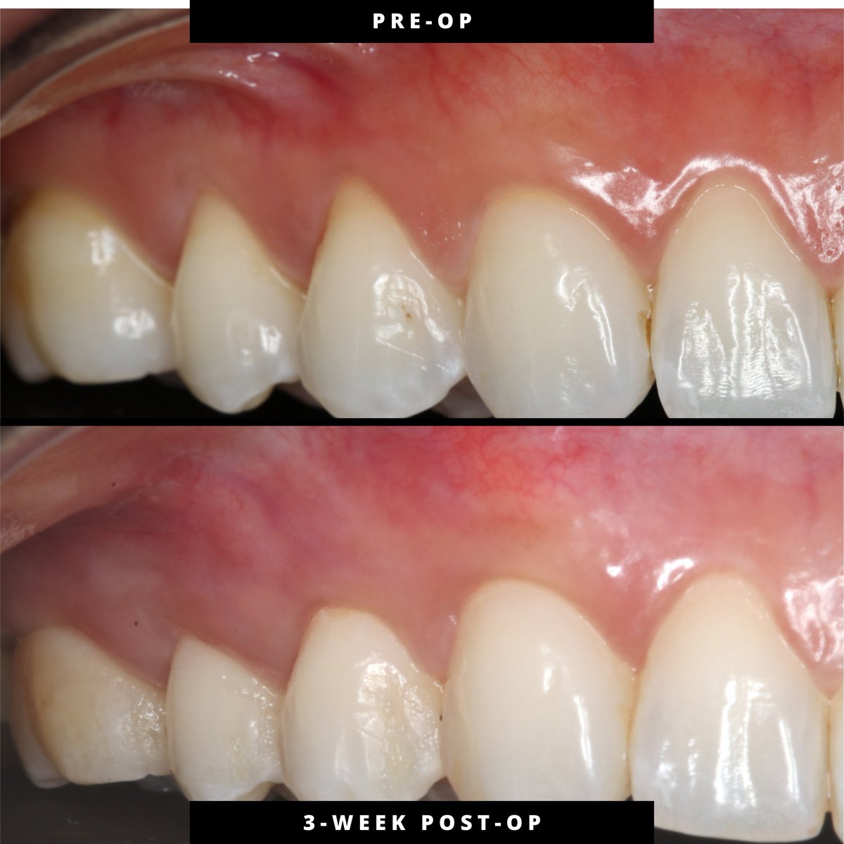
3 weeks post-op following treatment of maxillary gingival recession (#3, 4, 6–12).
Controlled overcorrection supports tissue thickness and biologic remodeling. Gingival margin position continues to evolve as healing progresses.
#PinholeSurgicalTechnique#ClinicalObservation