How does the retina distinguish an object moving through the world from the world moving around us? In work led by Pratyush Ramakrishna (w. Josh Morgan), we discover motion-coherence selectivity—and show how single-cell and population geometry create it. https://t.co/7LkrdtFWG9
Delighted to see our latest test of Active Space-Time Visual Encoding published!
Saccades do more than select where we look: their amplitude also helps determine what information we extract from a scene, as predicted by the transients they generate.
https://t.co/qfbdIrUT2l
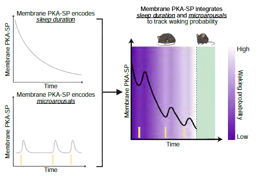
How does the brain track how much sleep has occurred, and gauge the likelihood of waking at each moment? Check out our new preprint: “A molecular integrator of sleep duration and interruption” https://t.co/YqP6ISz91W (1/10)
Bruce Carlson of Biology @washuartsci and grad student Marty Jarzyna found that in electric fish, the MCA brain region is a hub for corollary discharge, the universal process of distinguishing sensory inputs from self vs from others. @CurrentBiology
https://t.co/yTsynrooo3
Very excited that our paper is now out at @arxiv🎉
As a neuroscientist🧠, I wand to understand how (single) neurons encode the visual world. But how to do this in an automated yet interpretable way when we record thousands of neurons to complex natural stimuli?
Here, we use vision language models, specifically @GeminiApp, to convert neural selectivity in monkey visual cortex into language, i.e. a semantic hypothesis, which we verify with text-to-image generation!
Check out the below post and this beautiful homepage by @vedanglad: https://t.co/Q9IOwlP7cd
Great team effort with @AToliasLab@naturecomputes@SuryaGanguli@TamarRottShaham Nikos Karantzas & star first author @vedanglad 😍
This work was done at @StanfordMed and funded through The ENIGMA Project by @jamesfickel 🙏
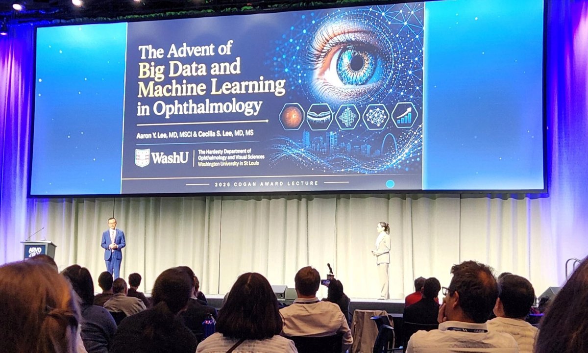
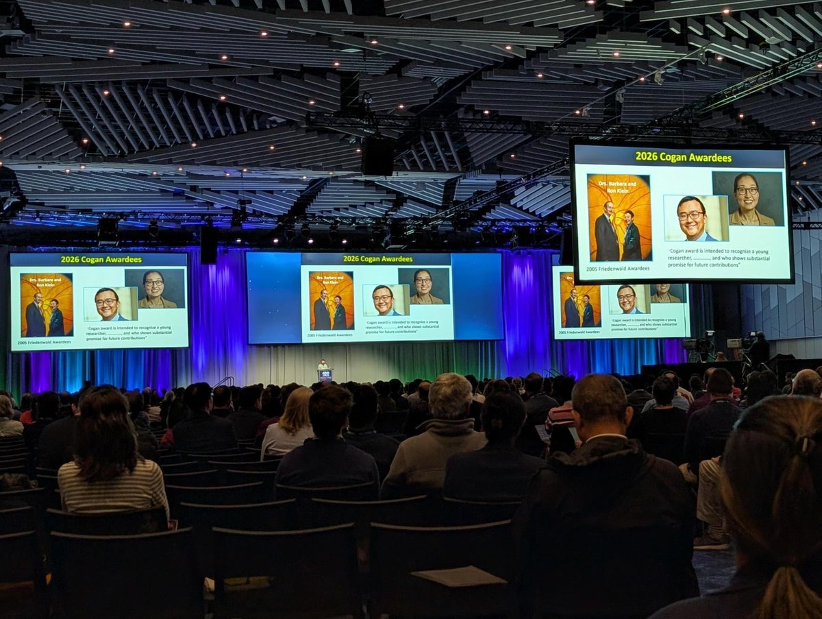
Congratulations to Aaron Y. Lee, MD, MSCI, Arthur W. Stickle Distinguished Professor and Chairman of Ophthalmology and Visual Sciences, and Cecilia Lee, MD, MS, Jane Hardesty Poole Distinguished Professor of Ophthalmology and Visual Science, on the #ARVO2026 Cogan Award! 👏
Every experimental neuroscientist knows the feeling: you have a hypothesis, but running the experiment takes months👩🔬
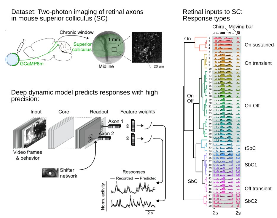
In our new preprint @biorxiv_neursci , we present an openly available functional ''digital twin'' of the retinal input to the mouse superior colliculus that lets you test hypotheses in the model first — try it out yourself using the link below 🧠
We combined chronic two-photon imaging of >200k retinal ganglion cell axonal boutons in the mouse superior colliculus (SC) with deep dynamic models that predict neural responses to parametric light stimuli and natural movies.
Key findings⚡️
▸ Retinal inputs to the SC form functionally distinct, laminar-organized response types, identified via Gaussian mixture model clustering
▸ The functional diversity of retinal output matches that of retinal input to the SC. We show this by aligning our dataset with a retinal reference dataset using a variational autoencoder with adversarial training
▸ Our deep dynamic digital twin learns stimulus–response transformations and generalizes to stimuli it was never trained on, including parametric stimuli used for cell type identification
The model functions as a virtual lab bench: feed in any stimulus you're curious about and generate predicted neural responses. As a proof of concept, we fed in a looming stimulus — known to trigger defensive behavior in mice — and identified putative response types selective for this stimulus
Try it in our Colab notebook with your own stimulus and see what the model predicts
📄 Preprint: https://t.co/5CpYx9lc4j💻
Colab: https://t.co/pfbl24MiGA📂
Code: https://t.co/tNI86Nt5i3
Huge thanks to an incredible cross-institutional team spanning @StanfordMed , @uktuebingen , @uniGoettingen , @bcm_neurosci & many more
@YongrongQ, @lisa_schmors, Na Zhou, Mels Akhmetali, Dominic Gonschorek, Cameron Smith, Anton Sumser, Marie Vallens, @crcadwell, Fabrizio Gabbiani, Maximilian Joesch, @AToliasLab, Philipp Berens, Thomas Euler, @sinzlab, @viajake 🙌
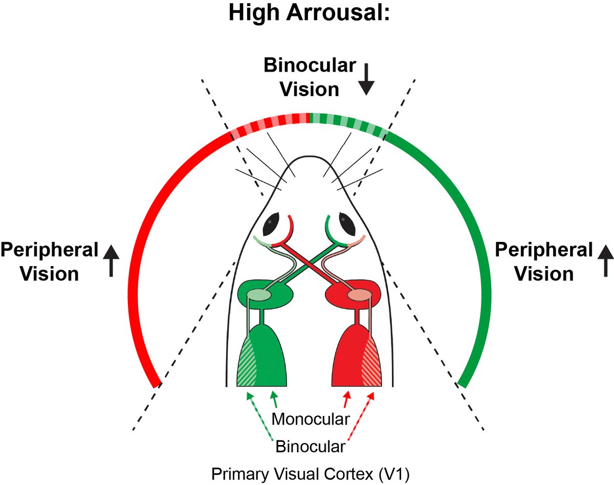
🚨Paper Alert🚨 After 4(!!!) years of revisions, the amazing @KetiCohen & team pushed out this beauty💪 Unlike what the textbooks say, V1 binocularity is not fixed: at high arousal, binocularity decreases while the strength of peripheral vision increases. https://t.co/mK3KEw7jbs
Paper alert!
Cognitive ethology of nest building in a shell-dwelling cichlid
Swantje Grätsch et al. found that L. ocellatus builds nests through an innate behavioral sequence, which is orchestrated by a series of stimulus-response loops.
https://t.co/rdFqZYpgJ2
The Bright Center for Human Vision is now hiring! We seek a skilled researcher to support translational vision research @WashUMedDOVS! More info below🧵
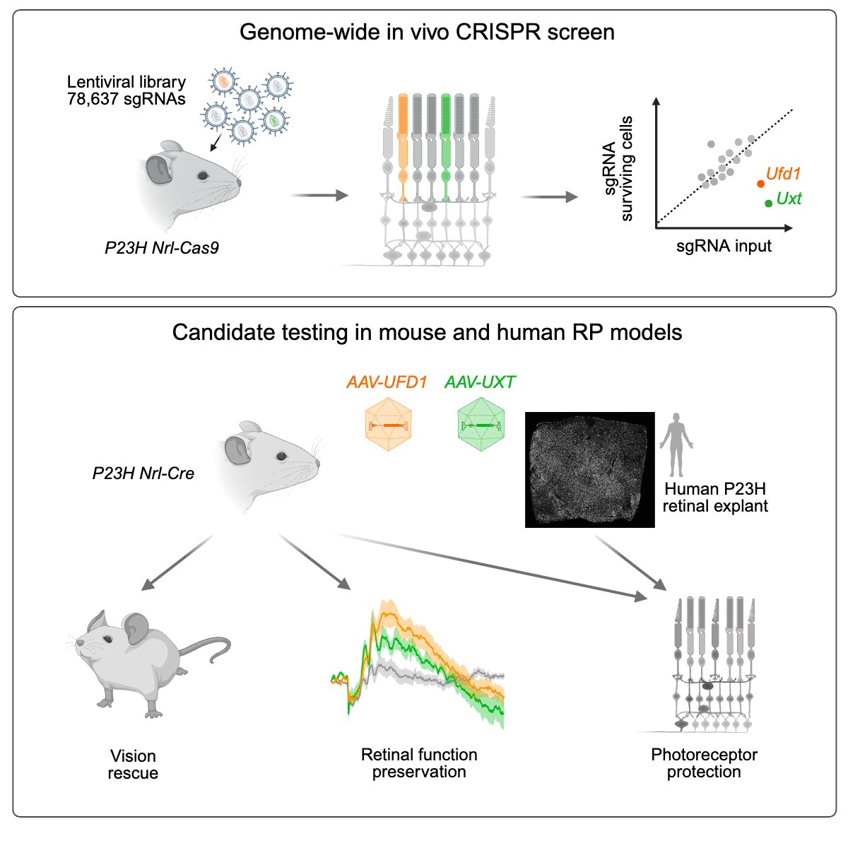
To bridge the gap to translation, we developed an RP model in adult human retinal explants and show that both gene therapies protect photoreceptors, highlighting their therapeutic promise as mutation-agnostic strategies against proteotoxicity. https://t.co/2rLQ7H3IuD
New study led by @NingShenStella w/ @phruzycki: a genome-wide in vivo CRISPR screen in a retinitis pigmentosa mouse model identifies genes whose loss accelerates photoreceptor death. Overexpressing two, UFD1 and UXT, preserves photoreceptors, retinal function, and vision. 🧵
New paper out in @NeuroCellPress 🎉
What determines contextual modulation in primary visual cortex (V1)?
The key result ⚡ V1 neurons are facilitated by surrounds that complete their optimal center feature according to natural scene statistics, and suppressed by surrounds that disrupt it — a principle explained by hierarchical Bayesian inference and conserved across mouse and macaque.
These results converge with Deveau et al. in @NeuroCellPress (https://t.co/THpNjjVuvT) from the lab of @HistedLab, who show that recurrent circuits in V1 filter temporal input sequences to selectively boost natural dynamics, and Lange et al. in @ScienceMagazine (https://t.co/j8EW7SnGUO) from the lab of @haefnerlab, who show that perceptual learning increases population redundancy as predicted by generative inference. A consistent picture is emerging: (early) visual cortex actively infers the statistical structure of the natural world.
Amazing collaboration with @AToliasLab@haefnerlab@sinzlab Antolik Lab & many more — led by @jiakunfu, with co-authors @suhas_shrinivas & @LuchinoBaroni & many more
The paper is open-access and available here: https://t.co/d1tg4MPFlm
More detailed approach:
We trained CNN digital twins on large-scale two-photon recordings from mouse V1 and used them to synthesize, for each neuron individually, the surrounds that most strongly facilitate or suppress its response to its optimal center stimulus. Closed-loop in vivo inception loop experiments confirmed the predictions.
Key qualitative finding:
Surrounds that complete the optimal center feature under natural scene statistics → facilitation
Surrounds that disrupt it → suppression
We verified this with an independent generative diffusion model (blind to our CNN): statistically likely continuations of the optimal center feature were significantly more similar to facilitatory surrounds in V1 representational space. The same principle holds in macaque V1 despite major differences in receptive field organization.
We formalize these results in a hierarchical Bayesian inference model — V1 neurons represent posterior beliefs about local features, with feedback from higher areas encoding global scene structure — and find like-to-like excitatory connectivity in the MICrONS dataset as a candidate circuit mechanism.
A @NeuroCellPress paper led by Kushal @UCBerkeley, Nicole&Victoria @UCSF in full collaboration with @shekharlab, Molecular and spatial analysis of ganglion cells on retinal flatmounts identifies perivascular neurons resilient to glaucoma https://t.co/QjeNMbQwWZ #retina#MERFISH
Finally it's out! https://t.co/ZfPTLWFRDO Thanks to Felix Baier, Karl Farrow and Hopi Hoekstra! And a big thank you to all the co-authors, reviewers, host institutes and funding sources! @BramNuttin, Arnau Sans Dublanc, Chen Liu, Victoria Tong, Julie Murmann and Keimpe Wierda
We found that endogenous ligand for GPR139 is opioid peptide dynorphin. With this, GPR139 may not be an orphan any more but the 5th opioid receptor. Heavy lift by talented Xiaona Li with great thanks to all who contributed!
https://t.co/OabhjeWV1q
Join us in congratulating professor Susan K. Dutcher, PhD, on her election to the National Academy of Sciences. Her groundbreaking research on cilia is unlocking insights into chronic disease and continues to elevate WashU Medicine���s legacy of scientific excellence and impact.
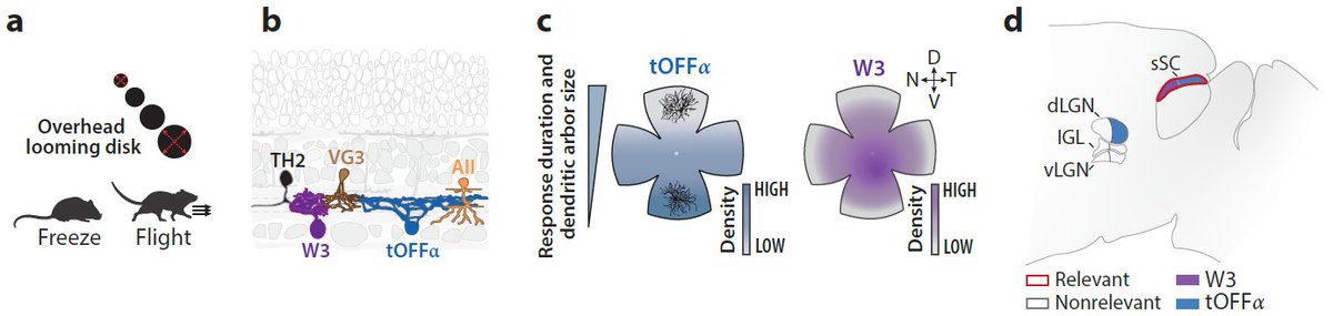
65 years after Lettvin’s bug detector neurons in the frog retina, we revisit how the retina drives behavior—from reflexes to prey capture to brain-state modulation
New review with @AnnaIntegrated & Serena Riccitelli in Annual Review of Vision Science 👇
https://t.co/xfwUGtpXkF