Want to know how to make use of #lightsheetmicroscopy as a quantitative technique? Register for our free live #webinar on July 16 or July 18 and learn all about it. https://t.co/YvxjPDIkEF
Greetings from #glia2019 in Porto! Don’t miss our workshop today at 12:30 p.m. on “Adult neural cells from healthy and diseased brain – challenges and opportunities”. See you there! https://t.co/45UNKjXFw4
We are proud to be part of the #CiMT project. Together with FH Bielefeld, @unibielefeld, @Miele_Presse and CNC Speedform we investigate how surfaces are affected by mechanical, biological and chemical impacts in order to develop high quality and long-lasting materials.
Why do we overeat? Researchers @UNC have discovered a brain circuit about hedonic versus homeostatic feeding in mice. We are proud to have contributed to this excellent work with our #UltraMicroscope II light sheet microscope. https://t.co/uMtbSOd3PD
Exciting discovery from our Innovation Impact Grant recipient @UNC_SOM@UNC on the hedonic vs homeostatic brain circuit in the central amygdala. https://t.co/orZdYVNBb5
We are proud to support the course on "Tissue clearing & 3D Imaging" @InstVisionParis. Register now and learn more about immunostaining of whole organs, 3DISCO/iDISCO clearing, 3D Imaging with #lightsheet#microscopy and
data analysis/3D reconstruction: https://t.co/z6MCa2ElTs
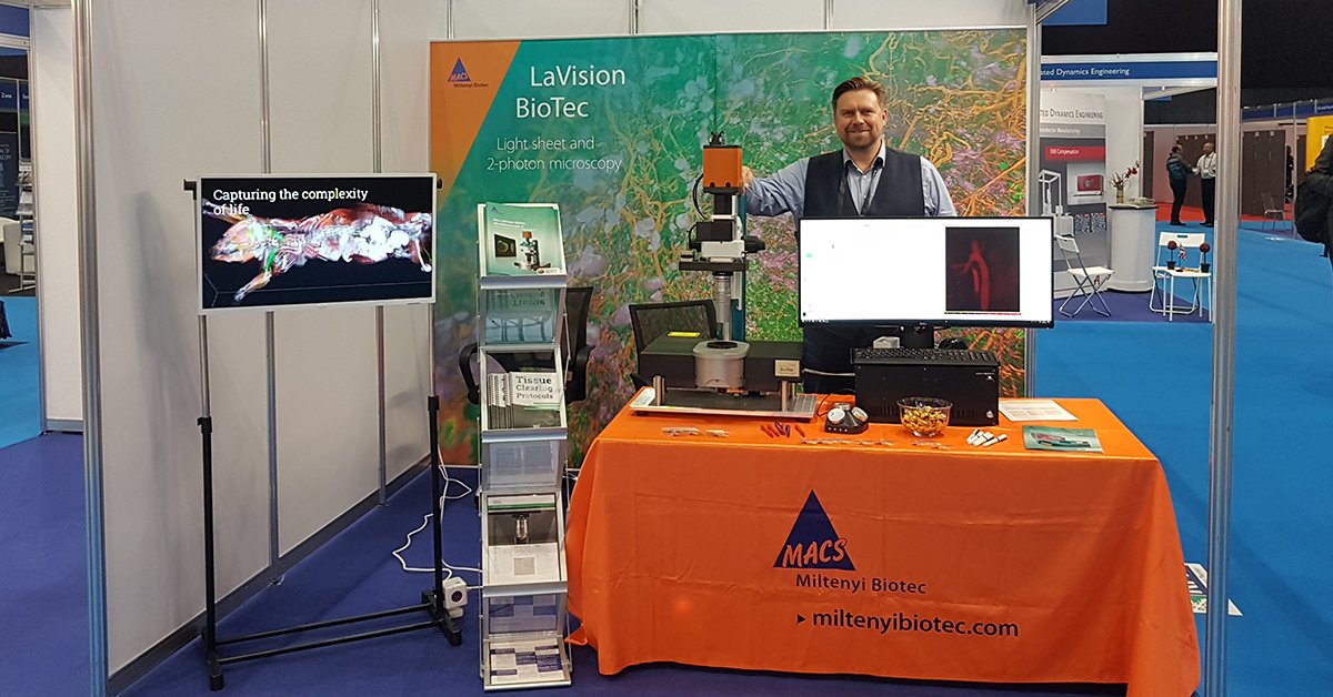
Attending #ELMI2019? Come by booth No. 18 @miltenyibiotec and explore 3D microscopic samples via our virtual reality headset.
...And don't miss Uwe's workshop on June 5 and 6, 14:30 -15:30 pm, directly at the booth. #lightsheet#microscopy
Immune responses in the gut and associated draining lymph nodes differ between tolerogenic and inflammatory depending on their anatomical location, according to a study published in Nature. Read the paper: https://t.co/rUdTcEt4dv
The @DeLIVER_INT project brings together an excellent group of scientists and companies exploring liver cell function with #superresolution#microscopy.
The art of science. This xenopus eye was triple-stained for visualization of photoreceptors and imaged with the #UltraMicroscope II. Find out all the benefits here https://t.co/kA9SnVDlqL
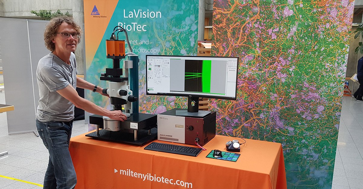
Want to explore the architecture of biology? Learn more about our new imaging portfolio, including the @LaVision_BioTec UltraMicroscope II light sheet #microscope. https://t.co/7F3YAObef9
Nice to see the emerging potential for #3Photon imaging beyond neuroscience! High resolution with high SNR at depth. Multimodal lymph node imaging of GFP, tdTomato, SHG and THG at 1320nm from Raluca A. Niesner #drfz, @LaVision_BioTec
And so with the feedback forms sent out, we reach the end of the beginning! One final time we would like to publicly thank our sponsors who helped to make this first meeting happen! @Sigvarisgroup_B @HaddenhamHealth @LaVision_BioTec@mediUKLtd @ICVS_UoB @Co_Biologists@TheBHF
Greetings from Yokohama, where we are presenting our #Lightsheet#microscope at the 41st Annual Meeting of the Molecular Biology Society of Japan #mbsj2018.
| #Dermatology | Just out our paper 3D optical clearing and imaging of pruritic atopic dermatitis and #psoriasis skin reveals downregulation of epidermal innervation @JIDJournal@astar_research
https://t.co/NuqFdDNx7F