We've released a #new State-of-the-Art Review, "The Effect of General Anesthesia and Mechanical Ventilation on the Echocardiographic Evaluation of Cardiac Function!"
Read it now: https://t.co/cbmJ46fd5C
📄 Can we finally measure RV volume accurately with simple echocardiography?
🔗 DOI: https://t.co/cHII0FgTLH
🫀 The right ventricle (RV) remains one of the most challenging chambers to assess—especially in congenital heart disease (CHD).
👉 Gold standard? Cardiac MRI
❗ But: expensive, time-consuming, and often requires sedation
✨ This study proposes a simple, fast, and accurate 2D echo-based method for RV volumetry—bringing us closer to true bedside quantification.
✨ The key idea:
👉 Model the RV using a cone-based geometric approach
➡️ Using only 2 standard echo views:
Apical 4-chamber (A4CH)
Parasternal short-axis (SAX)
📐 With just a few parameters:
✔ Cross-sectional areas (A4CH, ASAX)
✔ Tricuspid valve diameter
📊 Performance vs MRI:
🔥 Excellent agreement:
Systolic volume → ICC 0.98
Diastolic volume → ICC 0.96
📉 Minimal bias:
Δ systolic volume ≈ 0.1 mL
Δ diastolic volume ≈ 5.2 mL
➡️ Clearly outperforms traditional 2D models
💡 Why this matters clinically:
👉 Enables:
✔ Rapid bedside RV assessment
✔ Reduced need for repeated MRI
✔ Easier follow-up in paediatric & CHD patients
👉 Particularly valuable in:
Post-operative monitoring
Serial evaluations
Resource-limited settings
🚀 Key innovation:
👉 A mathematically robust yet practical model
➡️ Balancing accuracy + simplicity
➡️ Adaptable to different RV shapes
🚨 Bottom line:
2D echocardiography—when combined with smart modelling—can approach MRI-level accuracy for RV volumetry.
#Cardiology #Echocardiography #RightVentricle #CongenitalHeartDisease #CardiacImaging #CMR #Innovation #MedTech #PediatricCardiology 🫀📊
So proud of incoming Mayo Clinic cardiology fellow, @ayeshasinghmd, on her publication documenting a 40% rate of residual angina in ISCHEMIA patients following complete revascularization as adjudicated by angiographic core lab. https://t.co/96sDCwhbob
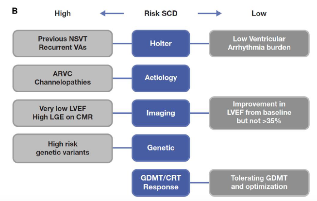
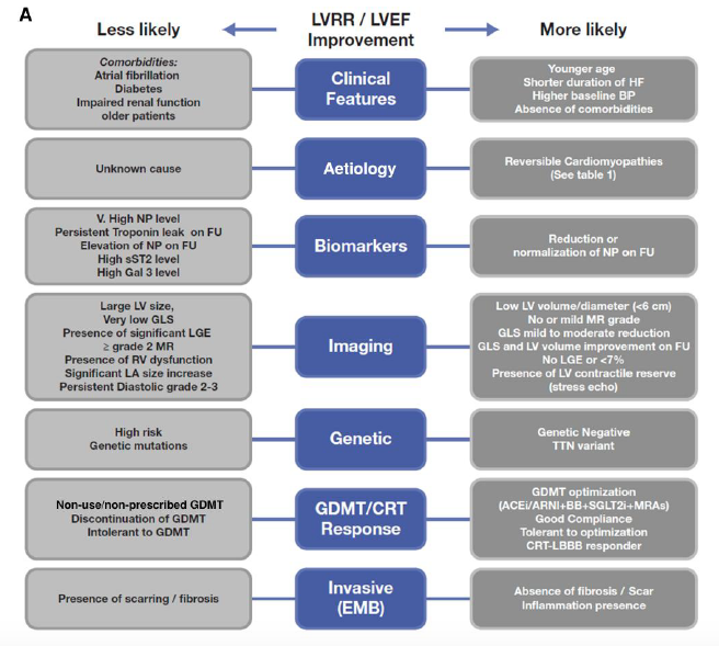
Ischemic cardiomyopathy remains a leading cause of HF and CV death. This review highlights the evolving role of imaging, viability, and revascularization, while reinforcing GDMT and device therapy as the foundation (https://t.co/AzWtCLBga9). #HREV@GianluSava@NicolasGirerd
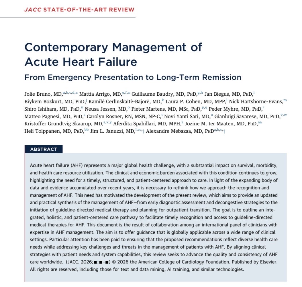
🚨 Managment of AHF is advancing!
Glad to share our review, aimed at:
🔄 Bring together management algorithms across different healthcare settings and resources
🏥 Translate contemporary evidence into practical bedside management
#HeartFailure#AcuteHeartFailure#JACC
It’s well known that there is a certain delay between electrical activity and a mechanical phenomenon at the myocardial level. However, the temporal relationship between myocardial electrical activity and venous return flow has never been studied, so we will probably see these discrepancies between the Doppler spectrum of venous flow and the ECG. But it is only a theory; there is no evidence.
I know the S wave seems to start before the QRS complex. But it's not the first time I've observed that. I don't have an explanation for this discrepancy between the Doppler spectrum and the electrical activity recorded on the ECG. They're probably temporally related phenomena, but not occurring simultaneously. 🤷🏻♂️
Continuous-wave Doppler interrogation in valvular heart disease: pearls and pitfalls | European Heart Journal - Imaging Methods and Practice | Oxford Academic https://t.co/xAQPqRgbjt
What is best formula to estimate mPAP on #echofirst?
📝 finds minimal end-diastolic PR pressure best correlation (R = 0.92) & diagnostic accuracy AUC 0.96 https://t.co/MCbDFf6aSN in almost 600 pts referred for RHC for PH dx, integrates both PR & TR signals
At 24.5 mm Hg cutoff, mPAPDPmin 89% sensitivity, 94% specificity
At 20 mm Hg, mPAPDPmin sensitivity⬆️99% w/ net reclassification driven by more accurate downgrading of pts w/o PH
ASE and the @accpchest recently partnered on a project to develop two educational webinars to improve the understanding of cardiovascular ultrasound's application in pulmonary hypertension.
You can find them on our #new Right Heart Resources web page! https://t.co/aHA1IniTS3
Very interesting image of IVC!
Any guesses on what the red flow is? (clue: congenital anomaly) - See thread for source/full case.
#POCUS#echofirst#FOAMed
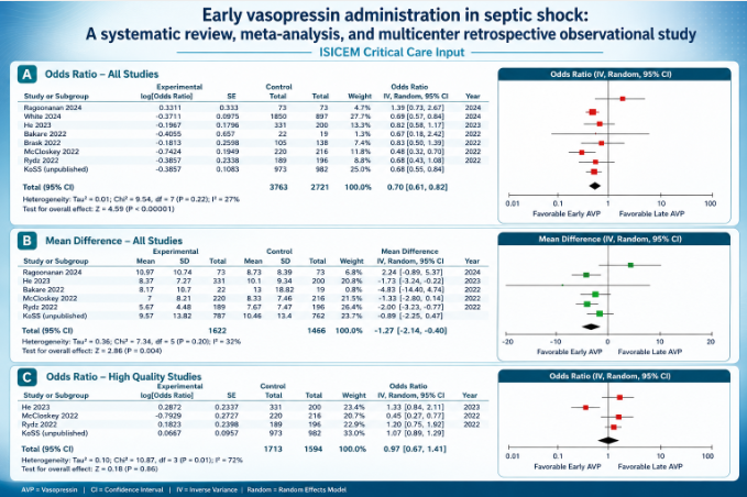
@ISICEM Critical Care Input
Early vasopressin administration in septic shock: A systematic review, meta-analysis, and multicenter retrospective observational study
https://t.co/iAyvOZji9e
#ISICEMelearning#Meded@Sciencedirect
#JADEL
I hope you enjoy reading my CASE editorial: Blow Hard!
https://t.co/CDR9j3hO8d
I discuss Antonio Maria Valsalva, the complex physiology of the Valsalva maneuver, & include a link to learn about goal-directed methods: https://t.co/SoUZbpMuFI
@CASEfromASE@ASE360
🫀In cardiogenic shock, we still focus heavily on MACROcirculation:
📉 blood pressure
📉 cardiac output
📉 LVEF
📉 vasopressor dose
But what if the real battle is happening deeper?
🩸 At the microcirculatory level.
This excellent ATS viewpoint highlights one of the most important evolving concepts in shock physiology:
⚠️ normalization of macrocirculation does not necessarily mean restoration of tissue perfusion.
Despite advances in cardiogenic shock management, mortality remains >40%.
Even more striking, up to 45% of deaths occur in patients with normalized cardiac index.
That disconnect may be explained by persistent:
🩸 microvascular dysfunction
🩸 impaired capillary perfusion
🩸 endothelial dysfunction
🩸 tissue hypoxia despite “acceptable” hemodynamics
The review reinforces that: Microcirculation is not a passive bystander.
It may be a central driver of:
• organ dysfunction
• lactate persistence
• shock progression
• mortality
Particularly interesting is the emphasis on simple bedside tools.
We often think microcirculation requires advanced devices, yet:
📌 capillary refill time (CRT)
📌 mottling
📌 ΔPCO₂
📌 lactate trends
still carry strong prognostic value.
A CRT >3 seconds at ICU admission was associated with worse outcomes, and combining CRT with the CardShock score achieved an impressive AUC of 0.93 for outcome prediction.
The article also reviews modern technologies:
🔬 handheld vital microscopy
🔬 sublingual microcirculation imaging
🔬 NIRS
🔬 laser Doppler assessment
bringing “real time” bedside microcirculatory monitoring closer to clinical practice.
One of the strongest physiological messages:
⚠️ Shock is not only about flow. It is about effective tissue level oxygen delivery.
The review beautifully summarizes the four major mechanisms of microvascular dysfunction:
• heterogeneity
• hemodilution
• congestion
• edema
Particularly relevant for intensivists:
📌 venous congestion itself may worsen microvascular flow
📌 elevated filling pressures impair driving pressure
📌 edema increases oxygen diffusion distance
This is highly relevant in:
• advanced heart failure
• VA ECMO
• mixed shock states
• fluid overloaded patients
Another important takeaway: Persistent microcirculatory dysfunction after VA ECMO initiation was associated with increased mortality, even when macrocirculation improved.
Perhaps the key message of this paper is:
🩸 Microcirculation should no longer be considered a secondary endpoint in cardiogenic shock.
It may become one of the most important physiological targets of the next decade.
📖 Merdji H, American Journal of Respiratory and Critical Care Medicine. 2026, 212(3), 410–413 https://t.co/81Zp3aj274.
Unexplained dyspnea? Here’s the new ESC framework for diagnosing HFpEF — and it goes way beyond a basic echo 🧵
From Landsteiner et al., Eur Heart J 2026, a stepwise domain-based approach