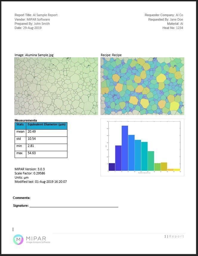
MIPAR enables titanium grain analysis using automated segmentation of microstructures. Recipes extract grain boundaries, ASTM grain size numbers, and morphology metrics under variable conditions. More at https://t.co/003hEBm0ge
MIPAR quantifies grain size and morphology in metals via automated ASTM-compliant segmentation, intercept methods, and EBSD-informed workflows, enabling high-throughput microstructural statistics. Learn more at https://t.co/uO5ngB9q5X
MIPAR enables materials analysis using hybrid deep learning segmentation to measure grains, phases, defects, and critical dimensions. Learn more at https://t.co/zh2kU7sfFJ
MIPAR performs sub-pixel CD metrology using adaptive edge extraction to measure linewidth, pitch, and critical features in SEM images. Learn more at https://t.co/8LyQZ5AzPn
Within MIPAR you can construct a fully automated “recipe” mixing cleanup, FFT-normalization, convolution filters and ML-enabled segmentation — then export it via API into a Python pipeline. Learn more at https://t.co/ABGuSyjeF5
MIPAR automates radial unwrapping of Petri dish images for illumination-corrected colony segmentation and morphology-based enumeration. Learn more at https://t.co/SlrBbQessO
Automating microstructural workflows, MIPAR integrates deep learning segmentation with rule-based recipes for reproducible quantitative analysis. Learn more at https://t.co/Sfn41BfNBi
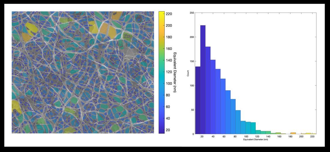
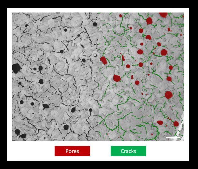
MIPAR quantifies pore size distribution by segmenting pores, extracting morphology metrics, and generating cumulative and differential distributions. Learn more at https://t.co/RrgS4jbnoO
MIPAR enables automated quantification of whole-slide pathology images, integrating deep learning segmentation with reproducible pipelines for histological feature extraction. Learn more at https://t.co/qVIXV76vxs
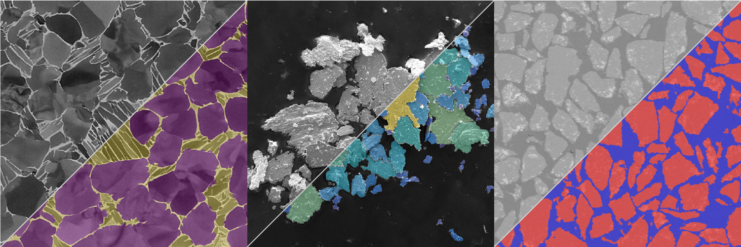
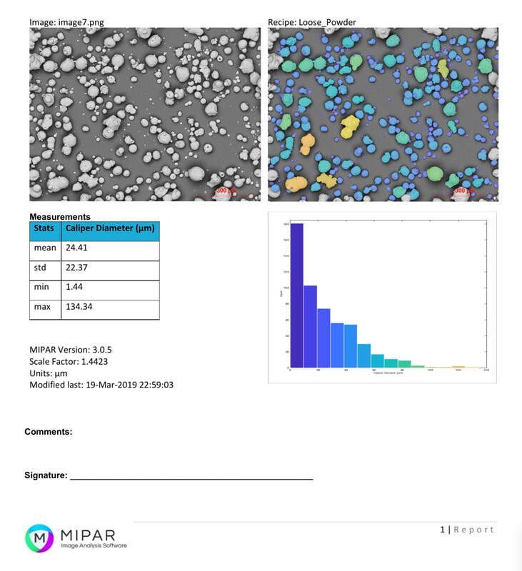
Quantify powder morphology with MIPAR: automated particle size and shape analysis yields reproducible metrics for distribution, aspect ratio, and sphericity. Learn more at https://t.co/nEdC9zGH3w
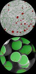
MIPAR automates phase segmentation in metallography, quantifying phase fractions and morphologies directly from micrographs. Learn more at https://t.co/CTljrM9YPu
MIPAR deep learning models segment contaminants across diverse substrates, enabling precise quantification of inclusion morphology and spatial distribution. Learn more at https://t.co/2ishlW4ox4
Automate segmentation of overlapping colonies in Petri dish images using thresholding, watershed, and circularity filters. Learn more at https://t.co/SlrBbQessO
Cell segmentation with MIPAR leverages local contrast and shape-based filtering to isolate overlapping, irregular, or faint cell boundaries in complex biological images. Learn more at https://t.co/NSJHZsv8dT
Quantify porosity and pore size distribution across scales using MIPAR's automated segmentation and measurement tools. Ideal for membrane, scaffold, and foam analysis. Learn more at https://t.co/duhwN2LmQT
AI-powered segmentation in MIPAR enables high-throughput phenotypic quantification of cell morphology across diverse imaging modalities. Learn more at https://t.co/nj04MYjUbr
Automated segmentation in MIPAR accelerates digital histology by quantifying structures in high-resolution slides with reproducible precision. Learn more at https://t.co/oQNyfsdnWc