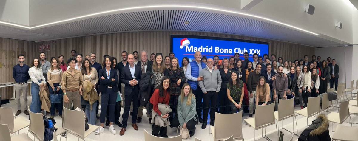
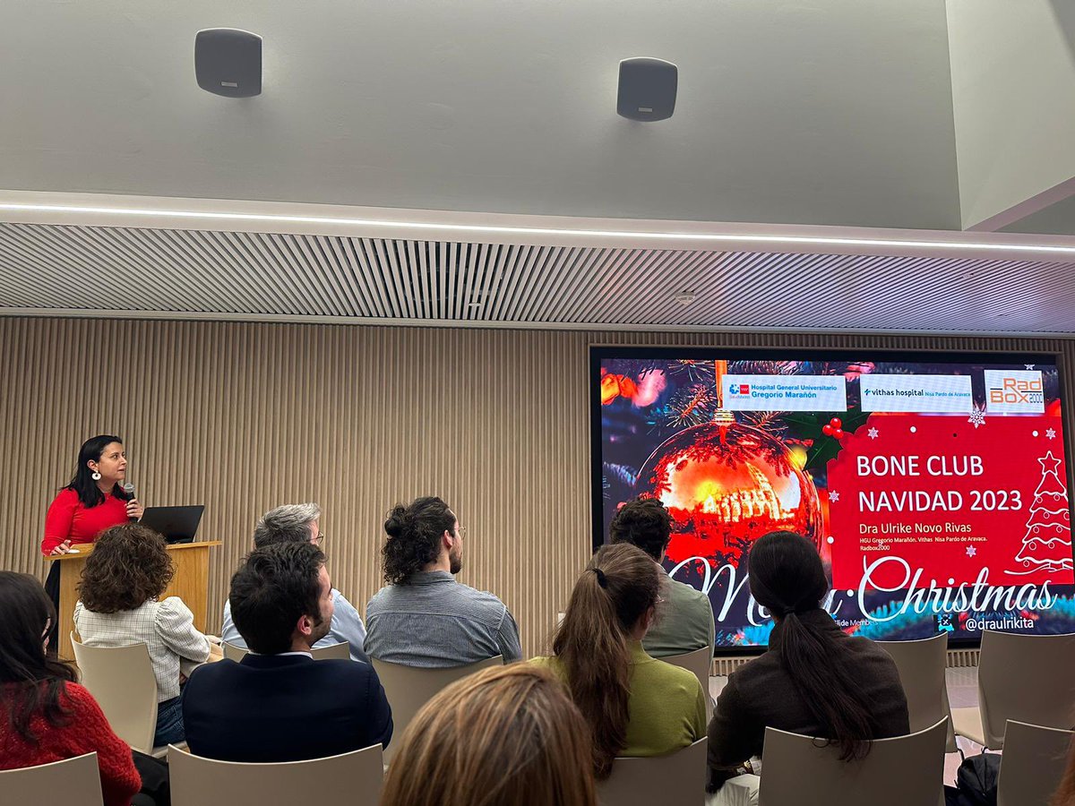
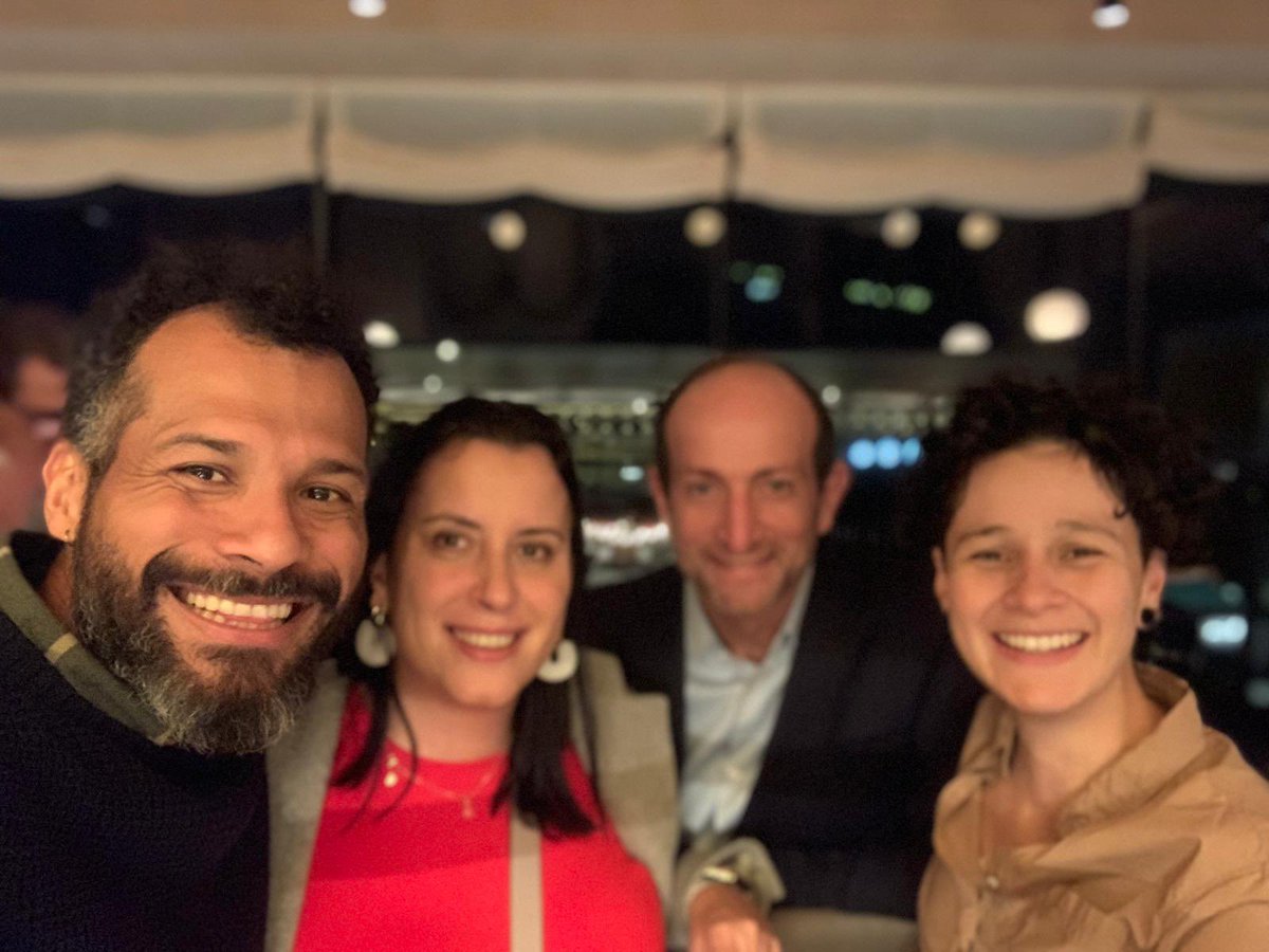
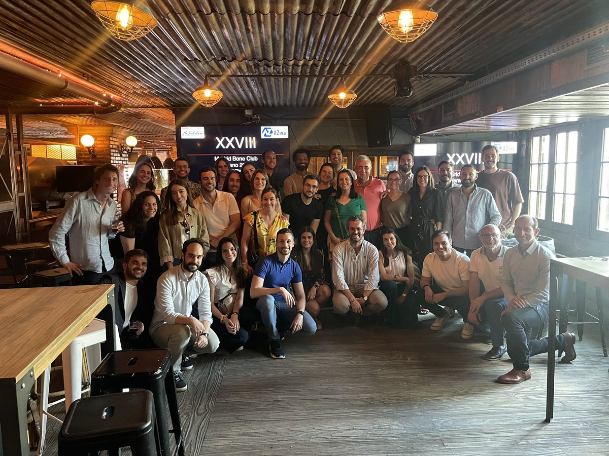
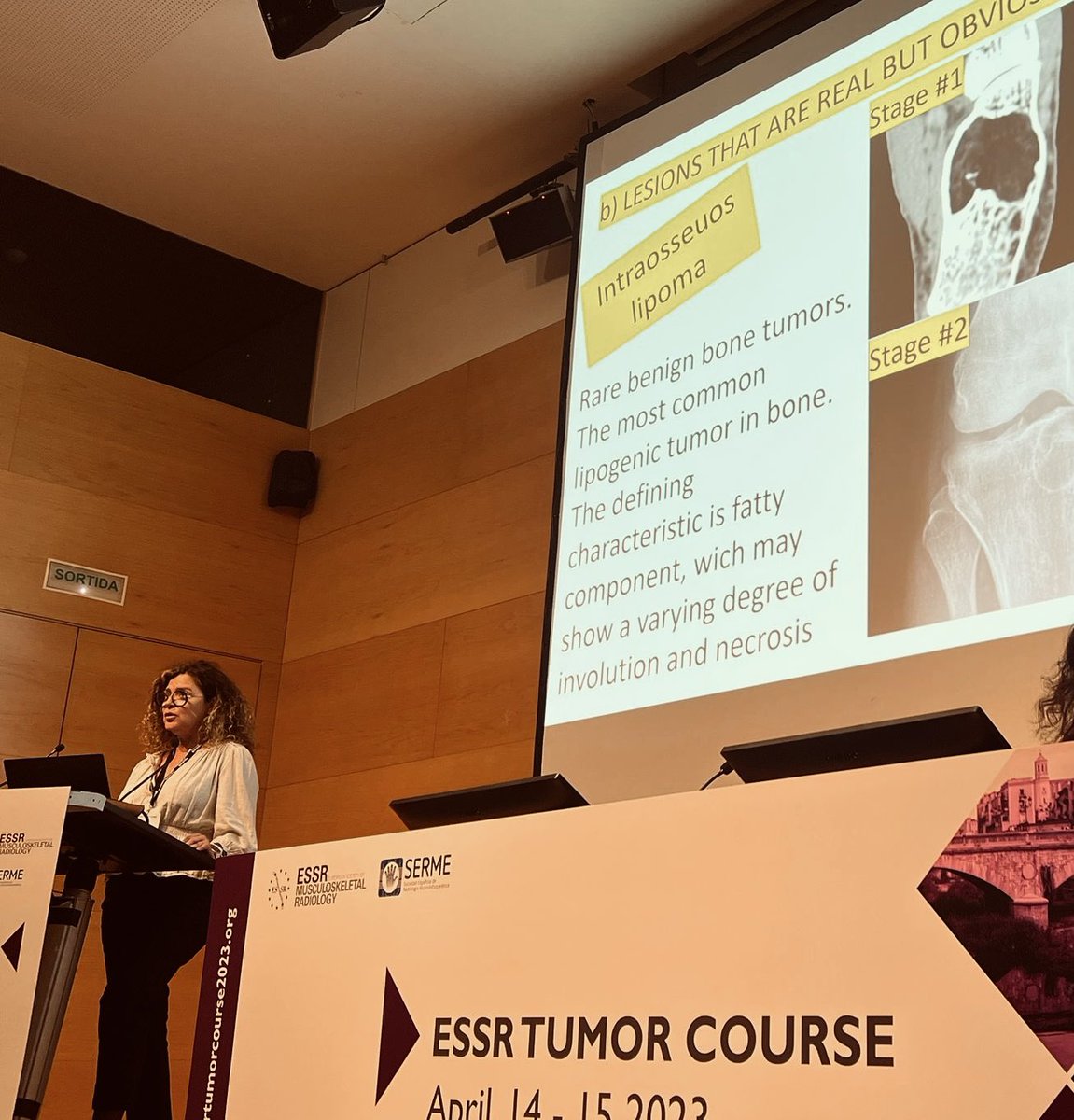
Emotiva 30 edicion del @MadridBoneClub en @Olympia_GrupoQS y estupendo cocktail en restaurante la maquina de torre caleido. Increible el nivel cientifico y humano!genial veros como siempre.
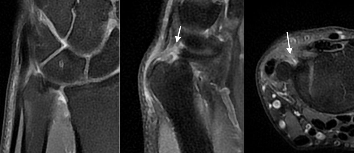
Complete tear of the dorsal capsular insertion of the TFC (Non-Palmer injury). Remember to always evaluate the sagittal views. Dorsal detachments are common and easily overlooked. See the normal TFC disk in coronal images. Previous MRI reported as normal.
Tarde de artroRM y punciones/infiltraciones en @Vithas Pardo de Aravaca. Realizamos punción lavado de calcificaciones, polectomía de dedo en resorte, infiltraciones ecoguidas, drenaje de quistes y hematomas entre otros procedimientos.Infórmate y pide tu cita!
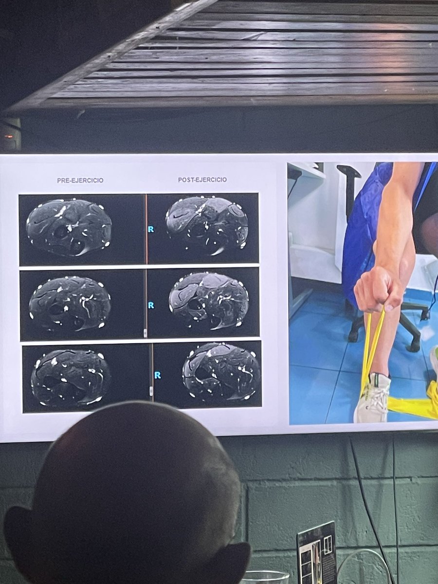
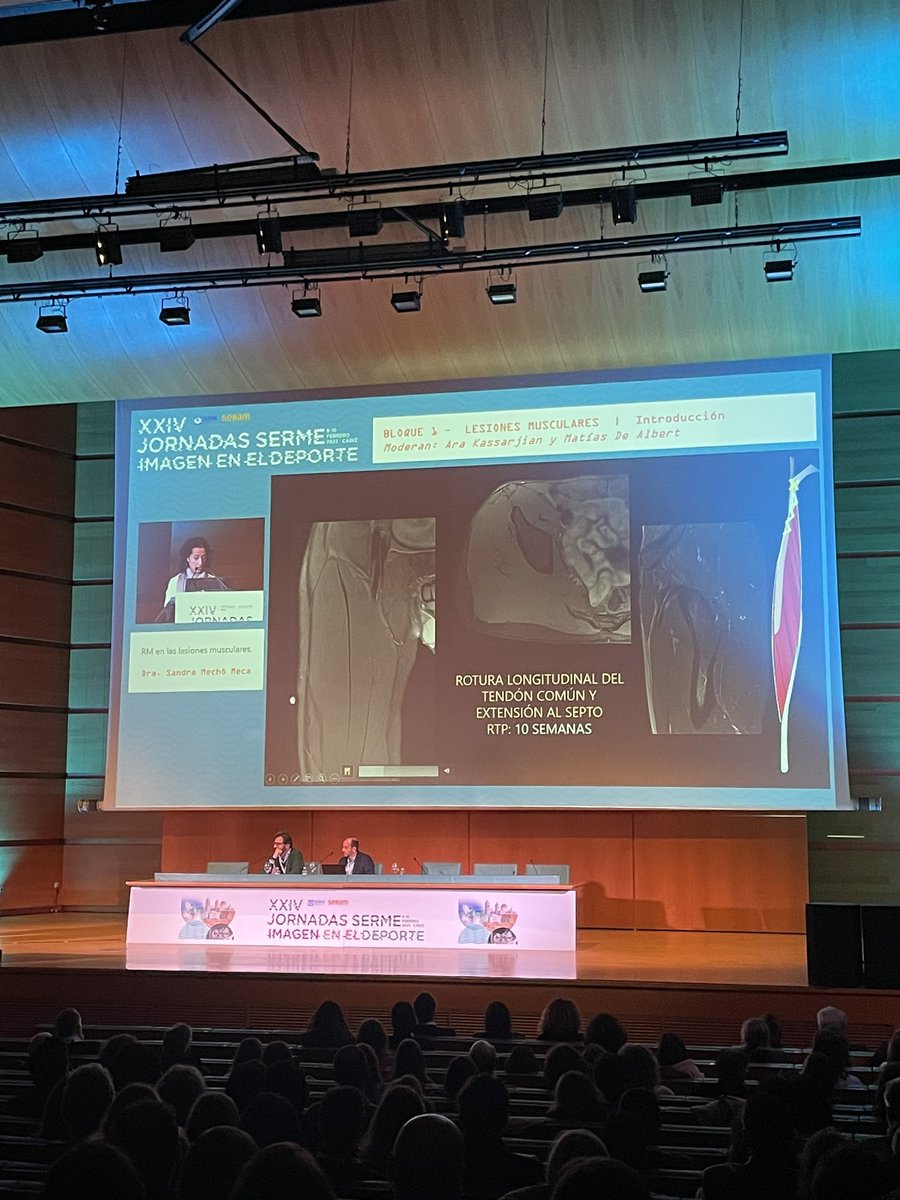
Check out our Webinar on Imaging of Muscle injuries in Sports! Exciting discussion with great experts. @DrMWLaupheimer @IsernKebschull@akassarjian and Michel Crema.
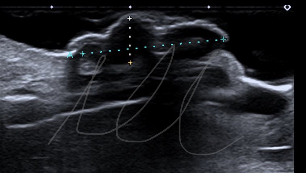
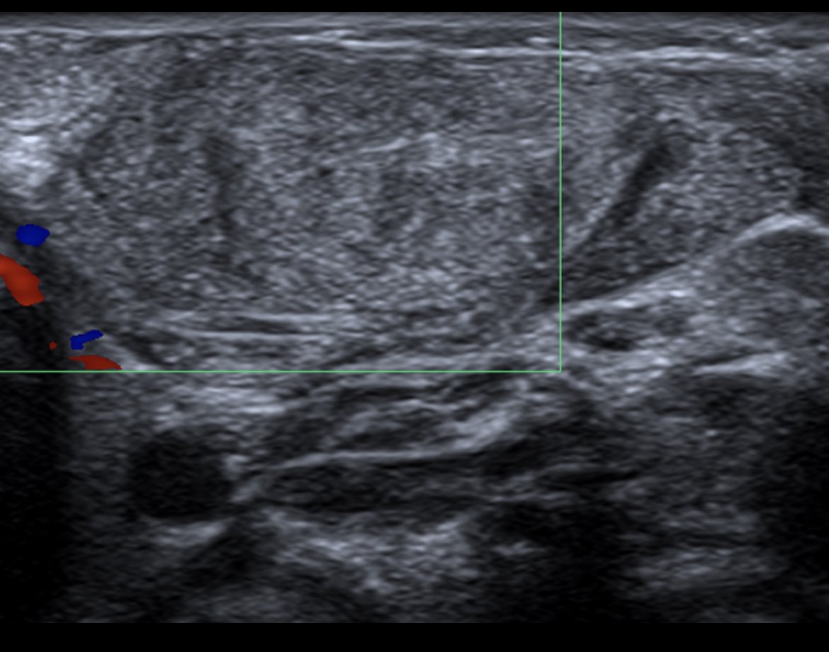
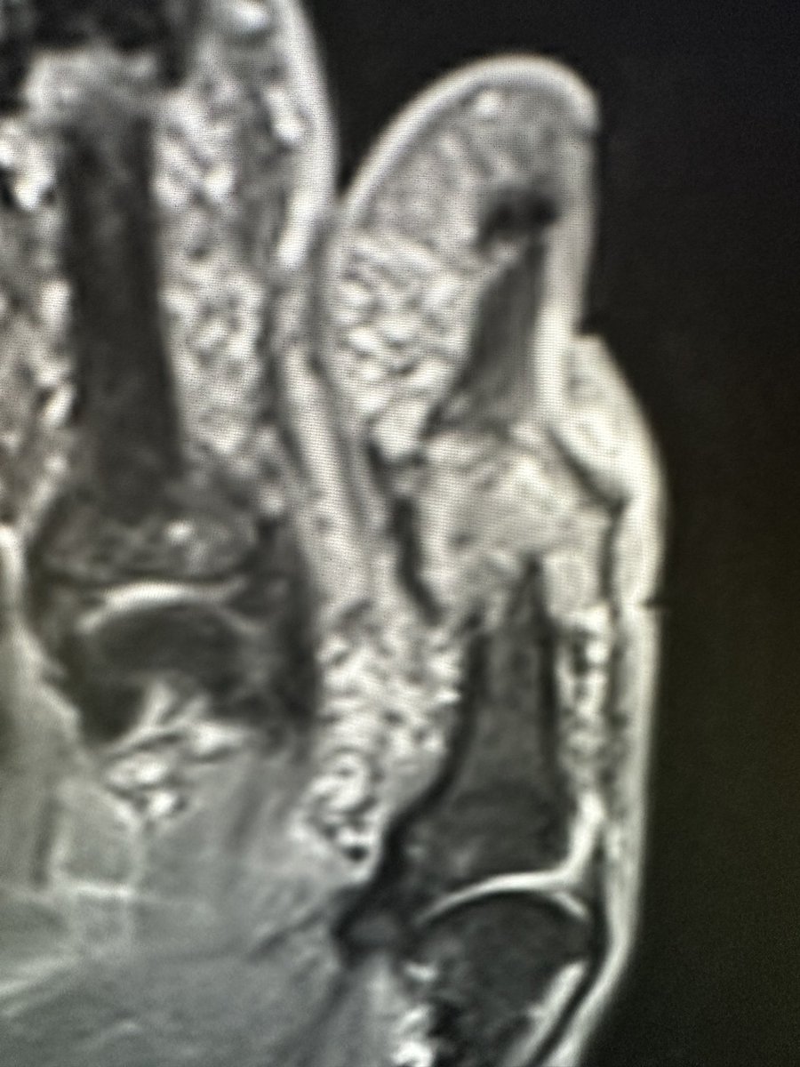
Hoy un casito un poco diferente a lo habitual pero que cada vez vemos más los radiólogos de msk. Ecografía de lesión nasal en varón de 70 años. ¿Qué os parece? @MskSerme@SERAM_RX@2000Radbox
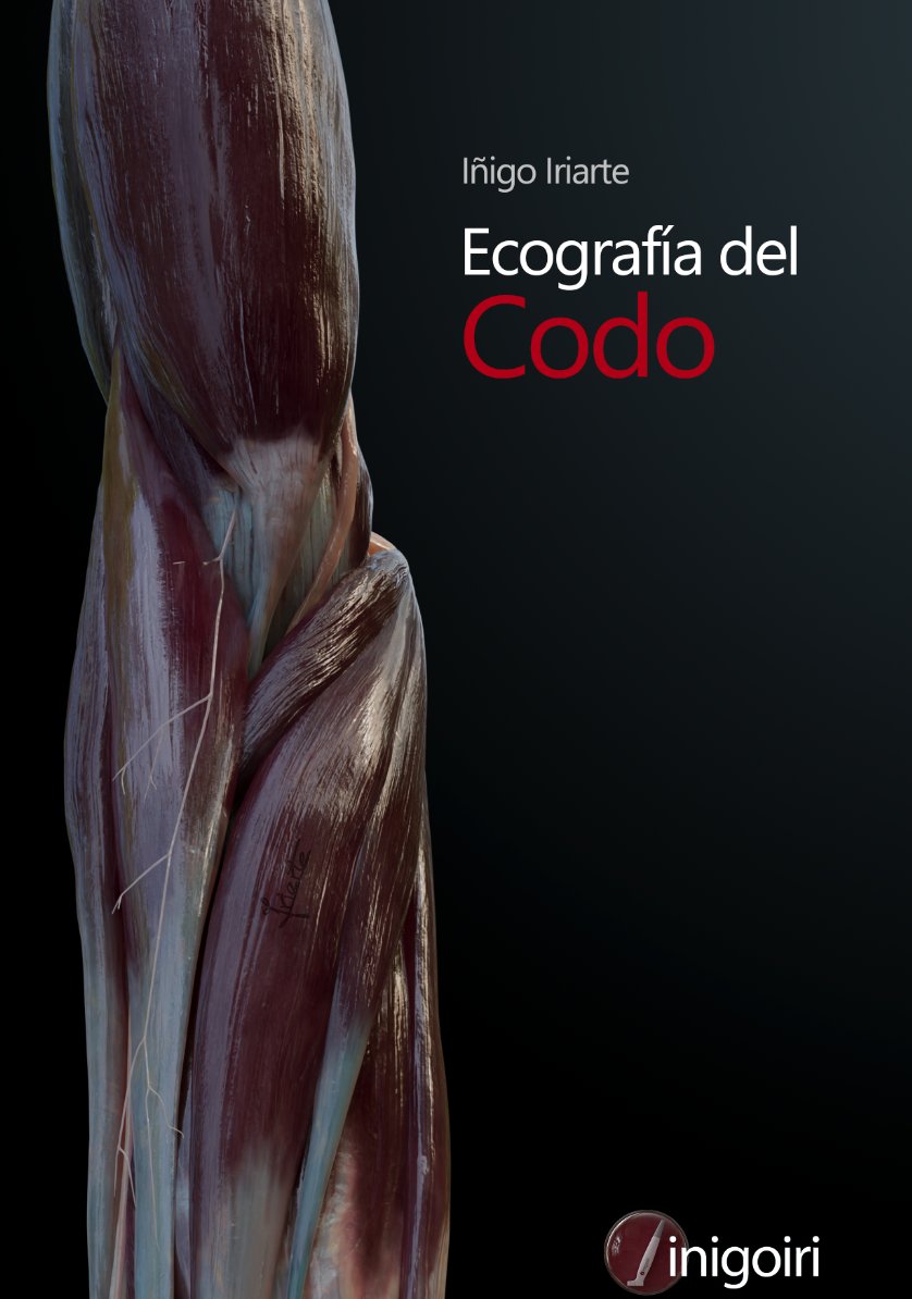
Por fin he acabado el capítulo "Ecografía del codo" que podréis ver en la nueva web de formación de ecografía https://t.co/9AOYBCT2F4 que estará disponible pronto (espero en mayo).
Me encanta el resultado pero yo no soy muy imparcial que digamos... esperaré vuestras opiniones 😅
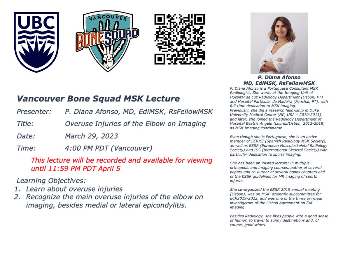
Want to receive the event zoom links and the 7 day links to view the recordings? Sign up at https://t.co/LekMb73c5e
@Afonso1Diana
For charitable donations:
https://t.co/iJ9mSR6p4F
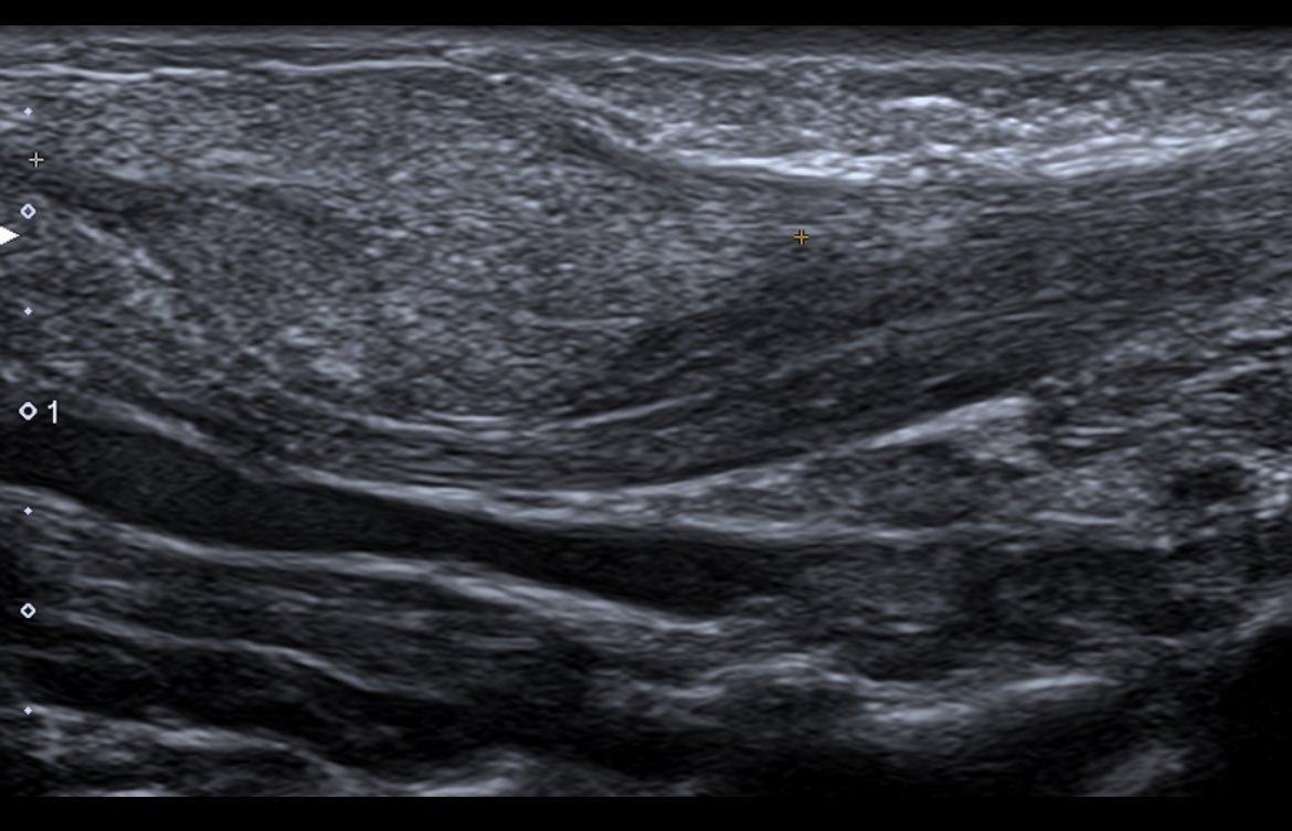
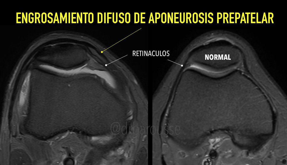
La aponeurosis prepatelar está constituida por la aponeurosis proveniente de los tendones vasto lateral y medial y las fibras superficiales del recto femoral. En este caso futbolista con antecedentes traumáticos crónicos de rodilla. Podría ser un engrosamiento secuelar.
It's time for @BIDMC_MSKImg's interesting case of the week!
⬇️ ⬇️ ⬇️
Case # 121:
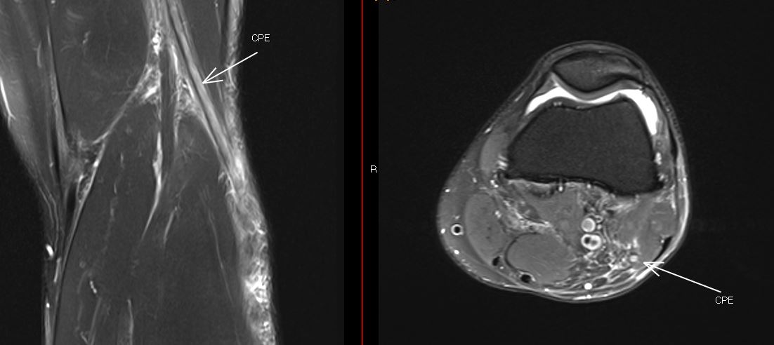
Posterior knee pain in 20-year-old.
Diagnosis?
Reply by Friday, March 17!
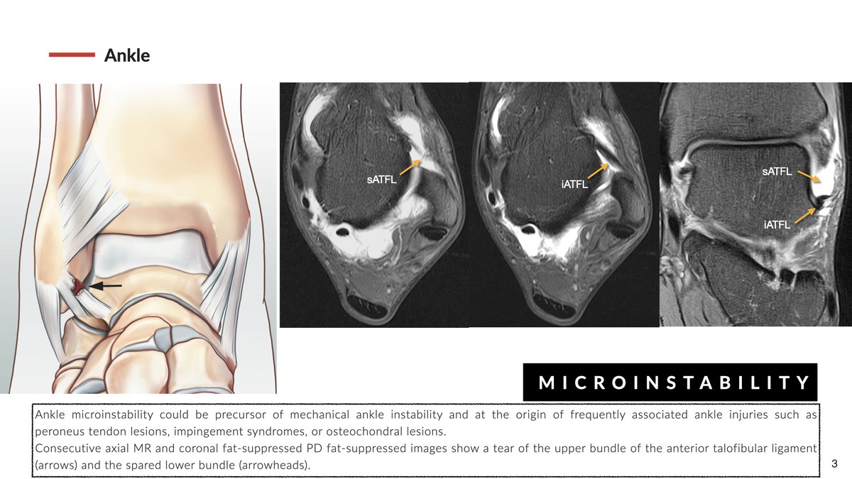
Ankle Microinstability might be the precursor for classic mechanical instability. It is secondary to the lesion of the superior fascicle of the anterior talofibular ligament. MRI and MR-arthrography, are key in precisely diagnosing ligament lesions and associated injuries
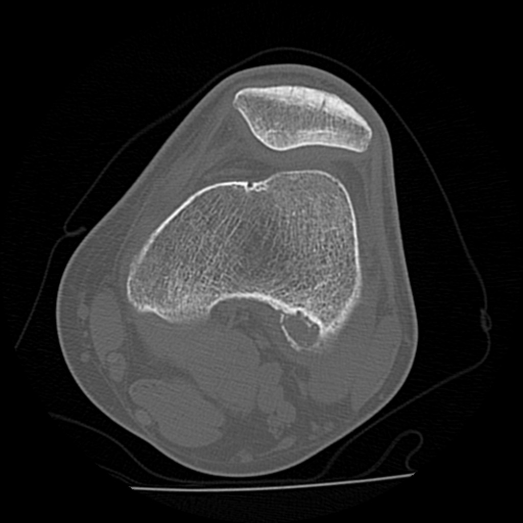
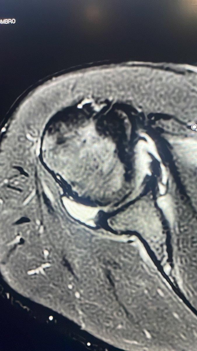
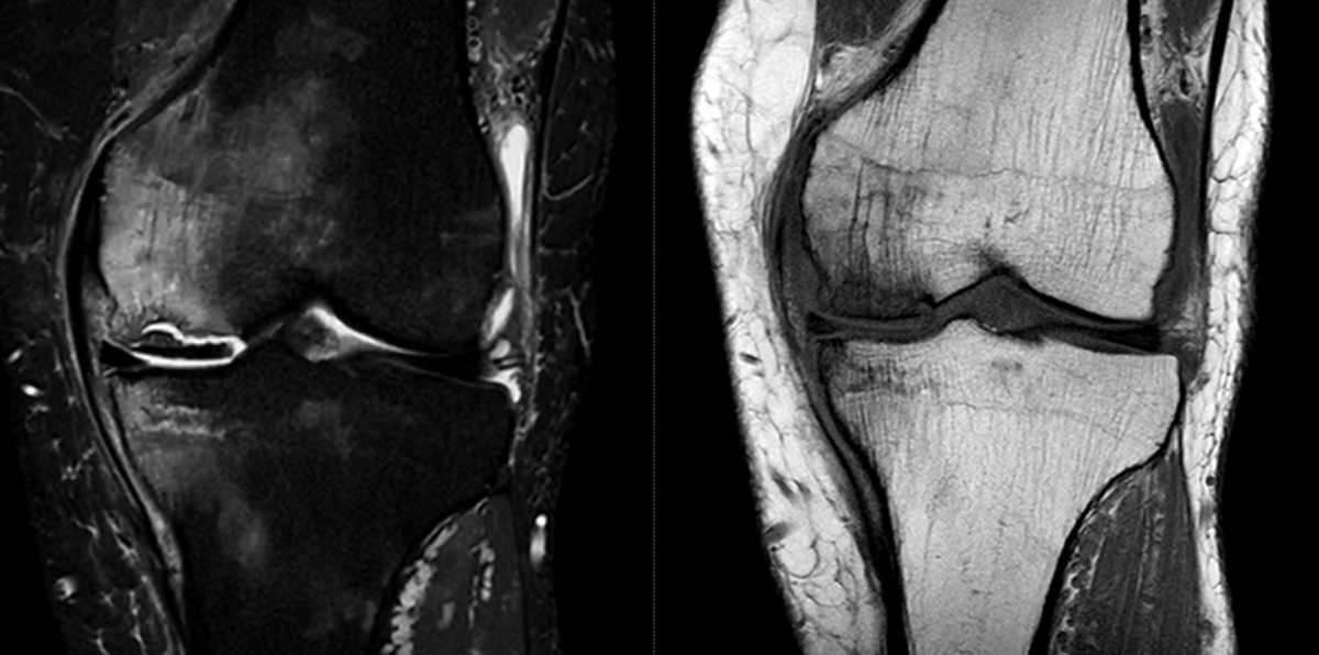
End Stage of SIF in medial condyle. Established osteonecrosis with signs of instability.
Concomitant insufficiency Fx tibial plateau.
MM root tear (not shown).
#msk#mskrad#ortho#orthotwitter
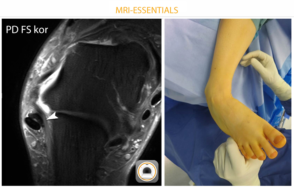
In cases of chronic ankle instability (esp. subtalar instab.), MRI can be misleading. In this case, edema along the calcaneofibular lig. was the only finding. The surgical image shows significant instability (thanks to Dr. Martin Jordan!).
#mskrad#orthotwitter#radres#radiology