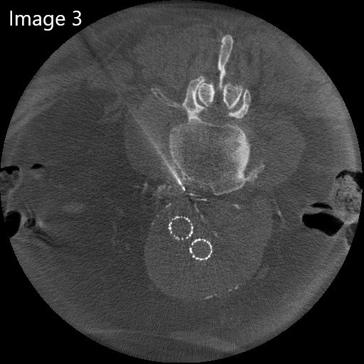
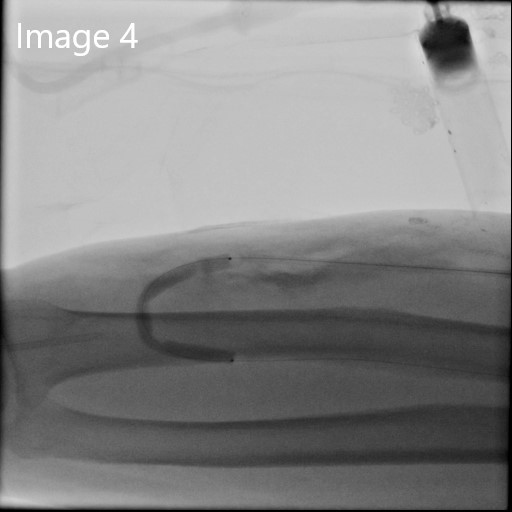
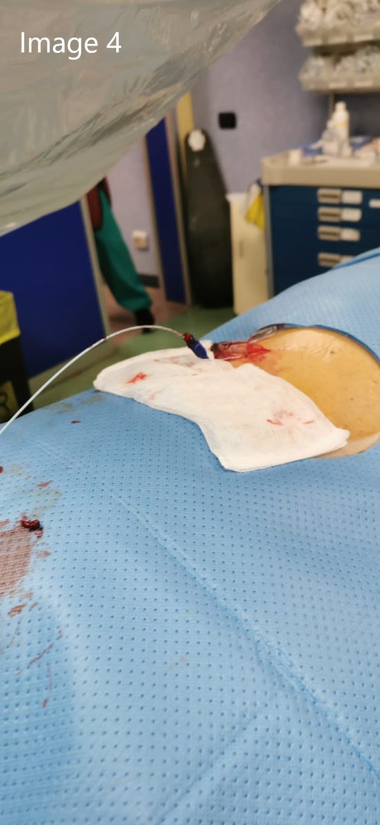
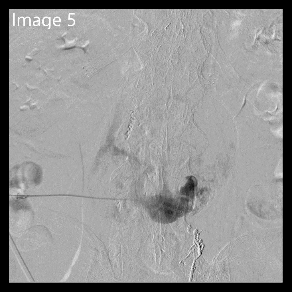
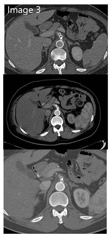
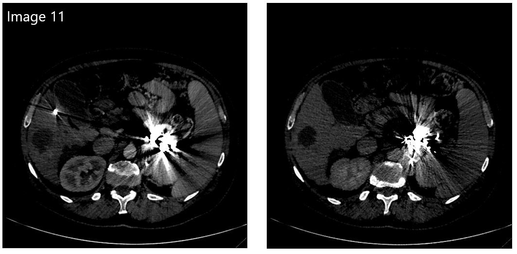
The patient was in a prone position, and mild sedation was performed. Using a combination of fluoroscopy and cone beam CT, the aneurysmatic sac was punctured using a 21 G needle. (Image 3) Afterward, a 6F coaxial system was advanced. (Image 4) (4/7)
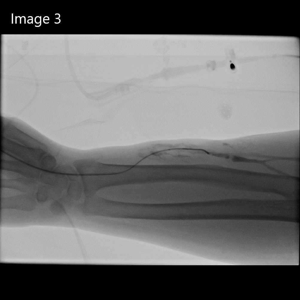
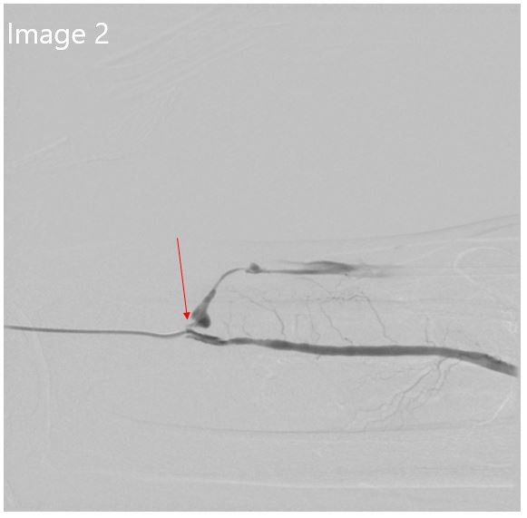
3. After failed attempt to perform retrograde recanalization, a distal radial arterial puncture was performed, & anastomotic stenosis was confirmed. Rendezvous w/ the venous side was performed & then angioplasty (3,4 & 5 mm) through the venous access was performed...
#CIRSEtakeover
Day 4 Case 2 by @PLucatelli
1. A 70-yo male patient w/ chronic renal insufficiency on a hypertensive basis also suffers from dyslipidemia & type II diabetes.
#CIRSEtakeover@PLucatelli
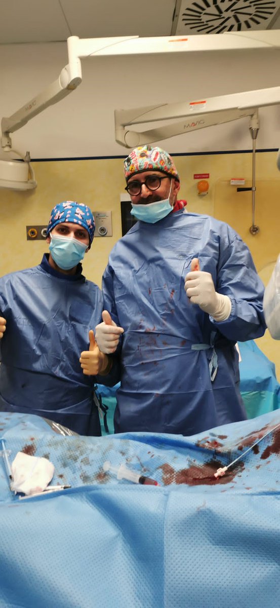
Day 4, Case 3 by Dr. Nardis & resident Dr. Vetri
85-yo patient w/abdominal aortic aneurysm treated with an infrarenal bifurcated stent graft five years ago. Type II endoleak was observed during follow-up, but the aneurysm sac remain stable in size(1/7)
#CIRSEtakeover@PLucatelli
Day 3 Case 3 Dural Arterio-Venous Fistula (DAVF)
72-yo patient w/ a history of headache & postural instability, assessed as idiopathic endocranial hypertension for which underwent ventriculoperitoneal shunt w/out significant clinical improvement. (1/5)
Young man arrived in the ER following car accident; the CT examination was urgently performed & revealed several fractures & large periaortic hematoma due to hemodynamic instability. The patient is transferred to the operating room to perform an angiography. (Image 1) (2/5)
Then a retrograde embolization of the origin of the celiac tripod was performed; a plug and an aortic stent were placed at the origin of the celiac tripod until the bleeding stopped. (Image 2) (4/5)
#CIRSEtakeover@plucantoni
Day 3 Case 1 performed by Doctor Mario Corona @Mario30855921
Trauma represents a mainstay of IR everyday practice - have a look at this tricky case!!! (1/5)
Superselective embolization of the tumor with a mixture of lipiodol and indocyanine green, thanks to its fluorescence using intraoperative infrared light, can easily allow visualization of the tumor through the kidney surface. #indocyaninegreen#embolization (2/4)
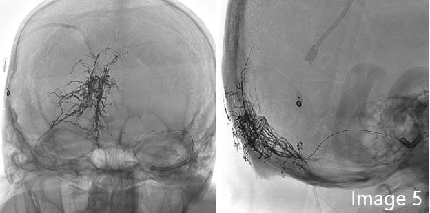
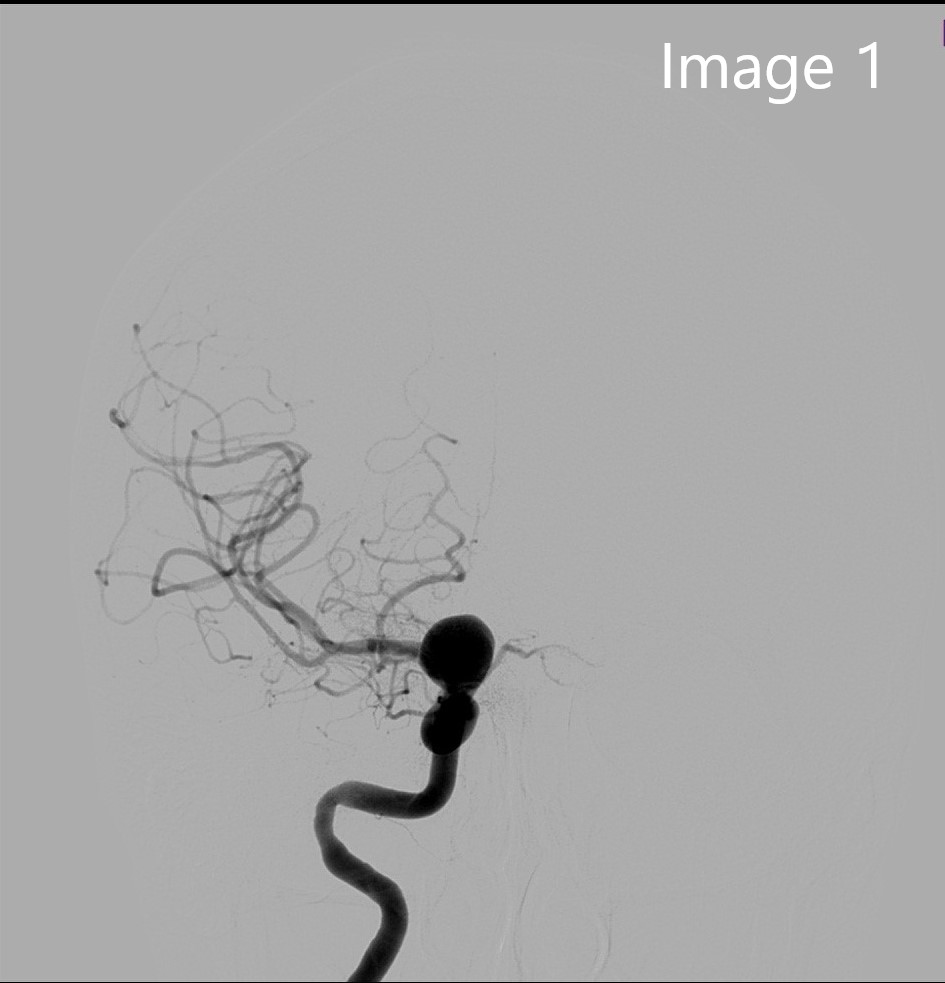
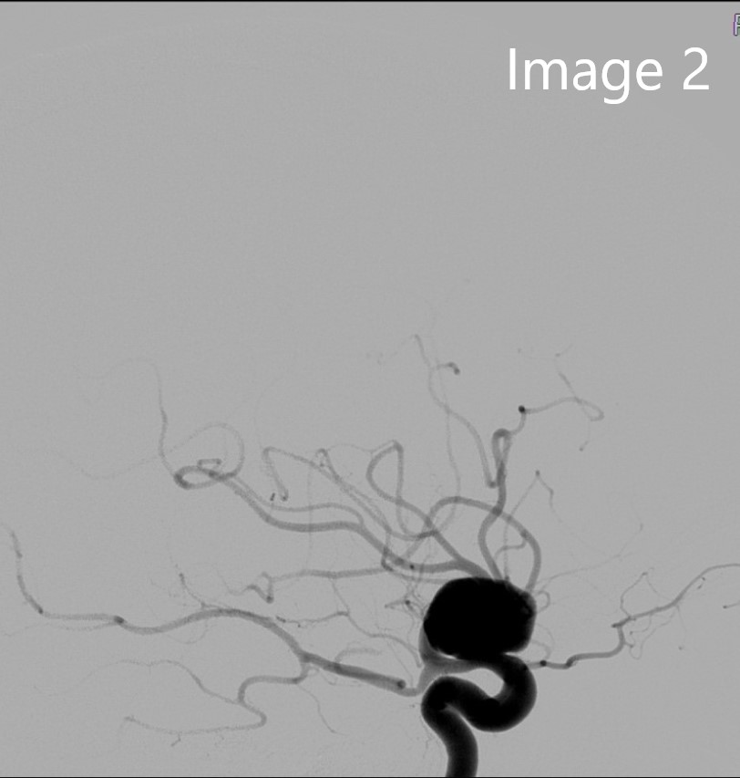
Fifty-four-year-old patient with headache, with a finding of 16x11mm right internal carotid-ophthalmic artery aneurysm, treated by placement of flow diverter (images 1-2). (2/4)