Medicine, Explained.
This account has one simple purpose: to help as many people as possible better understand their health.
After more than 25 years practicing medicine, I know that many people do not always have the time, the means, or the opportunity to see a doctor for every health concern.
Some topics can also be difficult or embarrassing to talk about.
So here, I’ll try to explain common health problems in simple terms and provide a few useful points of reference.
Modestly. Without unnecessary jargon. And without creating unnecessary fear.
Of course, no post can ever replace a proper medical consultation, a physical examination, or advice tailored to your individual situation.
But if a few simple explanations can help you better understand your body, know when to seek medical care, or simply ask yourself the right questions, then this account will have served its purpose.
Take care of your health — and the health of those around you.
DOES CREATINE SLOW DOWN ALZHEIMER’S DISEASE?
Maybe.
But we are nowhere near proving it yet.
In 2025, researchers gave 20 people with Alzheimer’s disease 20 g/day of creatine for 8 weeks.
Brain creatine increased by about 11%.
Several cognitive scores also improved, including global cognition, working memory and attention/inhibitory control.
That sounds impressive.
But there is one major limitation:
There was no placebo group.
This was a small, open-label pilot study.
So we cannot say that creatine caused the cognitive improvement.
And we definitely cannot say that it “slowed cognitive decline by 30%.”
There was no control group and no measurement showing a 30% reduction in disease progression.
A follow-up analysis published in 2026 added another interesting signal.
ATP and ADP increased in blood lymphocytes, and several measures of mitochondrial respiration improved in women.
But these were blood cells, not the brain.
Brain markers including NAA and glutathione did not significantly change.
And as of 2026, there are still no randomized controlled trials testing creatine in people with dementia.
So where are we?
Creatine reaches the brain: yes.
Interesting biological signal: yes.
Possible cognitive benefit: worth investigating.
Creatine slows Alzheimer’s progression: not demonstrated.
Creatine is a treatment or prevention for Alzheimer’s: far too early to say.
Promising pilot study ≠ proven treatment.
Study:
https://t.co/a4e9kuWA5D
Medicine, Explained.
VITAMIN D: ARE WE UNDERESTIMATING WHAT IT CAN DO?
Most people think of vitamin D as something we need for strong bones.
But vitamin D receptors are found throughout the body — including in many immune cells.
And low vitamin D levels have been associated with a surprisingly large number of diseases.
But remember:
association does not necessarily mean causation.
So what benefits are actually supported by evidence?
1. Bones and muscles
Vitamin D helps us absorb calcium.
Severe deficiency can cause rickets, osteomalacia, muscle weakness and bone fragility.
Correcting a true deficiency is clearly beneficial.
But high-dose vitamin D does not appear to prevent fractures in people who are already sufficient.
2. Pregnancy
Trials suggest supplementation may modestly reduce risks such as:
– pre-eclampsia
– preterm birth
– small-for-gestational-age babies
– neonatal mortality
This is one reason pregnancy is considered a group that may benefit from supplementation.
3. Prediabetes
In people with high-risk prediabetes, vitamin D may slightly reduce progression to type 2 diabetes.
The effect is modest — and it does not replace exercise, nutrition or weight loss.
4. People over 75
Trials suggest a very small reduction in overall mortality.
The effect is small, but consistent enough for the Endocrine Society to suggest supplementation in this age group.
5. Autoimmune diseases
This is one of the most interesting areas.
In the large VITAL trial, 2,000 IU/day was associated with fewer newly diagnosed autoimmune diseases during supplementation.
Interesting? Yes.
Proof everyone should take vitamin D to prevent autoimmunity? Not yet.
6. Multiple sclerosis
Low vitamin D has long been associated with a higher risk of MS.
In 2025, a randomized French trial went further:
In very early MS, high-dose vitamin D reduced overall disease activity, mainly new MRI lesions.
This does not mean vitamin D replaces disease-modifying treatments.
But MS is probably one of the most interesting examples of a possible benefit beyond bone health.
7. Depression? Maybe.
People with depression often have lower vitamin D levels.
Some meta-analyses suggest a small improvement in symptoms, especially in people who are already depressed.
But other analyses become negative when weaker studies are removed.
Correcting a deficiency? Reasonable.
Calling vitamin D an antidepressant? No.
8. Fertility? Caution again.
Women with low vitamin D often have poorer outcomes during assisted reproduction.
But that does not prove causation.
A large 2026 trial in women with PCOS undergoing IVF showed something very instructive:
Vitamin D corrected the deficiency…
but did not increase live births.
A perfect example of the difference between:
a marker associated with disease
and
a treatment that changes the outcome.
So what is the “right” vitamin D level?
A level around 30 ng/mL (75 nmol/L) is generally considered adequate.
But we still do not know the optimal level for many possible extra-bone benefits.
And there is no strong evidence that pushing everyone from 30 to 40–50 ng/mL provides additional protection.
Many large trials also included people who were already vitamin D sufficient — which may partly explain why supplementation often showed little benefit.
The bottom line:
Vitamin D is essential.
Deficiency can have real consequences.
Correcting it may improve specific outcomes in certain situations.
But once your needs are covered, more is not necessarily better.
ERECTILE DYSFUNCTION IN YOUNG MEN
Contrary to what many people think, this is actually a fairly common reason for consultation.
And in a man in his 20s or 30s, erectile dysfunction does not necessarily mean there is something physically wrong with the penis.
An erection is actually a fairly complex process.
The brain has to generate sexual arousal.
The nerves have to transmit the signal.
The blood vessels have to open.
And blood then has to remain trapped inside the penis.
Stress can interfere with almost every step.
One of the first useful questions is therefore:
Does it happen all the time?
If erections are normal in the morning or during masturbation, but become difficult with a partner, a psychological component — especially performance anxiety — becomes much more likely.
And a vicious cycle can quickly develop:
one episode of difficulty
→ fear that it will happen again
→ stress and adrenaline
→ even more difficulty getting or maintaining an erection.
But saying “he is young, so it must be psychological” is too simplistic.
Other possible causes include:
• stress, anxiety or depression
• relationship difficulties
• certain medications, particularly some antidepressants
• alcohol or recreational drugs
• nicotine
• diabetes
• high blood pressure or high cholesterol
• hormonal problems, including low testosterone
• more rarely, neurological or penile disorders.
That is why persistent erectile dysfunction deserves a medical assessment — even at 20.
The initial evaluation is usually quite simple:
understand when the problem occurs, ask about morning erections, assess libido, review medications and substances, perform a clinical examination and, when appropriate, check blood glucose, cholesterol and morning testosterone.
And treatment is not necessarily just “take Viagra.”
Sometimes understanding what is happening and being reassured already helps a lot.
Psychological or sex therapy can help break the performance-anxiety cycle.
Any underlying medical or lifestyle factors should also be addressed.
And medications such as sildenafil can be very effective — sometimes only temporarily, to help restore confidence while the underlying cause is being addressed.
Most importantly: do not hesitate to talk to a doctor about it.
Erectile dysfunction is common, including in younger men, and in many cases it can be significantly improved or treated.
It is not something you should simply accept and allow to affect your relationships, confidence or quality of life for years.
Erectile dysfunction is a symptom.
The important part is understanding why it is happening.
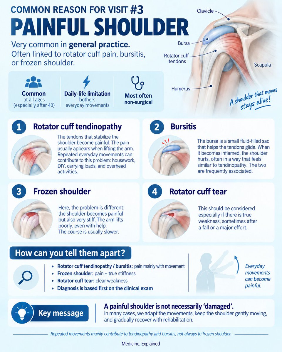
COMMON REASON FOR VISIT #3 — SHOULDER PAIN
After low back pain and kidney stones, another very common reason to see a GP: a painful shoulder.
The shoulder is one of the most mobile joints in the body.
Every time you lift your arm, reach overhead, get dressed, carry something or put your hand behind your back, several muscles and tendons have to work together.
So what usually hurts?
Rotator cuff tendinopathy
The rotator cuff is a group of tendons that stabilizes the shoulder and helps move the arm.
With repeated movements, overhead work, sport, housework or simply age-related changes, these tendons can become painful.
Typical clue: pain when lifting the arm, often worse at night.
“Tendinopathy” does not necessarily mean the tendon is torn.
Bursitis
The bursa is a small fluid-filled sac that helps the tendons glide smoothly.
It can become irritated and painful.
Bursitis and rotator cuff tendinopathy often occur together, which is why the symptoms can look very similar.
Frozen shoulder
This is different.
The shoulder becomes painful AND progressively stiff.
The key point: movement is limited even when someone else tries to move your arm for you.
It can develop without any obvious cause and is more common in people with diabetes.
Recovery can take months.
Rotator cuff tear
A tendon can partially or completely tear, either gradually over time or after a fall or sudden effort.
A major loss of strength, especially after trauma, should raise suspicion.
How do we tell them apart?
• Tendinopathy / bursitis → pain mainly with movement
• Frozen shoulder → pain + true
Could epilepsy beginning later in life sometimes reveal early Alzheimer-type changes?
A new study explored this possible connection.
When epilepsy begins after age 55, doctors usually look for a cause such as a stroke, tumour or previous brain injury.
But sometimes the MRI shows no clear explanation. This is called late-onset unexplained epilepsy.
Researchers studied 85 people with this condition.
They measured:
• memory and cognitive performance
• brain activity during 24 hours, including sleep
• p-tau217 in the blood
p-tau217 is a blood marker associated with the brain changes seen in Alzheimer’s disease. An elevated level does not diagnose Alzheimer’s on its own.
The researchers also examined what happened during sleep.
During deep sleep, the brain produces slow waves and short bursts of activity called sleep spindles. These rhythms need to coordinate properly to help the brain store new memories.
People with higher p-tau217 levels had:
• poorer cognitive performance
• less coordination between these sleep rhythms
The link with cognitive difficulties was strongest in people whose seizures remained uncontrolled despite medication.
This suggests that, in some people, late-onset epilepsy, disrupted sleep and memory difficulties may be connected to the same underlying neurodegenerative process.
But the study does not prove that epilepsy causes Alzheimer’s disease, or that a seizure after age 55 means someone has Alzheimer’s.
It also does not prove that better sleep can prevent the disease.
What we do know is that sleep is essential for memory and brain health, while chronic sleep disruption is associated with a higher risk of cognitive decline.
This was a small observational study involving only 85 people. More research is needed to understand which condition comes first and whether treating sleep problems could change the outcome.
The takeaway:
When epilepsy begins later in life without a clear cause, memory and sleep may deserve closer attention.
Study:
https://t.co/ony9jbMdc3
Kidney stones — when “back pain” is not really back pain
For this second episode of our series on common reasons people see a doctor, we stay close to back pain — but from a different angle.
In episode 1, we talked about low back pain.
But pain felt in the back or side does not always come from the spine.
Doctors call this a differential diagnosis:
the same symptom can have several possible causes.
One classic example is a kidney stone.
A kidney stone is a small hard deposit that forms when substances normally dissolved in urine crystallize and stick together.
A stone can sit silently inside the kidney.
Pain usually starts when it moves into the ureter — the narrow tube connecting the kidney to the bladder — and interferes with urine flow.
This can cause the intense, wave-like pain known as renal colic, often felt in the side or back and sometimes radiating toward the groin.
Step 1 — Confirm the diagnosis
Symptoms can strongly suggest a kidney stone, but imaging helps confirm it and answer three important questions:
Where is the stone? How big is it? Is it blocking urine flow?
Ultrasound uses sound waves and involves no radiation. It can detect many kidney stones and show whether the kidney is becoming obstructed.
A CT scan — often called a uroscanner — gives a more detailed view and can precisely show the stone’s size, location and degree of obstruction.
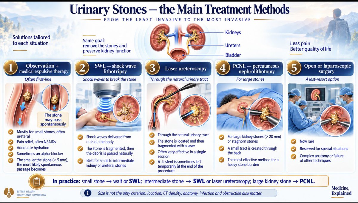
Step 2 — Choose the treatment
Treatment mainly depends on the stone’s size, location and whether it is causing complications.
Observation + pain relief → many small stones pass naturally
SWL — shock-wave lithotripsy → shock waves break the stone from outside the body
Laser ureteroscopy → a thin scope travels through the natural urinary tract and the stone is fragmented with a laser
PCNL — percutaneous nephrolithotomy → used mainly for large kidney stones, through a small tract created in the back
Surgery → now rare and reserved for special situations
In practice
Small uncomplicated stone → often wait for it to pass
Stone requiring active treatment → SWL or laser ureteroscopy
Large kidney stone → often PCNL
And one important exception:
A blocked kidney + infection or fever is an emergency.
The goal is then to drain the kidney and treat the infection before dealing with the stone itself.
Can gum disease affect your brain?
A bacterium commonly found in gum disease — Porphyromonas gingivalis — has been linked to Alzheimer’s disease.
But what does that actually mean?
When you have periodontitis, your gums stay chronically inflamed.
Bacteria and bacterial products can sometimes enter the bloodstream, potentially increasing inflammation throughout the body.
Researchers have also found signs of P. gingivalis and some of its toxins in the brains of people with Alzheimer’s.
So one hypothesis is:
gum disease → chronic inflammation → possible effects on the brain over many years
Sounds convincing?
Not quite yet.
People with periodontitis do appear to have a higher risk of cognitive decline and dementia.
But that does not prove that gum disease causes Alzheimer’s.
Age, diabetes, smoking and many other factors can influence both.
And the relationship could partly work the other way too: someone developing cognitive problems may simply take less care of their teeth.
So today, the best conclusion is:
There is an association.
There is a plausible biological mechanism.
But causation is not proven.
Taking care of your gums is important for your health.
Just don’t believe headlines saying that brushing your teeth will “prevent Alzheimer’s.”
Medicine, Explained.
Source:
Association Between Porphyromonas gingivalis and Alzheimer’s Disease: A Comprehensive Review — 2025
Read the full article
DOI: 10.2147/IDR.S491628
Kidney stones — when “back pain” is not really back pain
For this second episode of our series on common reasons people see a doctor, we stay close to back pain — but from a different angle.
In episode 1, we talked about low back pain.
But pain felt in the back or side does not always come from the spine.
Doctors call this a differential diagnosis:
the same symptom can have several possible causes.
One classic example is a kidney stone.
A kidney stone is a small hard deposit that forms when substances normally dissolved in urine crystallize and stick together.
A stone can sit silently inside the kidney.
Pain usually starts when it moves into the ureter — the narrow tube connecting the kidney to the bladder — and interferes with urine flow.
This can cause the intense, wave-like pain known as renal colic, often felt in the side or back and sometimes radiating toward the groin.
Step 1 — Confirm the diagnosis
Symptoms can strongly suggest a kidney stone, but imaging helps confirm it and answer three important questions:
Where is the stone? How big is it? Is it blocking urine flow?
Ultrasound uses sound waves and involves no radiation. It can detect many kidney stones and show whether the kidney is becoming obstructed.
A CT scan — often called a uroscanner — gives a more detailed view and can precisely show the stone’s size, location and degree of obstruction.
Step 2 — Choose the treatment
Treatment mainly depends on the stone’s size, location and whether it is causing complications.
Observation + pain relief → many small stones pass naturally
SWL — shock-wave lithotripsy → shock waves break the stone from outside the body
Laser ureteroscopy → a thin scope travels through the natural urinary tract and the stone is fragmented with a laser
PCNL — percutaneous nephrolithotomy → used mainly for large kidney stones, through a small tract created in the back
Surgery → now rare and reserved for special situations
In practice
Small uncomplicated stone → often wait for it to pass
Stone requiring active treatment → SWL or laser ureteroscopy
Large kidney stone → often PCNL
And one important exception:
A blocked kidney + infection or fever is an emergency.
The goal is then to drain the kidney and treat the infection before dealing with the stone itself.
Welcome to the first post in our new series: the most common reasons people visit their doctor.
We’re starting with low back pain.
Low back pain is one of the most common reasons people see a doctor.
But why does the lower back hurt so easily?
Our lumbar spine is made of 5 vertebrae stacked on top of each other.
Between them are intervertebral discs: flexible cushions that help distribute loads and allow the spine to move.
Around them are muscles, ligaments and small joints called facet joints.
So your back is not a rigid column.
It is a strong, mobile structure designed to bend, rotate and carry weight.
So why can everyday movements hurt?
When you bend forward, lift something, twist, sit for a long time or arch your back, the forces acting on the spine change.
Some structures are compressed.
Others are stretched.
Muscles work harder to stabilize the area.
Most of the time, this is completely normal.
Pain does not necessarily mean that something has been damaged.
Sometimes muscles become irritated or tense.
Sometimes the small joints at the back of the spine become sensitive.
Sometimes a disc becomes painful.
And occasionally, a nerve can become irritated.
What happens when you arch your back?
When you extend your lumbar spine, the facet joints at the back of the vertebrae move closer together.
In some people, especially if those joints are already sensitive, this can trigger pain.
But again: extension itself is not a “bad” movement.
The spine is made to move.
And what about a “slipped vertebra”?
A vertebra can sometimes move slightly forward relative to the one below it.
This is called spondylolisthesis.
It can increase local mechanical stress and sometimes cause back pain or nerve symptoms.
But many people have spondylolisthesis on an X-ray or MRI and feel absolutely nothing.
That is an important point:
An abnormal scan does not automatically explain your pain.
The same is true for disc degeneration.
Discs naturally change with age, just like skin develops wrinkles.
These changes are extremely common — including in people with no back pain at all.
So the goal is not to protect your back from every movement.
It is to keep it moving.
Learn to use your whole body during everyday tasks.
For activities that keep you bent forward for a long time — such as bathing a baby, making a bed, picking things up from the floor or gardening — try not to stay folded over at the waist.
Bend your knees and hips, bring yourself closer to what you are doing, and change position regularly.
The goal is not to avoid bending altogether.
It is to avoid repeatedly loading your lower back in the same position when another movement strategy is available.
Stay active.
Change positions regularly.
Lift progressively.
Strengthen your muscles.
And gradually return to normal activity.
For most episodes of common low back pain, movement is part of the solution — not the problem.
A new Nature Medicine paper does not say “a virus causes MS.”
It says something more precise.
People who already have relapsing MS gave blood over time.
Up to 3 months before a flare, two things show up:
• B cells (where Epstein-Barr virus hides) look reactivated
• monocytes look primed, mostly via interferon
EBV is the mono virus.
Almost everyone with MS has had it. That part was already known.
What’s new: a viral-looking immune signature in blood often precedes the attack — it does not prove the virus started the disease, and it is not a clinic test yet.
Paper: https://t.co/JqxwaUpb1E
Can a blood test detect Alzheimer’s before symptoms?
Maybe — and this could become important.
Alzheimer’s does not start when someone begins forgetting things. Brain changes can begin years earlier.
Scientists can now measure signs linked to two proteins in blood: amyloid and tau.
A Lancet study looked at about 1,350 people in their 50s and 60s with no dementia.
Some already had blood results suggesting early Alzheimer-related brain changes. On average, they also scored a little worse on some thinking tests.
A positive result does not mean you will get Alzheimer’s. It means some biology linked to the disease may already be there.
Should everyone get tested? Not yet. Finding something early only helps if we know what to do next.
We are not there yet. But a cheap blood test could move detection much earlier.
Takeaway: we may soon see signs of Alzheimer’s in blood before dementia.
Detecting it early and knowing how to stop it are two different problems.
Source: The Lancet, May 2026 — CARDIA study, 1,350 adults aged 53–69 without dementia.