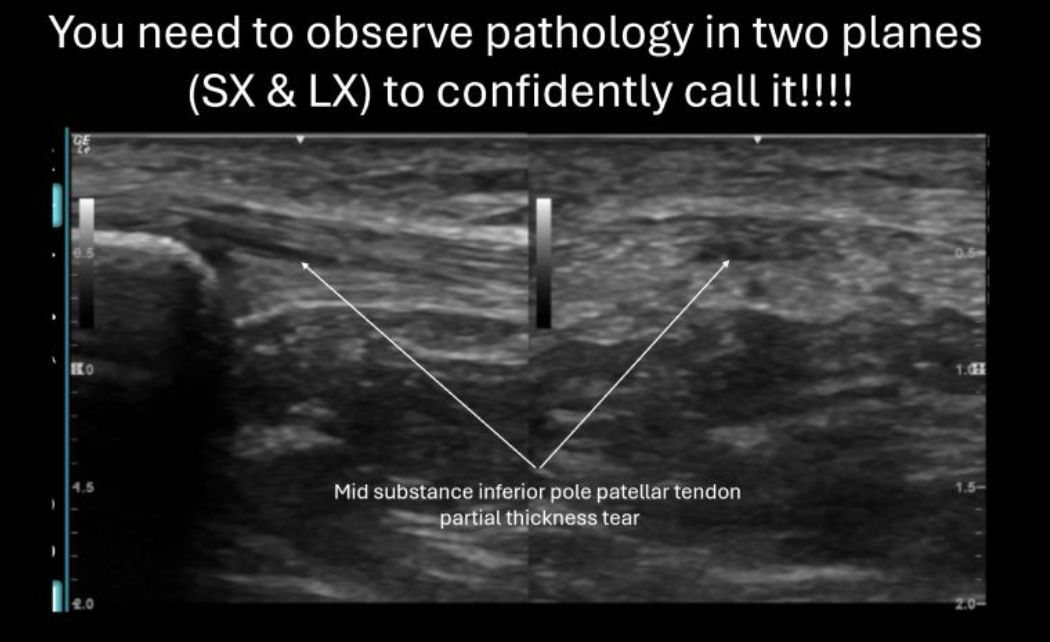
A common mistake I see when mentoring clinicians, on there ultrasound journey, is to call a tear without seeing it in only one plane. To be confident in calling pathology you need to see it in both long and short axis
#diagnosticultrasound#probeskills
Scanning a traumatic injury can lead to fascinating discoveries, often revealing uncommon pathologies not typically encountered in a routine clinic setting. This ultrasound image revealed a partially severed flexor carpi ulnaris tendon
#DiagnosticUltrasound#FCU#WristPain
This patient suffered from a non traumatic complete long head of bicep rupture. Imaging also shows an anechoic intraarticular effusion within the sheath (coming from the GHJ) and synovial proliferative sheath changes.
#shoulderpain#longheadofbiceprupture
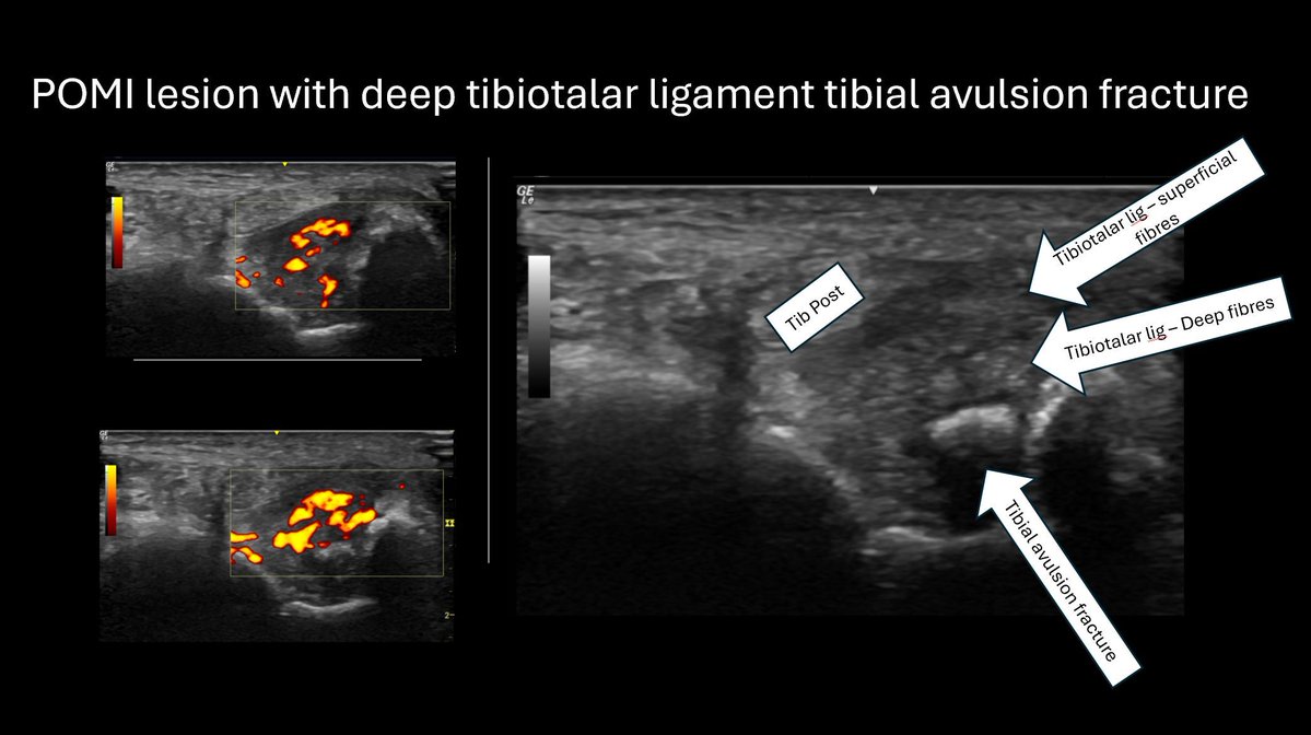
How often do you see a tibial avulsion fracture associated with a posteriomedial ankle impingement? Good candidate for a drop of steroid to calm the hyperaemia down followed by an onward referral to a podiatrist?
#diagnosticultrasound#footpain#avulsionfracture
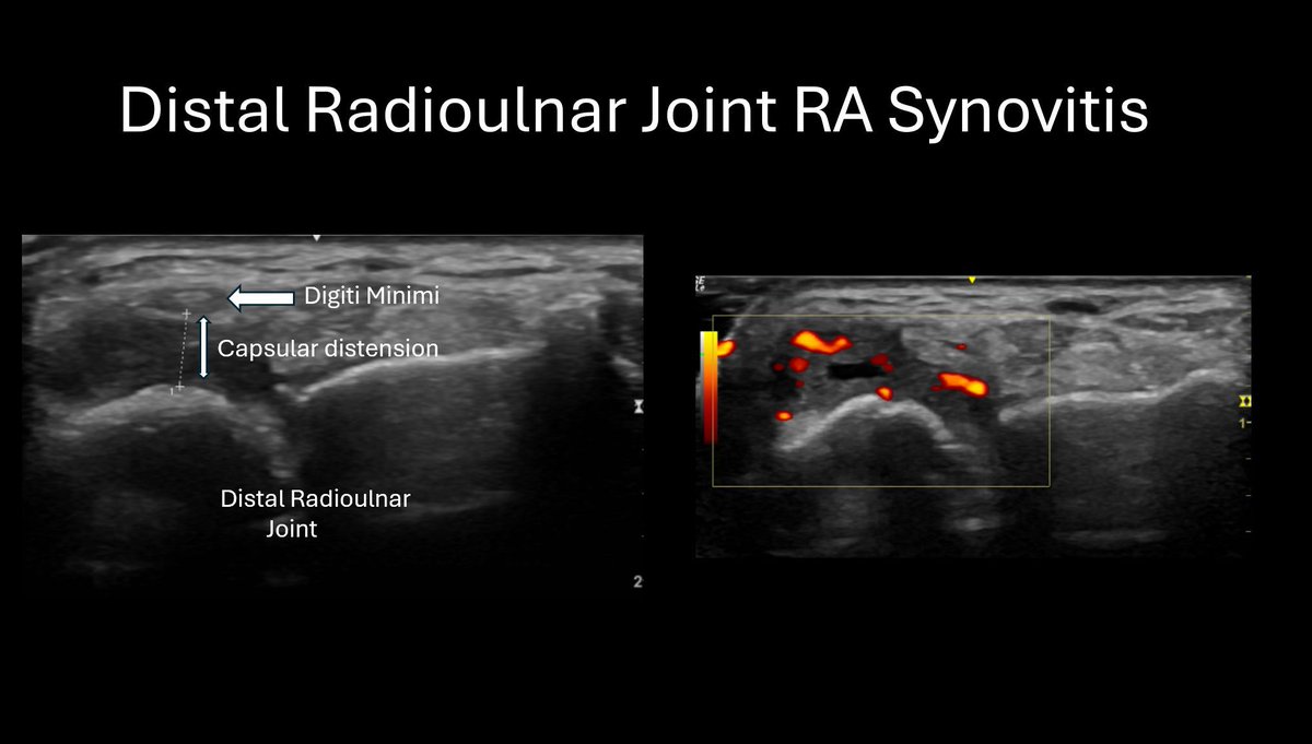
This distal radioulnar joint an essential place to scan for RA synovitis. You can often pick up subtle changes at this joint and is one not to miss.
hashtag#rheumatoidarthritis hashtag#distalradioulnarjoint hashtag#wristpain hashtag#synovitis hashtag#diagnosticultrasound
An interesting dorsal talonavicular joint injury! This image shows a shearing of an osteophyte which has also become displaced. Seen this before?
#diagnosticultrasound#footpain#avulsionfracture
Pathology affecting the 5th metatarsal region is a common clinical presentation. Ultrasound imaging is perfectly placed, alongside a clinical assessment for diagnosing pathology in the lateral foot.
Colloidal aggregates are clusters of small particles suspended in the synovial fluid within the cyst. These aggregates can develop for many reasons including;
1. Increased inflammatory markers
2. Mechanical Shear Forces
3. Chronicity
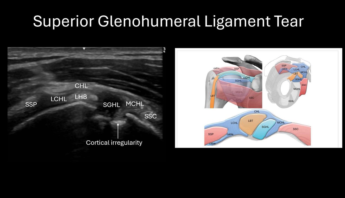
SGHL tear, cortical irregularity of the humeral head and intraarticular synovial proliferative changes within the joint space. Furthermore, there was a full thickness tear of supraspinatus and a large anechoic effusion within the bursal plane. Beautifully seen with ultrasound
50-year-old runner came into clinic after tripping over a thorny bush.
Ultrasound found an arrow shaped foreign body buried within the Achilles! She was send for emergency surgical removal as this was seen as a dirty wound. Amazing what you can find with ultrasound.
What could be more satisfying than a good Barbotage Lavage!
I see lots of patients with Calcific Supraspinatus Tendinopathy but it’s the ones where the calcific foci start to infiltrate into the subacromial bursal space that become super painful.
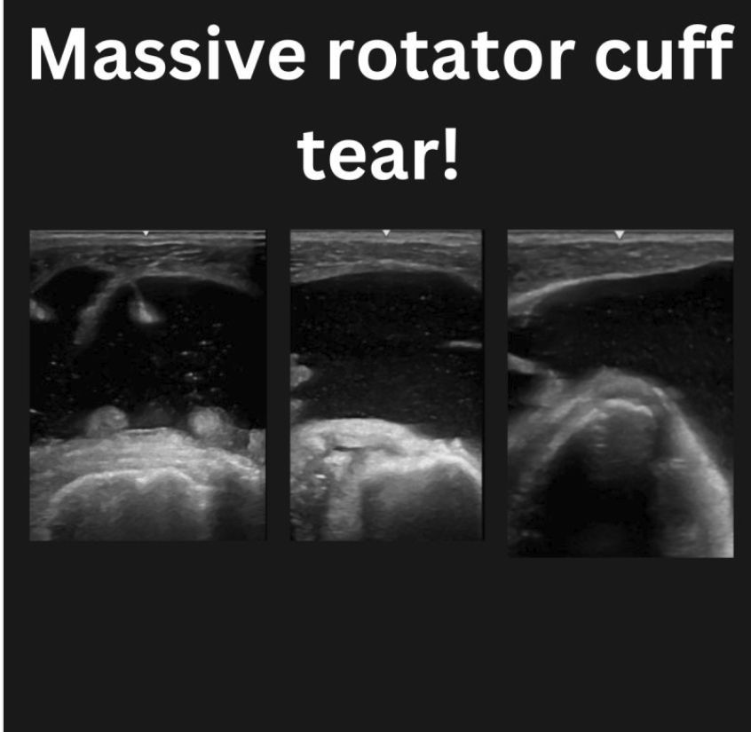
When your shoulder scan starts with a huge amount of anechoic fluid within the sub acromial space it can only mean one thing. A massive rotator cuff tear resulting in an effusion emanating from the glenohumeral joint collecting on the bursal plane!
Some joints, like the TMT joints of the foot, can be very difficult to inject using an in-plane needle approach. Using additional gel (stand-off technique) gives you space and allows you to get the angle right before entering the skin.
The key is to a successful barbotage is keep the needle within the calcific foci. Going straight through can cause you to lose the negative pressure necessary to suck out the calcific debris.
A reliable method of assessing for carpal tunnel syndrome is by measuring the diameter of the median nerve at the level of the pronator quadratus muscle and comparing it to the diameter of the median nerve at the level the lunate. diagnostic ratio of 1:4
Tibialis posterior pathology can be tricky to treat. Ultrasound imaging is excellent at helping us to unpick the underlaying causes of the patient’s pain. How would we treat this case?