It’s amazing to see the power of media in shaping our practice! Subsequent to this historic article in @WSJ in 1987, the US congress passed CLIA88 to strictly regulate the quality standards for lab testing. 👌🏼👌🏼(Thanks to Dr. Cibas for the photos.)
GU PATHOLOGY FELLOWSHIP! We have added a 2nd GU Fellowship position @BWHPath & have openings for July ‘22 & ‘23. Great academic opportunity for pathologists-in-training with a sincere GU interest Email/PM me if interested.🔬
@GU_Path_Society@IntSocUropath@Andres_M_Acosta
As we gather on the steps of HMS, we remember all the challenges of this past year but, most of all, the memories and contributions of the wonderful people of this department. Best wishes to all who are moving on to new positions and welcome to all who are joining us now!
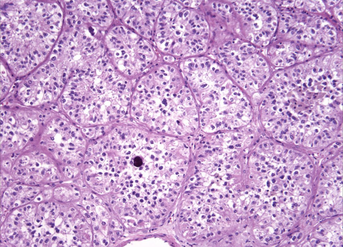
Rare example of verumontanum mucosal gland hyperplasia sampled in needle biopsy, a mimic of low grade #prostate cancer. Note the orange-red concretions, corpora amylacea and discernible basal cells. #GUPath
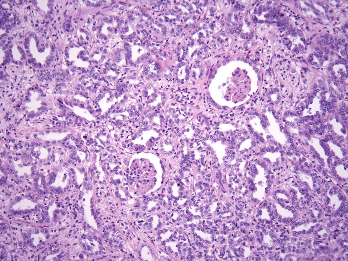
Invasive high grade papillary urothelial carcinoma of the ureter, pT3 with complete obliteration of the lumen. Elderly male with history of colon cancer #gupath
Just a "Perfect Example" of the hard-to-find and elusive orange "Parakeratotic-like Cell" (a term coined by Mac DeMay) in Malignant Mesothelioma (Pleural Fluid). On the right, you can see brisk mites.
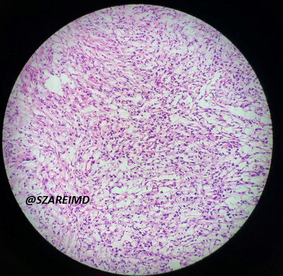
Lobectomy for a primary lung mass. Histology shows a poorly differentiated malignant neoplasm. What is the differential diagnosis? Which stains would you consider?
#BWHPathCotW#pathology#pulmpath
1/4
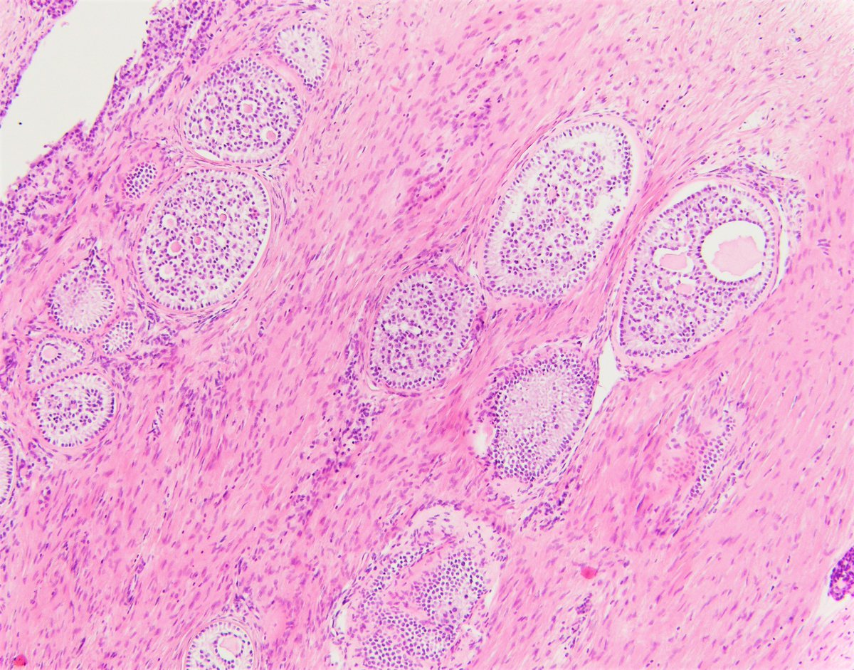
What do you think of these bilateral ovarian masses in a young female? What is the associated syndrome? Is the prognosis different between syndromic and non-syndromic cases?
#BWHPathCoTW#pathology#GynPath#PediPath 1/
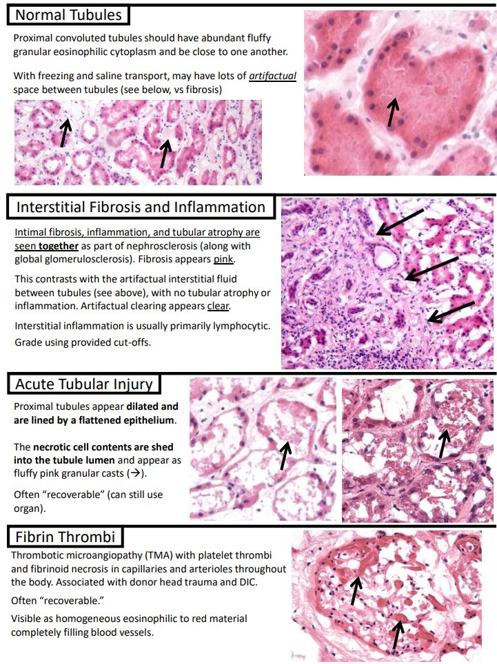
I made some handy "Kurt's Notes" (https://t.co/OKmaPvoHbB) for those late night donor organ frozen sections😴📟🤔
Full notes available here: https://t.co/L1OE3l2xRH
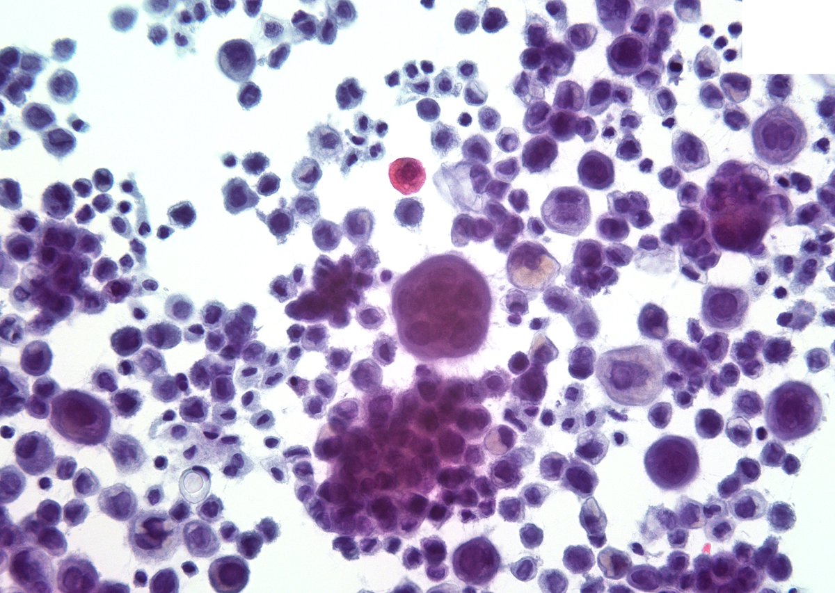
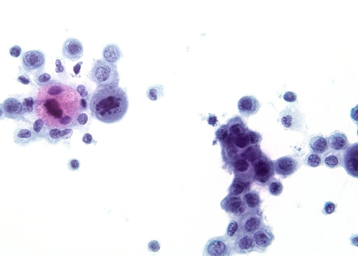
“Notorious” member of the solid Pancreatic Ductal Carcinoma variants, the “Undifferentiated Carcinoma with Osteoclast-like Giant Cells”. The reactive histiocytes often predominate the FNA picture with smaller malignant cells seen only in the background (CB).