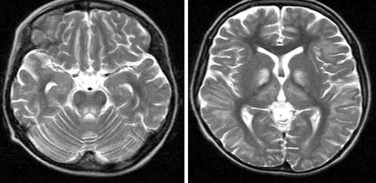
Hx of parotid surgery and chronic facial droop. This was perineural spread of a parotid tumour along the facial nerve resulting in this CP angle mass.
Case courtesy of Prof. E. Leon Kier.
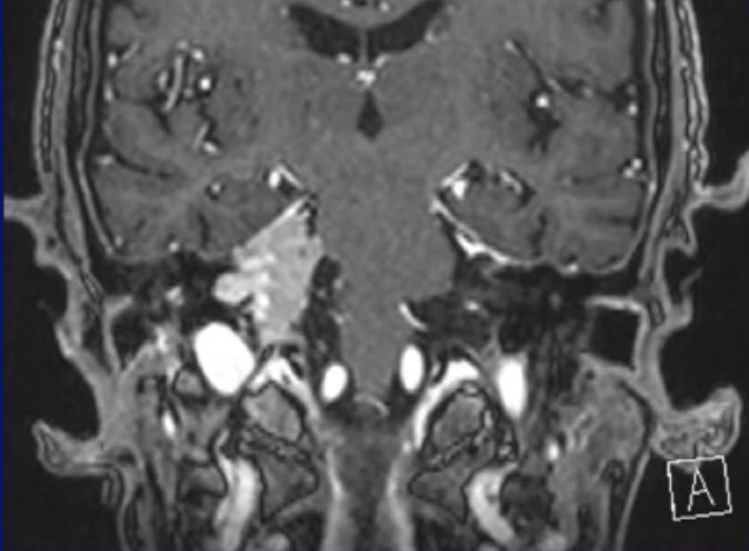
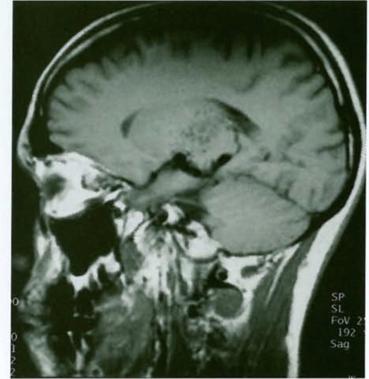
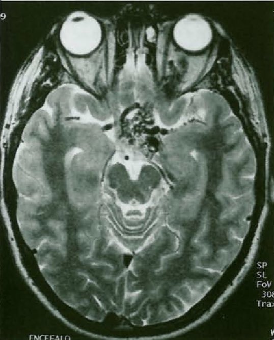
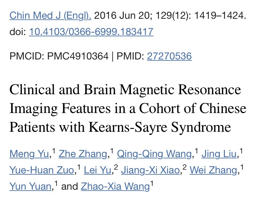
The most commonly reported brain MRI findings in KSS are cerebral and cerebellar atrophy with bilateral high-T2 signals in subcortical white matter, thalamus, basal ganglia, and brainstem.
KSS presents before age 20 with ophthalmoplegia and cardiac conduction defects. ⬇️⬇️
@BSIR_News@RCRadiologists@ClinRadiology article from the to be President of BSIR is based on false information and an anecdotal case sample of 1 physician associate.
Not good enough.
The 2,200+ jobless applicants to radiology in 2023 deserve better.
@r1chardf1tzg3r1
@TSM_Humanist@Tejzai6 HOA is a syndrome characterised by a periosteal reaction of the long bones. It can be primary (pachydermoperiosteitis) or secondary. Clubbing is the result of soft tissue proliferation associated with HOA. So HOA does not equal clubbing!
@drmankad Especially in supratentorials tumors I feel like an ancient astronomer looking at the sky with an optical telescope, while in the next room, the pathologists, are looking at the heavens with the Hubble 😅
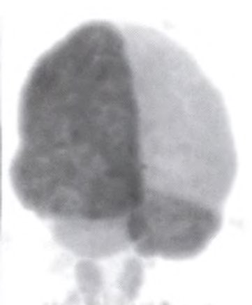
Crossed cerebellar diaschisis (CCD) is the result of reduced metabolism and blood flow in the cerebellar hemisphere contralateral to a cerebral lesion. Involvement of the cerebropontine-cerebellar pathway is thought to result in CCD.