The official account for the Life Science Solutions division of the Nikon Healthcare Business, with a focus on biological microscopy products and services.
🔬 ICYMI: We recently opened the doors to the new Nikon BioImaging Lab Europe at Plus Ultra Leiden. While the facility showcases the latest imaging technologies, its true value lies in the people, partnerships, and scientific discoveries.
Learn more 👉 https://t.co/pUeolGDAcL
Thank you for joining our #ExpansionMicroscopy webinar last week. Thank you, Aussie Suzuki, Ph.D., for sharing insights into the 3D-ExM workflow, its performance, and its potential applications.
Explore:
- AX / AX R + NSPARC https://t.co/IV0VVfZRuk
- NBIL https://t.co/MyFqRsLEyf
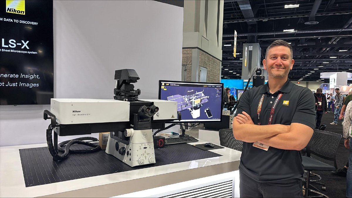
📍We're at #Neuroscience2026 in Washington, D.C. Stop by Booth 1217 and meet the Nikon team.
👀You'll also have the opportunity to get a first look at our recently launched LS-X Light Sheet Unit, on display at the booth.
See you in Washington, D.C.👋 @SfNtweets#Neuroscience
✨ NEW: Nikon AX MP Fiber Delivery
Why compromise between performance, cost, system footprint, and ease of setup?
A new approach to multiphoton imaging is here.
Contact your local Nikon representative to learn more 👉 https://t.co/qaTla4dV8v
🆕The LS-X Light Sheet Unit brings versatile light sheet imaging, enabling researchers to image living organoids, embryos, cleared tissues, and expansion microscopy samples within a single workflow.
To be available regionally from 30 November. Learn more: https://t.co/mL8cYLtx5D
🆕The LS-X Light Sheet Unit brings versatile light sheet imaging, enabling researchers to image living organoids, embryos, cleared tissues, and expansion microscopy samples within a single workflow.
To be available regionally from 30 November. Learn more: https://t.co/mL8cYLtx5D
Why choose between commercial software and open-source tools? NIS-Elements brings them together. Create analysis pipelines using intuitive visual workflow design and combine them with integrated Python libraries.
Contact your local Nikon representative to learn more.
🐁A recent Nature study shows that chemically induced totipotent-like stem cells can self-organize into embryo models that recapitulate key stages of mouse embryogenesis, from blastocyst formation through gastrulation and early organ development.
🔗 https://t.co/WRk5zwXX2B
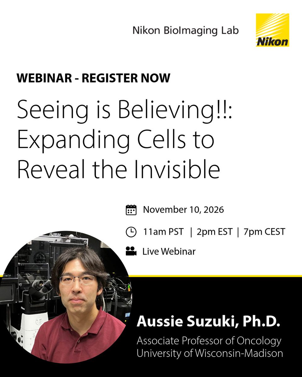
Tomorrow: Join Aussie Suzuki, Ph.D., Nikon, and Dojindo for a live discussion of the 3D Expansion Microscopy workflow, performance, and potential biological applications.
📅 TOMORROW: November 10, 2026
🕑 11AM PST | 2PM EST | 7PM CEST
Register Now 👉 https://t.co/TDgtVov3T4
🧠Check out the new ND Denoise⋅ai in NIS-Elements version 7! It uses information from neighboring images in z-stacks and time-lapses to distinguish signal from noise.
This software version is available regionally. Contact us to learn more 👉 https://t.co/YHtLAwn2Sb
💊 Drug responses can differ markedly in 3D versus 2D cell cultures.
A team at Uppsala University developed a scalable 3D Cell Painting workflow to profile spheroids at single-cell resolution.
📖 Read the paper: https://t.co/d2rSz6Capj
#NikonScienceSpotlight
Have you signed up yet? 🔬 Join Nikon & Dojindo live with Dr. Aussie Suzuki to discover how 3D Expansion Microscopy reveals the invisible.
📅 November 10, 2026
🕑 11AM PST | 2PM EST | 7PM CEST
Live Zoom Webinar - register now 👉 https://t.co/TDgtVov3T4
#NikonMicroscopy
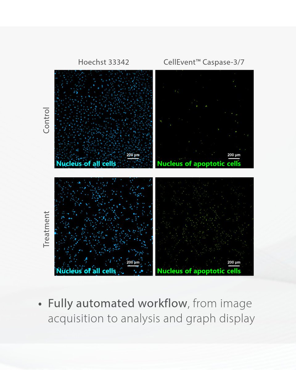
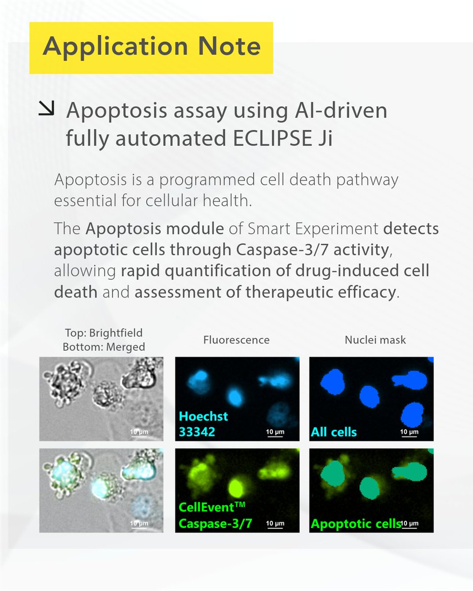
🧬Understanding #apoptosis is critical for studying cell health, drug responses, and disease progression. The ECLIPSE Ji enables you to streamline apoptosis assays using AI-driven imaging and analysis workflows.
📖 Read the application note: https://t.co/gH4kvw8B41
Introducing Detect ai in NIS-Elements v7 - Built on the Segment Anything Model, it turns hours of tedious manual annotation into simple clicks for both 2D and 3D images.
Contact your Nikon rep to learn more👉 https://t.co/sog1hI7mcY
*This software version is available regionally
Join Nikon and Dojindo for a live webinar featuring Aussie Suzuki, Ph.D., Associate Professor at the University of Wisconsin–Madison.
📅November 10
🕑2:00 PM ET | 1:00 PM CT | 11:00 AM PT
Register👉 https://t.co/TDgtVov3T4
#ExpansionMicroscopy#CellBiology#Bioimaging
🗣️"The ECLIPSE Ji-AX has transformed the way we work, combining intelligent automation with high-end imaging to make advanced microscopy accessible to everyone in the lab."
Read the full interview with Professor Sylvain Gabriele: https://t.co/JzlkXioXnY