Twitter account for the Optical Microscopy in Medicine lab. Under the direction of Dr. Melissa Skala at Morgridge Institute for Research.Tweets by @GINA_GALLEGO
Congratulations @rupsa Datta for her new paper that unravels neutrophil metabolism on a single-cell level 🎉 She showed early metabolic changes (<15 minutes) with stimulation and used machine learning to identify activation states.
https://t.co/YVP6GpD2eL
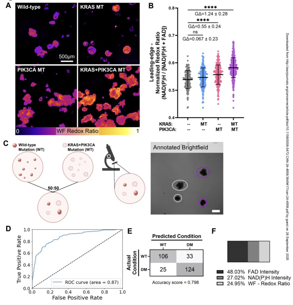
Congratulations to Amani Gillette for her paper, which identified accessible hardware and sensitive analysis methods to classify patient tumor organoids by mutation status and drug response. @UWCarbone@UWMadEngr@Morgridge_Inst@AACR 1/2
https://t.co/wWLiHkMRwl
Researchers are using advanced metabolic imaging to identify ways to improve CAR T cell therapy against solid tumors. Their findings were recently published in @natBME. @melissa_skala@sahakris@UWCarbone@UWMadEngr https://t.co/FgoPCoq33a
Are you searching for an end-to-end GUI to analyze single-cell data from fluorescence lifetime imaging microscopy (FLIM)? Wenxuan Zhao, @rupsa Datta, and Kayvan Samimi made FLIM Playground for you!
https://t.co/dLUzENnEPy
https://t.co/29qUZmRnXG
https://t.co/jYA54BiCFN (1/2)
Congratulations to Dan Pham for her paper published today! Dan showed that autofluorescence lifetime imaging can monitor single cell metabolism to improve CAR T cell metabolic fitness. @sahakris@UWCarbone@Morgridge_Inst@UWMadEngr
https://t.co/TnioBdYTy5
https://t.co/EEvzZrkGA0
"Neutrophils have been understudied, and our specific technique is underused — most people don't know about it yet. But our lab loves collaborating. It's exciting to share our technology with so many different branches of research." @rupsa@melissa_skala https://t.co/N9aN3934qf
Morgridge #postdoc Amani Gillette took the international stage @Falling_Walls to present her work on #CARTcell therapy, using imaging technology to screen the health of patients’ cells before manufacturing them into #cancer-fighting cells. https://t.co/ZGTBsoaoRN #FearlessScience
In a paper in @ActaBio, @UWMadison_BME’s Paul Campagnola and @melissa_skala show how the alignment of collagen fibers accelerates the movement of clusters of pancreatic cancer cells.
https://t.co/Pc7Tp98a3Q
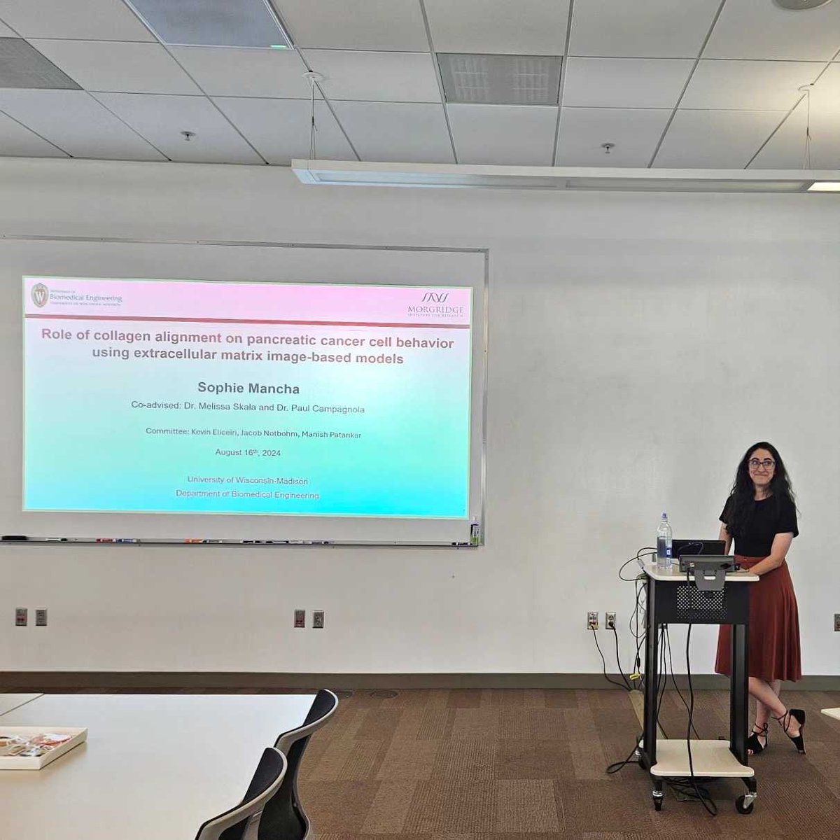
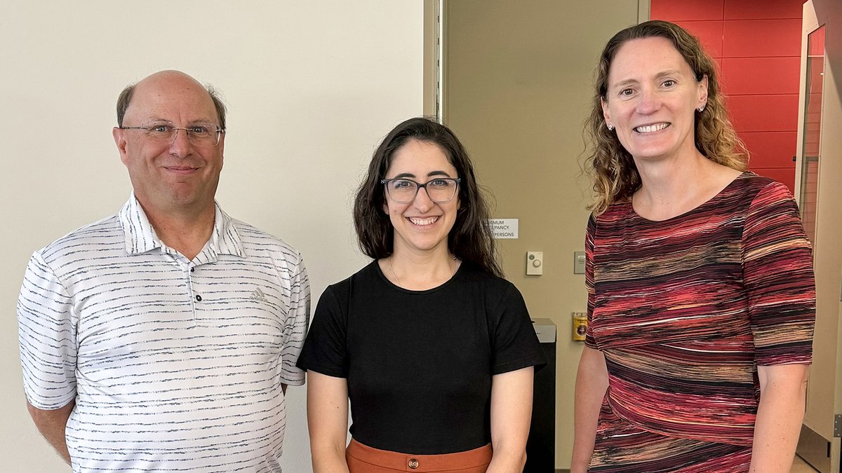
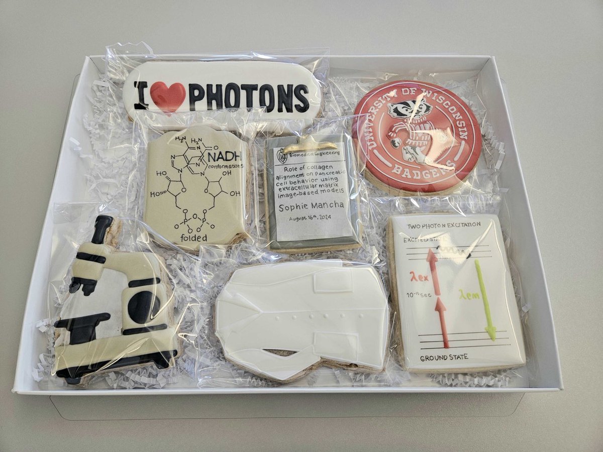
Congratulations again to Sophie Mancha who is a recent PhD graduate @UWMadison_BME for her fantastic work on pancreatic cancer! She made exciting discoveries in cell migration and tumor microenvironment. @UWMadEngr@Morgridge_Inst Story here 👇
https://t.co/jldpKwXplk
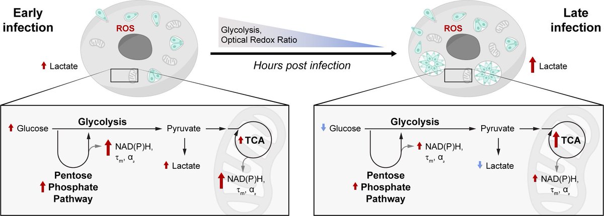
"We have a first-hand scenario of what is happening at the level of redox biology in cells infected with #Toxoplasma gondii.”
@GINA_GALLEGO in the @melissa_skala@OMM_lab uses advanced #imaging to study the how the parasite affects host cell #metabolism. https://t.co/fuN5eKBz1I
Happy to share our new preprint and Congratulations to @rupsa !🎉
Single cell autofluorescence imaging reveals immediate metabolic shifts of neutrophils with activation across biological systems https://t.co/YcI0bR1SMn
Meet @melissa_skala, professor of @UWMadison_BME@UWMadEngr & @Morgridge_Inst, who is developing biomedical optical imaging technologies for cancer research, cell therapy and immunology. https://t.co/LazpIAmkbD
Morgridge researchers harness the power of optical metabolic #imaging to study how the parasite #Toxoplasma changes host cell metabolism over the course of infection. https://t.co/fuN5eKBz1I
Congratulations @GINA_GALLEGO for her paper published today: "Metabolic changes in Toxoplasma gondii-infected host cells measured by autofluorescence imaging" https://t.co/BkHk2yyMbU