ECG From Scratch #31: When the Heart Chambers Enlarge
We’ve spent the first 30 posts building the ECG foundation.
Now we move from normal ECG patterns to abnormal ECGs.
One major topic is cardiac chamber enlargement.
What does cardiac enlargement mean?
A chamber can become enlarged because of:
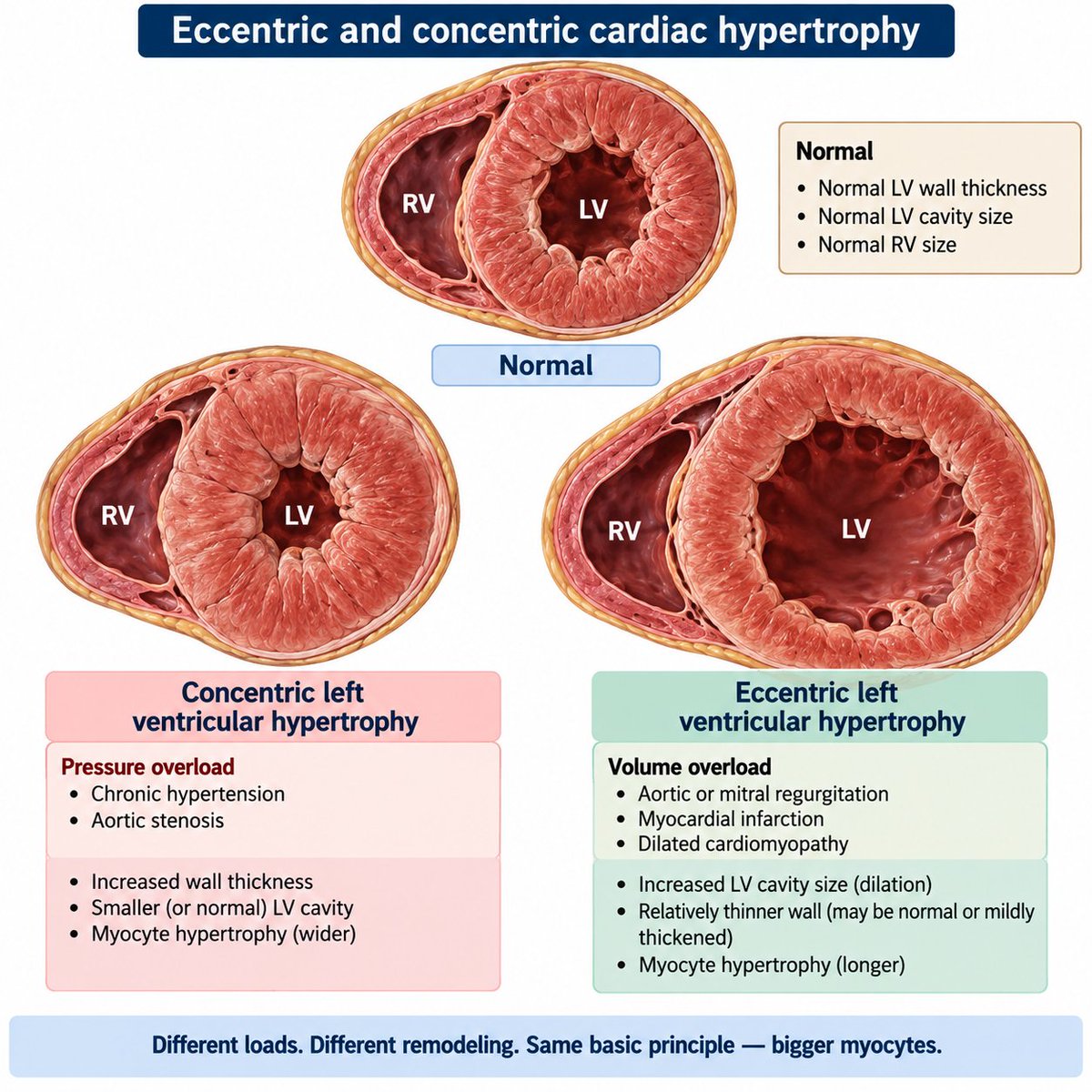
1️⃣ Increased cavity volume
The chamber dilates and the muscle fibers tend to become longer.
2️⃣ Increased wall thickness
The myocardium becomes thicker and the muscle fibers tend to become wider.
These processes are often described as:
⭕ Eccentric hypertroph: predominantly increased chamber volume
⭕ Concentric hypertrophy: predominantly increased wall thickness
And both can occur together.
What does this do to the #ECG?
More advanced hypertrophy can produce changes in the electrical forces generated by the heart.
The ECG may show:
- Increased P-wave voltage or duration
when atrial enlargement is present.
- Increased QRS voltage or duration
when ventricular hypertrophy is present.
But remember:
An enlarged chamber does not automatically produce a diagnostic ECG.
ECG findings are clues and should be interpreted with the clinical picture and, when appropriate, imaging such as echocardiography.
(Why do chambers enlarge)
A useful way to think about it is pressure load vs volume load.
Pressure load
Examples:
🔸Systemic hypertension
🔸Aortic stenosis
These tend to increase wall thickness.
Volume load
Examples:
🔹Significant valve regurgitation
🔹Dilated cardiomyopathy
These tend to promote chamber dilation.
Enlargement can have consequences
Pathologic hypertrophy and dilation may be accompanied by:
Fibrosis
⬇️
remodeling
⬇️
electrical instability
⬇️
dysfunction
These changes can contribute to:
*️⃣Atrial fibrillation
*️⃣Ventricular arrhythmias
*️⃣Heart failure