@petrogustavo Gracias x su dedicación y valentía al frente d 🇨🇴, x poner siempre en primer lugar a los sectores + vulnerables, x dfender la justicia social x trabajar incansablemente para q nuestro país sea "Potencia Mundial de la Vida". Su legado d cambioydignidad se graba en el ❤️del pueblo
Confidence in #glaucoma care comes from recognizing subtleties — when to escalate, when to monitor and how to avoid diagnostic traps that can delay treatment. #YOInfo#Ophthalmology https://t.co/fRWKjQSlYI
La miopía no se trata igual que antes.
Hoy se controla.
¿Sabes cómo hacerlo bien?
El Dr. Javier Prada te lo muestra en FRANJA 2026.
📍 Bogotá
📅 9 y 10 de julio
🔗 Inscríbete: https://t.co/hYba1NgATp
#Franja2026#SaludVisual#Optometría#Oftalmología#IndustriaÓptica
Nearly Half of Phakic Eyes Need Cataract Surgery within Two Years of PPV, National Study Shows
Risk factors included age, baseline cataract and vitrectomy for conditions related to abnormal vitreoretinal interface. Diabetes and PRP were protective.
https://t.co/SlgkrkXDoc
Study Describes Factors Associated with Endophthalmitis Following Anti-VEGF
Incidence is quite low—just 0.059%—but significantly associated with living in the southern US, prior intravitreal corticosteroid exposure, older age and history of smoking.
https://t.co/phctXY6S8x
An anterior segment photograph of a 1-week-old male infant presenting with the classic corneal haze characteristic of congenital glaucoma (A). The same eye after trabeculotomy, showing significant resolution of the corneal haze and improved clarity (B). A fundus photograph taken at 6 years old during routine examination (C). Three months later, a flame-shaped optic disc hemorrhage is visible (arrow), originating from the superotemporal neuroretinal rim and extending into peripapillary atrophy (D). At the time of hemorrhage, intraocular pressure was stable at 14 mmHg without medication. Although choroidal neovascular membranes were considered, the flame-shaped appearance was consistent with disc hemorrhage. While associated with glaucomatous progression in adults, disc hemorrhage is rarely documented in pediatric glaucoma. Consequently, the long-term implications of this finding in children remain to be determined.
https://t.co/GvqFQ7Zy8k
#ophthalmology #glaucoma
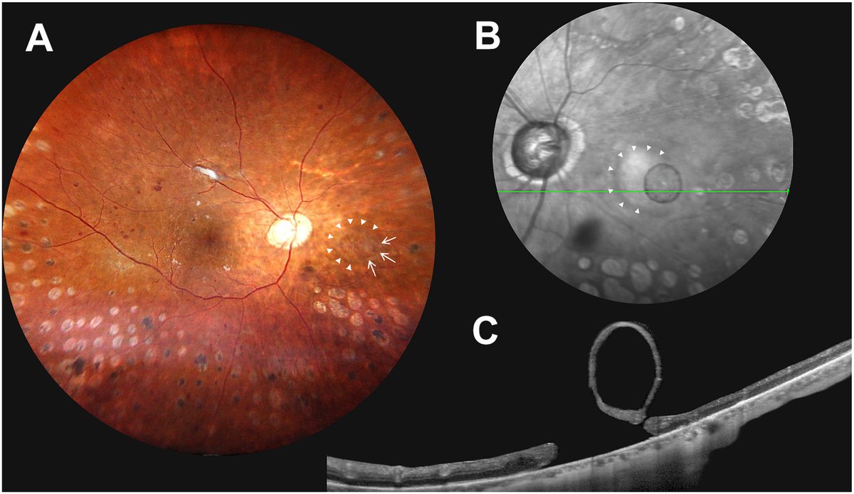
A 62-year-old woman presented for routine follow-up of proliferative diabetic retinopathy. A, B, Ophthalmoscopy and infrared autofluorescence revealed a retinal hole nasal to the optic disc (arrowheads) with an adjacent oval, translucent, nonpigmented cyst (white arrows) located anteriorly in the posterior vitreous cavity of the right eye. C, OCT demonstrated a full-thickness retinal hole measuring 1041 μm and a floating vitreous cyst with an isoreflective wall and hyporeflective lumen. Laser photocoagulation was applied to the margins of the retinal hole, and close follow-up was maintained. An acquired nonpigmented vitreous cyst associated with retinal hole formation was presumed.
https://t.co/K0mg4Gxc1u
#ophthalmology #retina
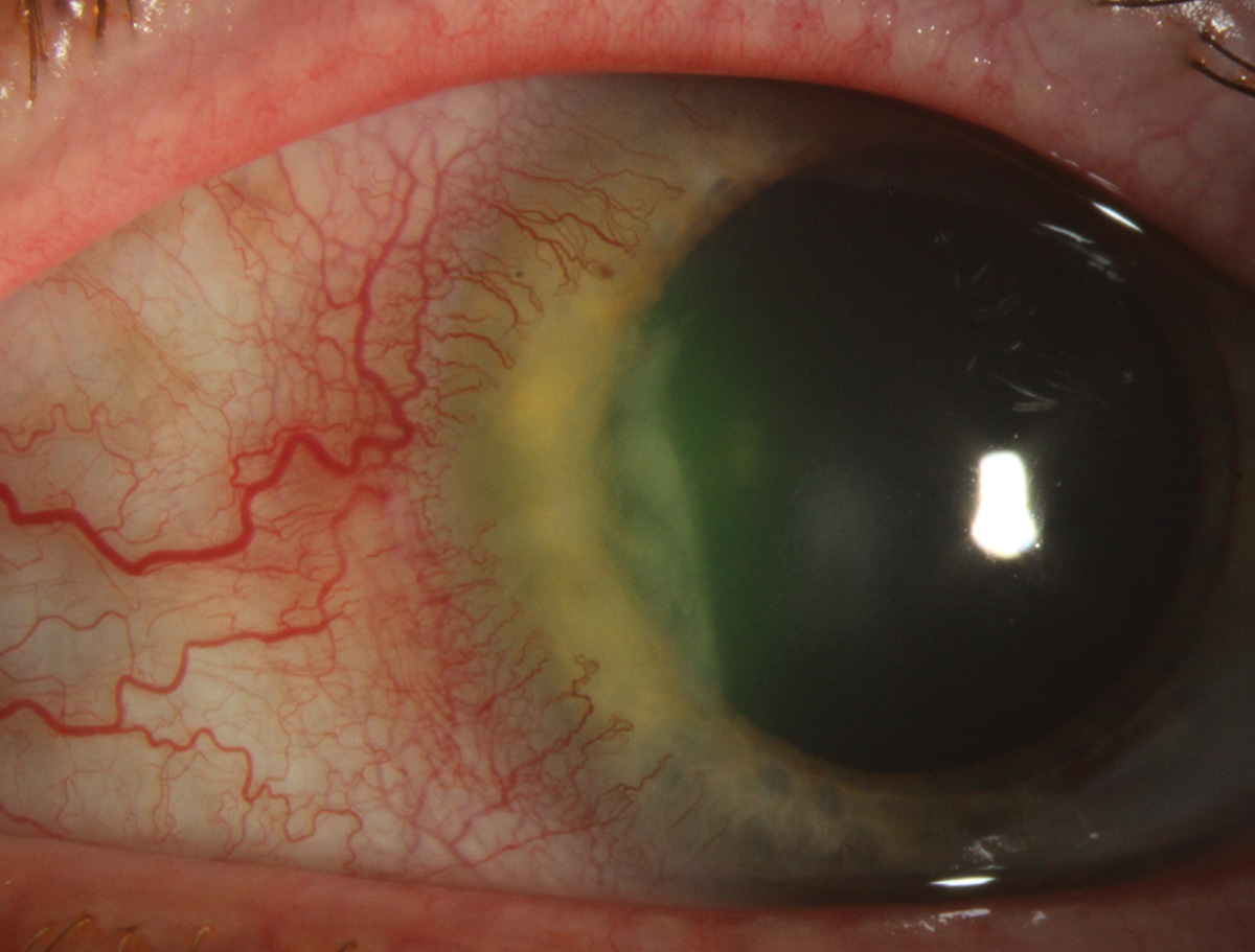
"What the Wind Brought" — When encountering this common corneal infection, time is vision.
A 73-year-old man presented with pain, photophobia and foreign body sensation in the left eye for two days. Full case: https://t.co/TTA87Z3sc4
#optometry#optometrist#eyecare#cornea
No merece la pena tratar una metástasis coroidea.
Son incurables.
El paciente está sentenciado.
No sobreviven más de 6 meses.
No puede hacerse nada.
La visión no tiene ninguna solución, lo mejor es dejar en paz al paciente.
Bien.
Mira este caso.
Es antes y después.
Over 30% of Glaucoma Patients’ Relatives Also Vulnerable
A literature review pinpoints prevalence rates, as well as problems with current screening methods.
https://t.co/0bR0ZT60L0
#glaucoma#eyecare#optometry
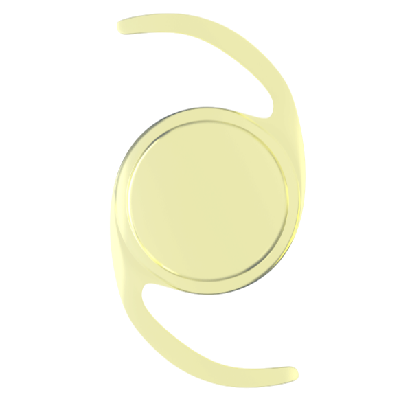
Study Challenges Protective Role of Blue-blocking IOLs
Yellow-tinted lenses were associated with a decline in macular pigment density over three years—the opposite of the intended effect.
https://t.co/UY4zR4huNr
#cataractsurgery#eyecare#optometry
Breach At the Surface: Recognizing Threats Behind the Red Eye
This presentation remains a frequent source of diagnostic error, despite its commonality. Understanding subtle differences will point the way toward appropriate mgmt.
https://t.co/uCjhXlvxOX
#redeye#conjunctivitis
Ocular Complications of Biologics: A Review for ODs
Learn about the potential risks associated with each medication class and how to manage side effects while maintaining effective treatment.
https://t.co/6791RZil3F
#optometry#optometrist#eyecare
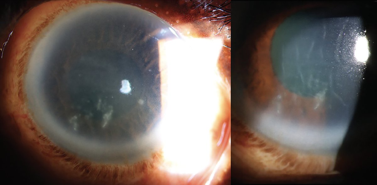
What is 'bread-crumb appearance'?
Multiple small, discrete, sharply demarcated white opacities in the corneal stroma.
Resemble scattered bread crumbs or snowflakes.
Typically located in the central anterior stroma.
Pathology:
Deposits of hyaline material within the stroma.
Clear stroma in between + discrete white crumbs = granular dystrophy
#MedTwitter #ophthalmology #neetpg
Image from google
Contact lens + most common cause for infectious keratitis → Pseudomonas
Contact lens + severe pain with minimal signs → rule out Acanthamoeba
# Pseudomonas may show ring infiltrate at later stages. Important is to look for perineuritis and symptoms out of proportion to signs.
#MedTwitter #ophthalmology #neetpg
Image from google
PUK
Pathophysiology:
Immune complex deposition in limbal vasculature.
Leads to vasculitis → ischemia → stromal melt.
Upregulation of collagenases → corneal thinning and perforation.
#MedTwitter#ophthalmology#neetpg
Image from google
Which corneal condition may mimic infective keratitis but is actually immune-mediated and associated with rosacea?
#MedTwitter#ophthalmology#neetpg
Image from google