🚨 The newest issue of POCUS Journal (Vol 11, Issue 1) is now live!
Explore fresh research, case reports, POCUS protocols, and insights from across the global POCUS community.
Start reading: https://t.co/AiqNn7VB2N
#POCUSJournal#POCUS#FOAMed#MedEd#Ultrasound#MedTwitter #EchoFirst
🔎 Think you know your POCUS scans? What do you see in this image?
This case series highlights a very unique yet deceptive ultrasound artifact that can mimic a life-threatening abdominal aortic dissection — even in otherwise healthy patients.
📍Recognizing this artifact can make all the difference in emergency settings, helping clinicians avoid unnecessary alarm, additional imaging, and potential misdiagnosis. Read through these cases to sharpen your POCUS interpretation skills and stay one step ahead in high-stakes clinical scenarios.
🔗 Article: https://t.co/o7W0hpTYvy
📌 This publication also serves as a corrigendum to the previously published article: https://t.co/z0Sj9OMeG6
🦋 Follow us on BlueSky: https://t.co/VkNvXOkHM7
#MedTwitter #POCUS #POCUSFocus #MedEd #POCUSJournal #Ultrasound #UltrasoundEducation #EmergencyMedicine
Point-of-care ultrasound (POCUS) is transforming bedside medicine.
Our latest blog + video introduces the essentials:
🔹 What POCUS is & how it works
🔹 From early sonar to handheld devices
🔹 Key clinical applications across multiple systems
🔹 Advantages & limitations
🔹 How POCUS Journal supports education & research
🎥 Watch & read: https://t.co/2KRAEaxyDN
#POCUS #MedEd #Ultrasound #MedicalEducation #HealthcareInnovation
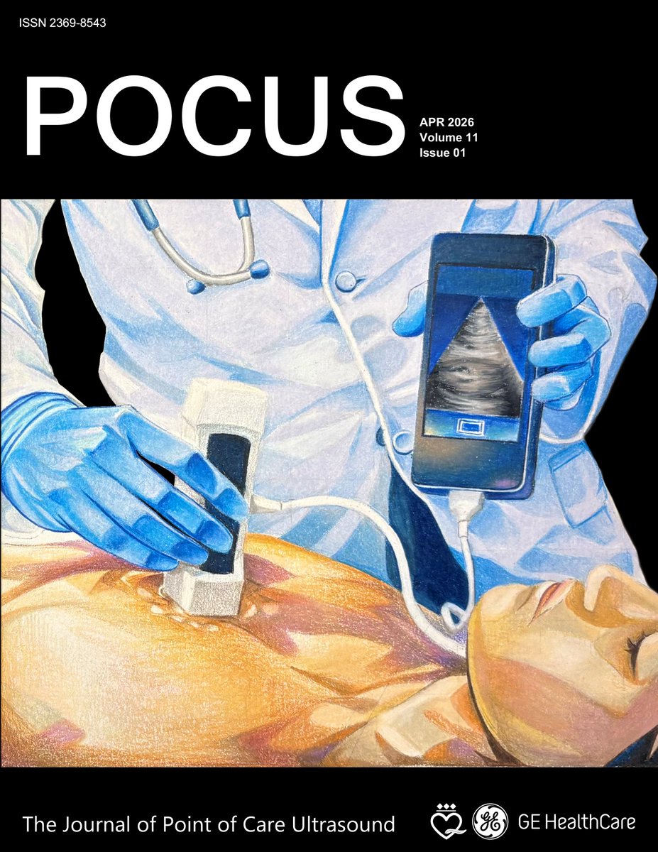
Picture this. A 3.5-year-old previously healthy boy presented with two episodes of non-bilious and non-bloody emesis to the pediatric emergency department six hours following a minor abdominal trauma. The patient had fallen from a standing height onto a seesaw, with the seesaw bar hitting the right side of his abdomen.
What was the first tool taken out of the toolbox? A POCUS FAST exam, later followed by a contrast enhanced ultrasound scan. The CEUS scan revealed a triangular area lacking enhancement in the pre-renal space, indicative of a large hematoma adjacent to the adrenal gland…how did the physicians approach the rest of this case?
Read more here to see the full story: https://t.co/UYeCRmFeoJ
#POCUSJournal #POCUS #FOAMed #FOAMus #MedEd #MedTwitter #echofirst #echo #ultrasound #anesthesiology #pocusfocus #UltrasoundEducation
POCUS isn’t just images on a screen. It’s the Presence, Observation, Connection, Understanding, and Story at the bedside. Every scan becomes a moment of shared clarity, compassion, and truly patient-centered care.
Read more here to see the full story: https://t.co/2J2cXLSUHV
#POCUSJournal #POCUS #FOAMed #FOAMus #MedEd #MedTwitter #echofirst #echo #ultrasound #anesthesiology #pocusfocus #UltrasoundEducation
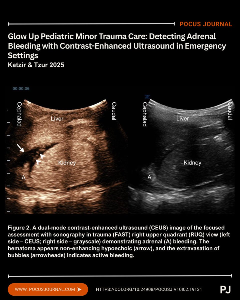
New POCUS Journal Podcast Episode! “Measurement of Systolic Blood Pressure Using POCUS With Color Doppler Compared to with an Intraarterial Line”
📺YouTube: https://t.co/YOK90tNjqj
🎧Spotify: https://t.co/Hx2LGFZ53e
🗂️All episodes: https://t.co/kQvWrmOQ1A
#POCUS#MedImaging #Ultrasound #MedTwitter #FOAMed
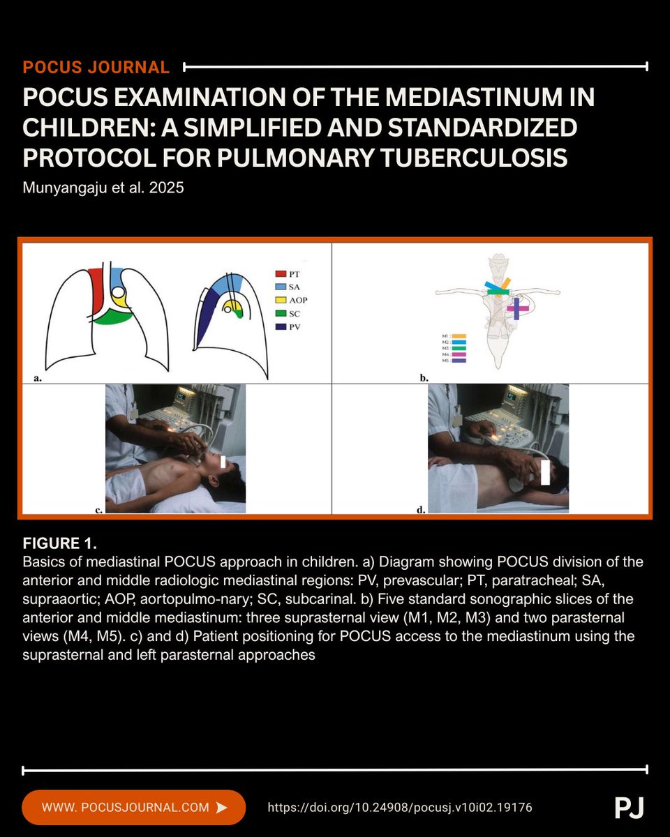
🌍 Pediatric TB is a global problem, and access to imaging remains a barrier.
This study presents a standardized mediastinal POCUS protocol to improve detection of TB-related lymphadenopathy in children, particularly in resource-limited settings.
💡 By using mediastinal POCUS to identify lymphadenopathy, clinicians can leverage a radiation-free, bedside tool that’s especially valuable when CT or advanced imaging isn’t available.
Learn more here: https://t.co/YtbDF7gTos
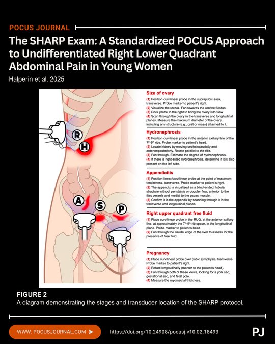
Teaching RLQ pain in young women? Make it SHARP, a high-risk differential that demands a structured approach.
The SHARP Exam is a POCUS-based protocol targeting:
S - Ovarian torsion
H - Hydronephrosis
A - Appendicitis
R - RUQ free fluid
P - Pregnancy
A practical framework that mirrors eFAST and RUSH, but for RLQ pain.
Learn the protocol here: https://t.co/dlC5OqChUg
#MedEd #FOAMus #POCUS #UltrasoundEducation #EMEducation
🩻Picture this: A 44-year-old battling brutal hand pain from acute limb ischemia (ALI) due to a self-inflicted artery crunch after a fentanyl injection.
Options:
Opioids? ❎
Surgery? ❎
POCUS? ✅
💡This first-of-its-kind case flips the script on refractory ALI pain. ED Doctors performed POCUS-guided radial and median nerve blocks at the mid-forearm. Agony pain relieved with integrative magic with POCUS! Another POCUS success
📖 Read this case to level up your regional anesthesia game: https://t.co/AGAYpixrnM
#POCUSJournal#POCUS #FOAMed #FOAMus #MedEd #MedTwitter #echofirst #echo #ultrasound #anesthesiology #pocusfocus #UltrasoundEducation
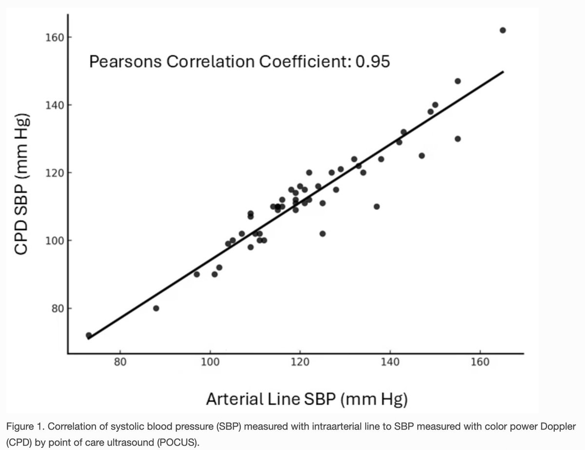
A new study in the @pocusjournal shows how you can use color power Doppler to measure blood pressure.
They placed the color Doppler box over the brachial artery and inflated the blood pressure cuff until the Doppler signal disappeared. They then slowly release the cuff until it reappears, mimicking how it is done with a stethoscope.
They compared the pressures to an arterial line, and the results were quite impressive. The Pearson correlation coefficient was 0.95, suggesting that a systolic pressure from this method or from an arterial line can be used interchangeably.
This is an interesting use case for color power Doppler, which can detect very low-velocity flows and is not angle-dependent like color Doppler.
The November 2025 issue of POCUS Journal is now live. Explore innovations in point-of-care ultrasound, new educational strategies, case insights, and more.
📘 Read the latest issue today on our website and stay at the forefront of POCUS practice.
Taking the probe away from an ultrasound learner is like taking the bike away from someone learning to ride — and showing them a video instead. We shouldn’t expect applicable skill without real hands-on practice. https://t.co/4vdz6xp57X @SimonOrlob@sono4you_graz@POCUSJournal
🔎 POCUS has become an extremely useful tool in the placement of temporary Transvenous Pacemakers. Emergency medicine specialists detail how using POCUS doesn’t just help with central venous access; Rather, it directly visualizes pacemaker wires as they advance into the heart’s chambers, and flags misplacement or complications in real time. Read more about the three series in this case study to learn exactly how emergency medicine physicians utilize POCUS in real time.
💡For anyone involved in emergent environments, whether in the ED, ICU, or procedural setting, this article highlights how integrating POCUS more deeply can boost safety, speed, and confidence.
📖 Read the full study: https://t.co/JEYTxioZNM
#pocus #POCUS #MedTwitter #Ultrasound #POCUS #POCUSJournal #MedEd #Cardiac #Echocardiography #pocusjournal
📊 A 26-day-old boy was referred to the pediatric emergency department for evaluation of a left-sided neck mass. Physical and POCUS examinations were performed to better visualize the anatomy of the mass. A discrete, homogenous, solid mass contained within the left SCM, consistent with fibromatosis colli was diagnosed.
💡POCUS is the preferred imaging modality for detecting neck masses and for differentiating and supporting the diagnosis of fibromatosis colli from other neck masses.
Read more about this amazing case here: https://t.co/0KBYEJ2TPb
🦋 Follow us on BlueSky: https://t.co/VkNvXOkHM7
#MedTwitter #Ultrasound #POCUS #POCUSJournal #PediatricUltrasound #MedEd #Pediatrics
🔎 Pediatric POCUS experts put 6 handheld ultrasound devices to the test — comparing image quality, ease of use, and overall performance across common pediatric POCUS scans.
For clinicians and institutions, this first-of-its-kind comparison offers practical guidance when choosing the right handheld device for use in pediatrics.
💡 Key insight: No single device excels at every application, but image quality and ease of use consistently led the way in expert preferences.
📊 Dive deeper into what experts suggest: https://t.co/XGp3HqOCzY
#pocus #LungUltrasound #EmergencyMedicine #Pediatrics #Echocardiography #pocusjournal #MedTwitter #UltrasoundEducation