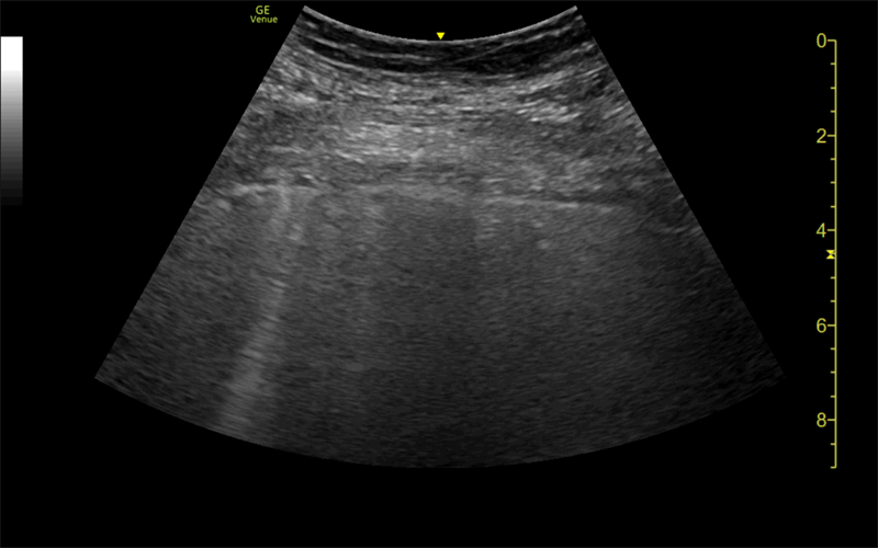
A Spanish team used ultrasound scans to document lung abnormalities in three patients who recovered from COVID-19 but still had persistent symptoms months later -- patients commonly known as "longhaulers." @yaletung#radiology https://t.co/XXKTsRgCyJ
Read "Usefulness of Lung Ultrasound Follow‐up in Patients Who Have Recovered From Coronavirus Disease 2019" by @yaletung et al. in the Journal of Ultrasound in Medicine #JUMultrasound
https://t.co/Wb8JoJlXk8
Usefulness of Lung Ultrasound Follow‐up in Patients Who Have Recovered From COVID-19. Easy to use, without side effects or radiation, helps monitor the disease resolution or assess early progression to lung fibrosis. #COVID19 https://t.co/Wy6k4MQmYA
Monitorización del tratamiento rehabilitador de la disnea de esfuerzo por COVID-19 | Enfermedades Infecciosas y Microbiología Clínica @yaletung https://t.co/dchrFD9llJ
Oficina de Transferencia de Resultados de Investigación. Gran trabajo colaborativo (Dasel, @unicomplutense, Hospital la Paz y @CSIC): ULTRACOV en detección temprana de #COVID19. Enhorabuena! https://t.co/PVcS28U7RK
The STONE (Sonography and Testing of a Nephrolithiasis Episode) randomized clinical trial. A Point-of-Care pathway to manage #renalcolic in the ED is effective and safe. Also reduces the amount of time spent in the ED 112 minutes vs 244 (P < .001) #POCUS
https://t.co/dYTS3Fn5n3
4) The annual risk of rupture of aneurysms larger than 5 cm is 25 to 41%.
To measure the diameter, it is important to include the mural thrombus so as not to underestimate the size.
3) In stable patients with an aneurysm greater than 3 cm in diameter, a CT scan should be performed to confirm it.
More than 90% of abdominal aortic aneurysms are located below the outlet of the renal arteries.
1) A 66-year-old man presented to the emergency department with a quite few weeks history of general discomfort.
A pulsatile mass was evidenced in the abdominal exploration, so an ultrasound study of the aorta was performed. #POCUS@yaletung
Young patient after a blunt chest trauma. Lung #POCUS revealing a nondisplaced rib fracture causing CHIMNEY PHENOMENON by reverberation artifacts occurring at the fracture margins. @yaletung@pocuslapaz
Patient with chest pain and shortness of breath. Lung #POCUS revealing a Lung Point at the mid axillary line (MAL). According to previous study MAL, represents the limit for predicting>than 15% of lung collapse when volume is measured at CT(S 83.3%/Sp 82.4%) @yaletung@pocuslapaz