Largest description of a recently described #bstpath entity, in @AJSPjournal. Proud to be part of this multi-institutional collaboration.
https://t.co/jSLeAyYKwa
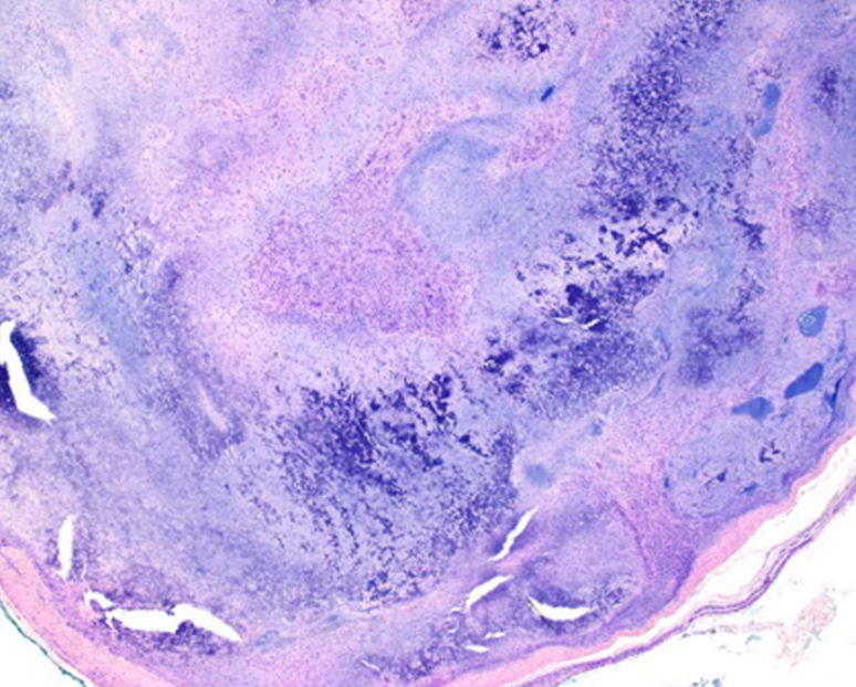
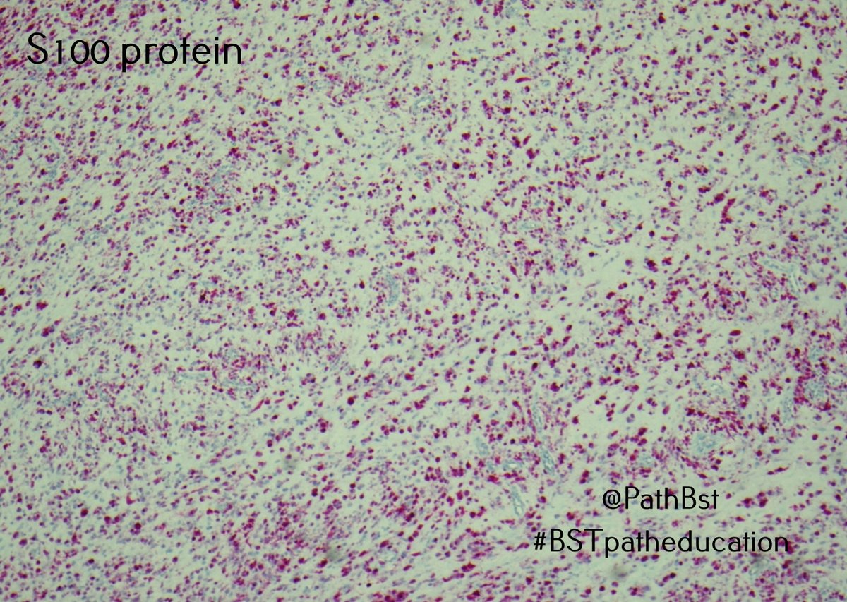
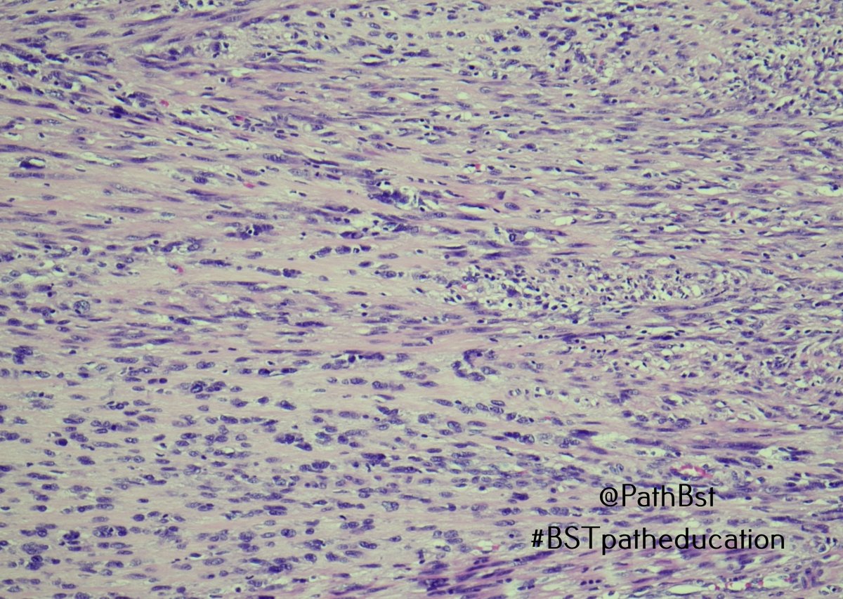
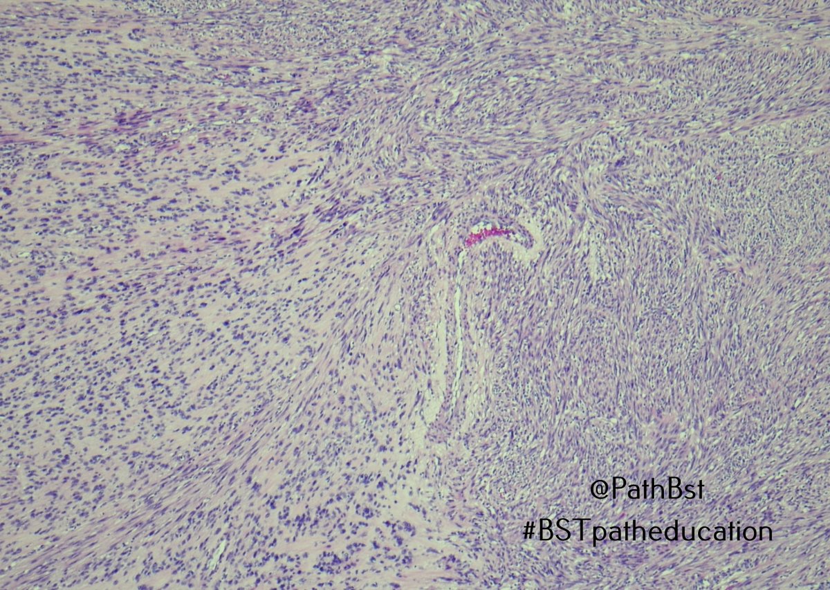
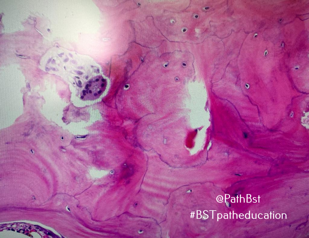
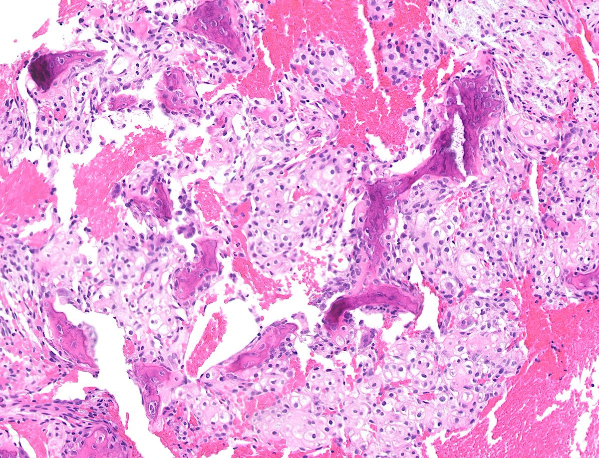
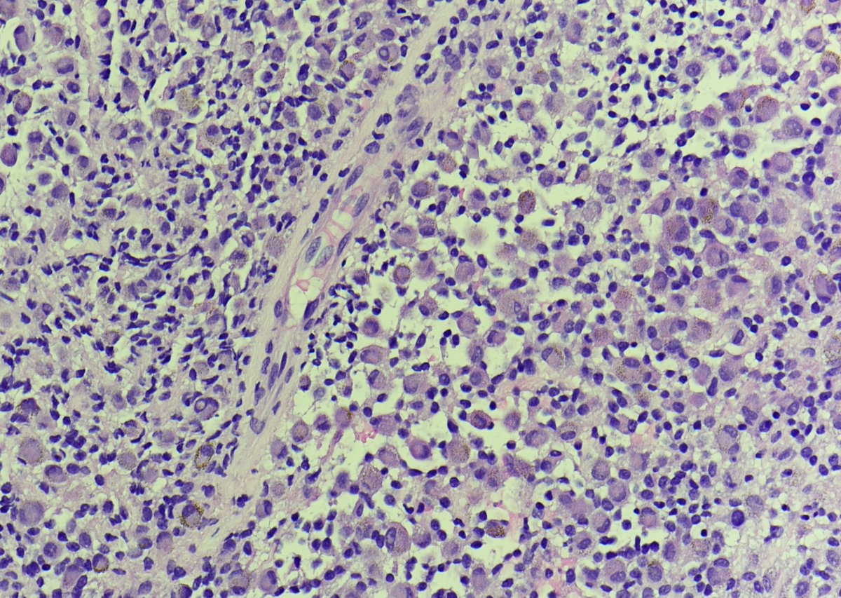
This is "cutaneous" syncytial myoepithelioma...of bone!
@JMcMahonG with @SintawatWang recognized that these morphologically distinctive tumors occur in diverse anatomic locations, including bone.
https://t.co/vWV8WU4FGk
They have recurrent EWSR1::PBX3 fusions too!
#BSTpath
Happy Memorial Day all!
Our Summer Seminar is Sat June 3 @ 8 am EST.
Bone and Soft Tissue theme.
All star line-up including Dr. Khin Thway, @JMcMahonG, @AndersMeyerMD
Register:
https://t.co/btzpO641Vu
Coming soon, don't miss out!
#BSTpath#PathTwitter
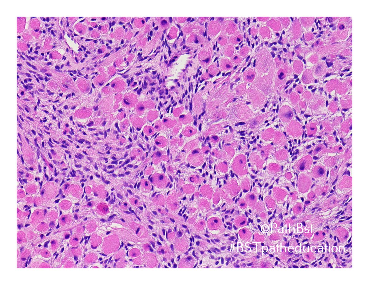
Excellent work all! Yes, this is embryonal rhabdomyosarcoma with chemotherapy-induced cytodifferentiation. Some say portends a better prognosis, perhaps controversial (and therefore twitter appropriate). Location is classic. Great responses. #BSTpath#PediPath
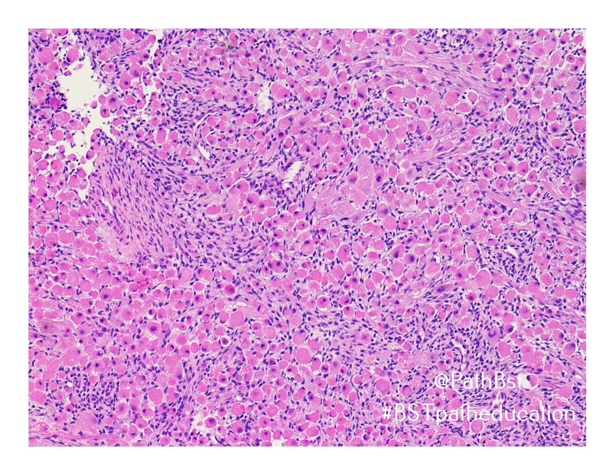
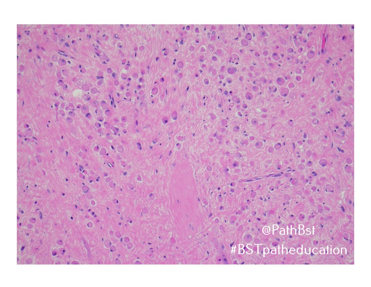
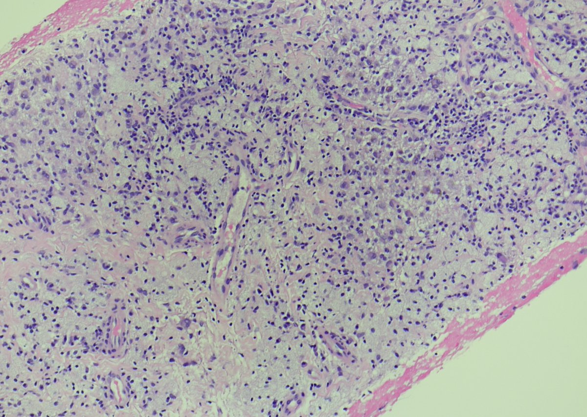
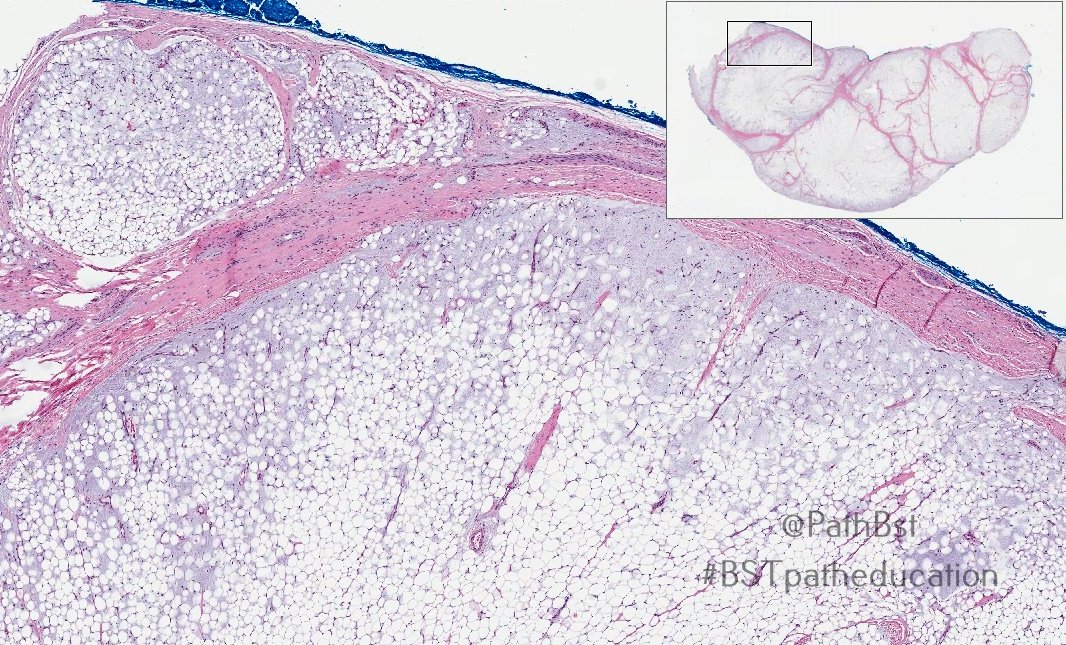
@JMGardnerMD @kells108 @JMcMahonG@MdSuster@AndersMeyerMD@Greg_Charville@MichaelMichal28 Excellent work all! This is lipoblastoma. Clues are lobular architecture and fibrovascular septae at low power, spectrum of adipocyte maturation w/immature cells in myxoid areas, and zonal architecture with immature cells in periphery / adjacent to septae.
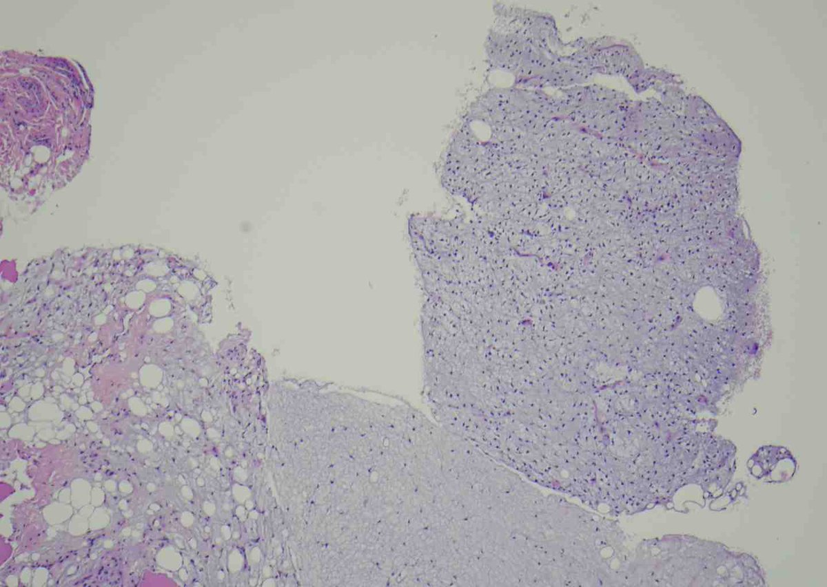
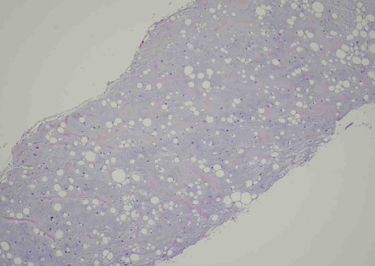
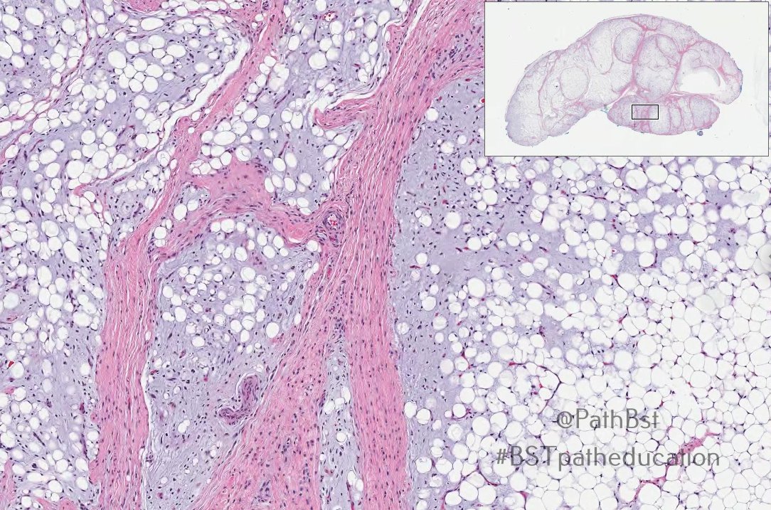
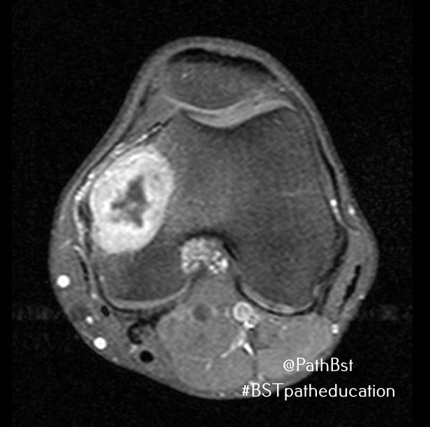
Since it's back to school season: this one's a foot mass from a toddler. Your diagnosis? Anything else in the differential?
#pedipath#pedspath#BSTpath#dermpath
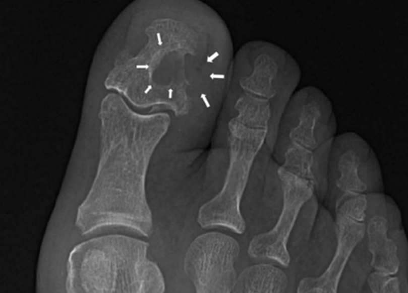
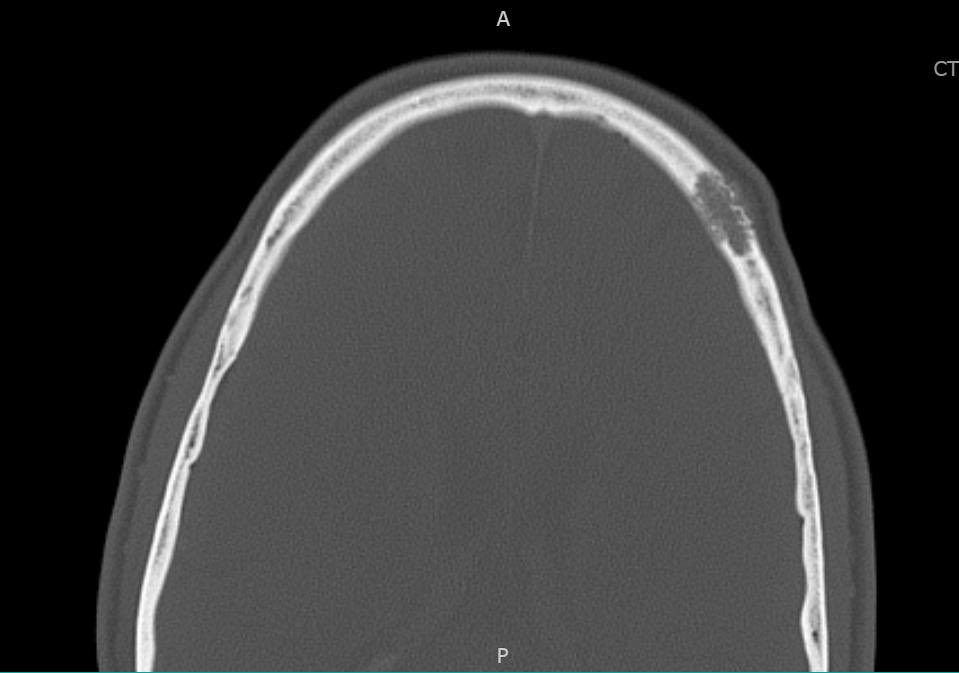
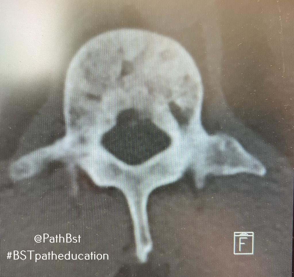
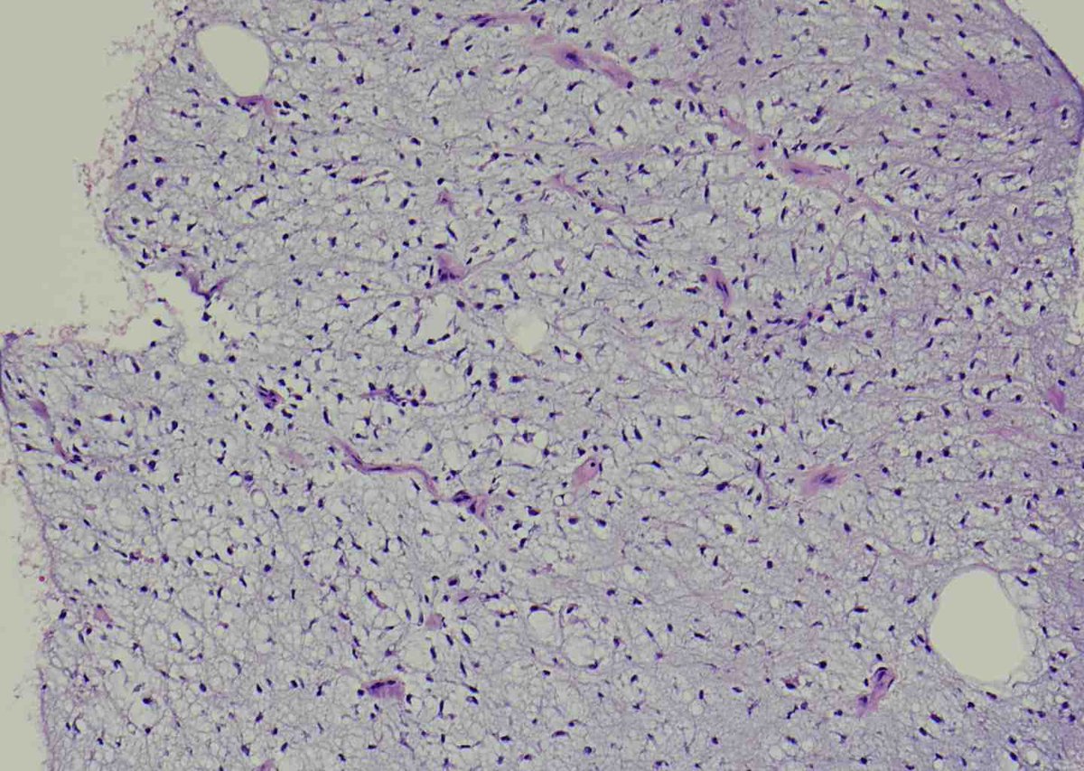
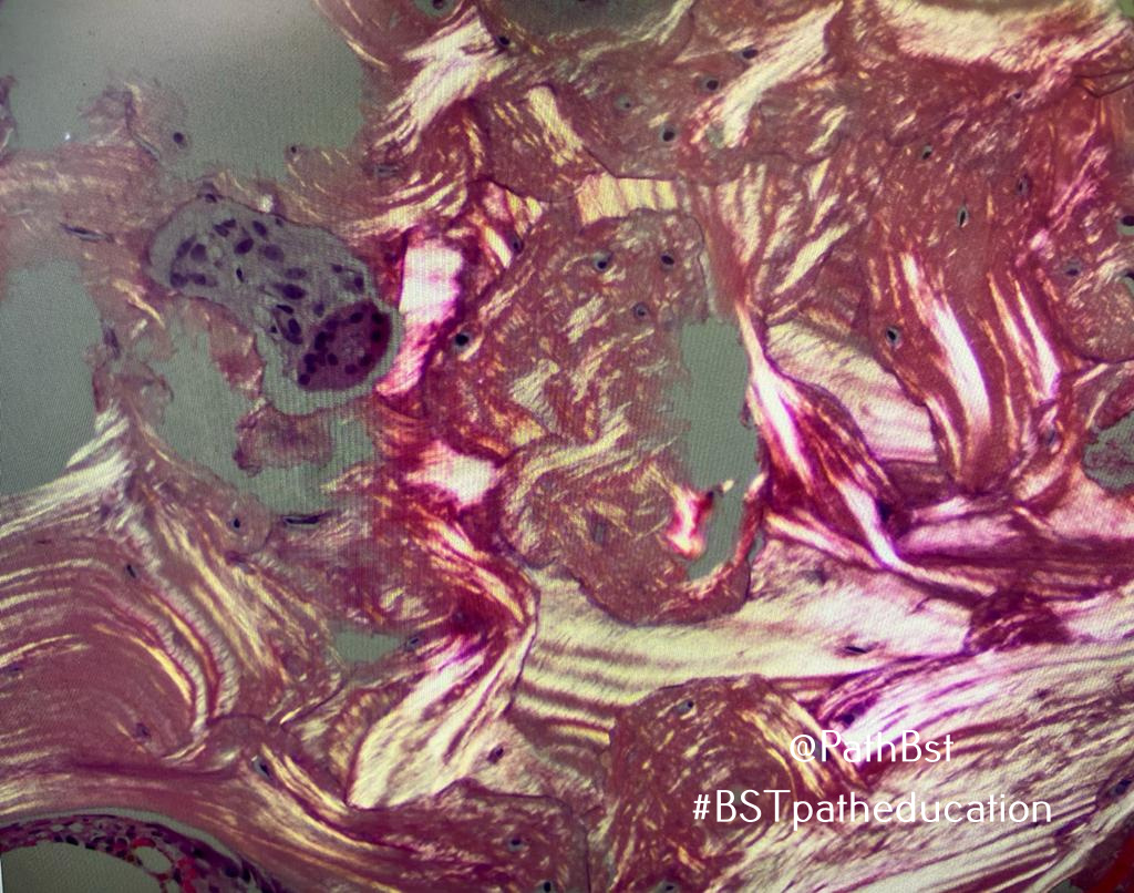
Radiolucency in frontal bone
sagittal + axial CT.
Here’s a clue: Keep a low power impression
Mild expansive remodeling, cortical thinning, radial (spoke) trabecular bone radiating around a central lucency
Diagnosis?
@reith16@ahlawat_shivani@LauraMFayad@LisaRooperMD
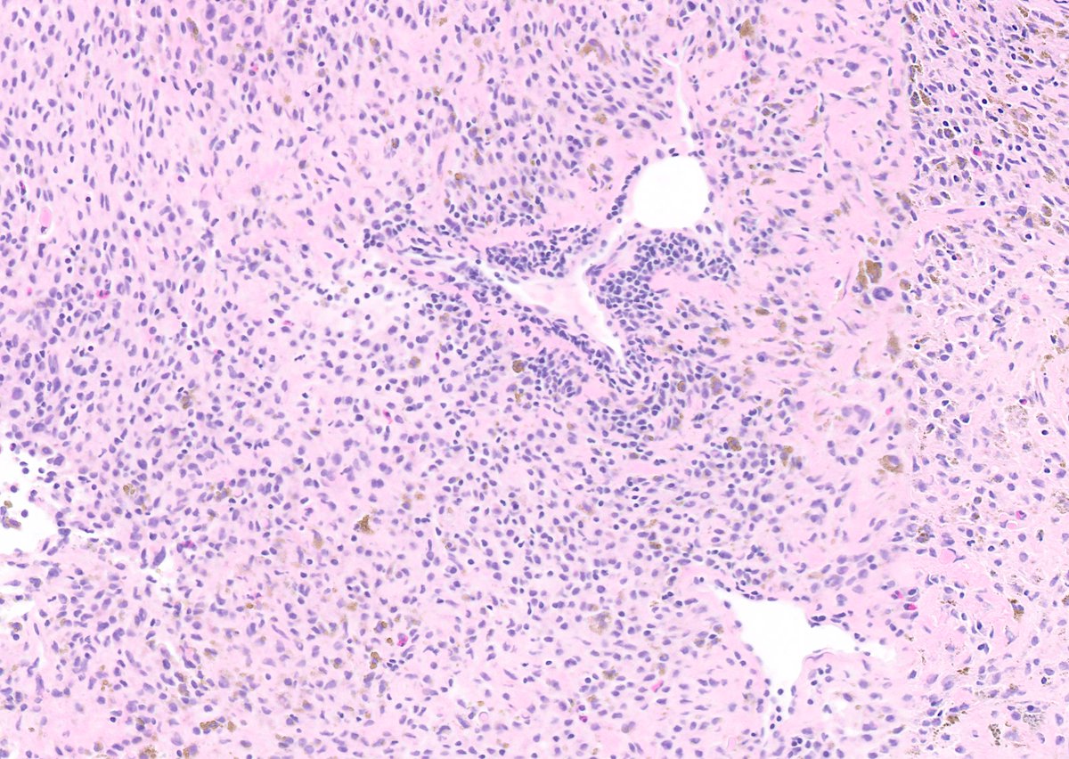
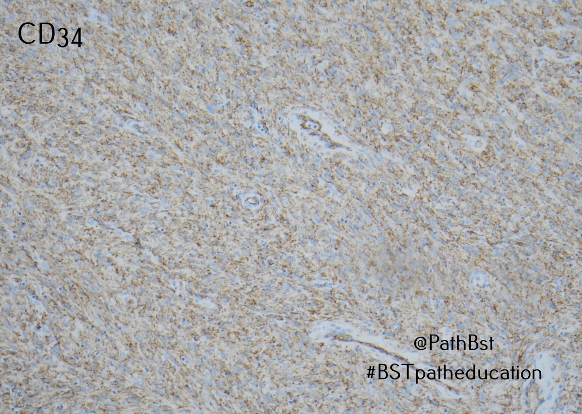
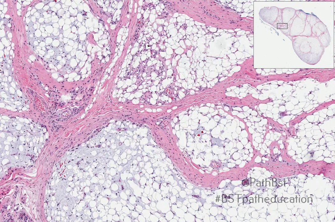
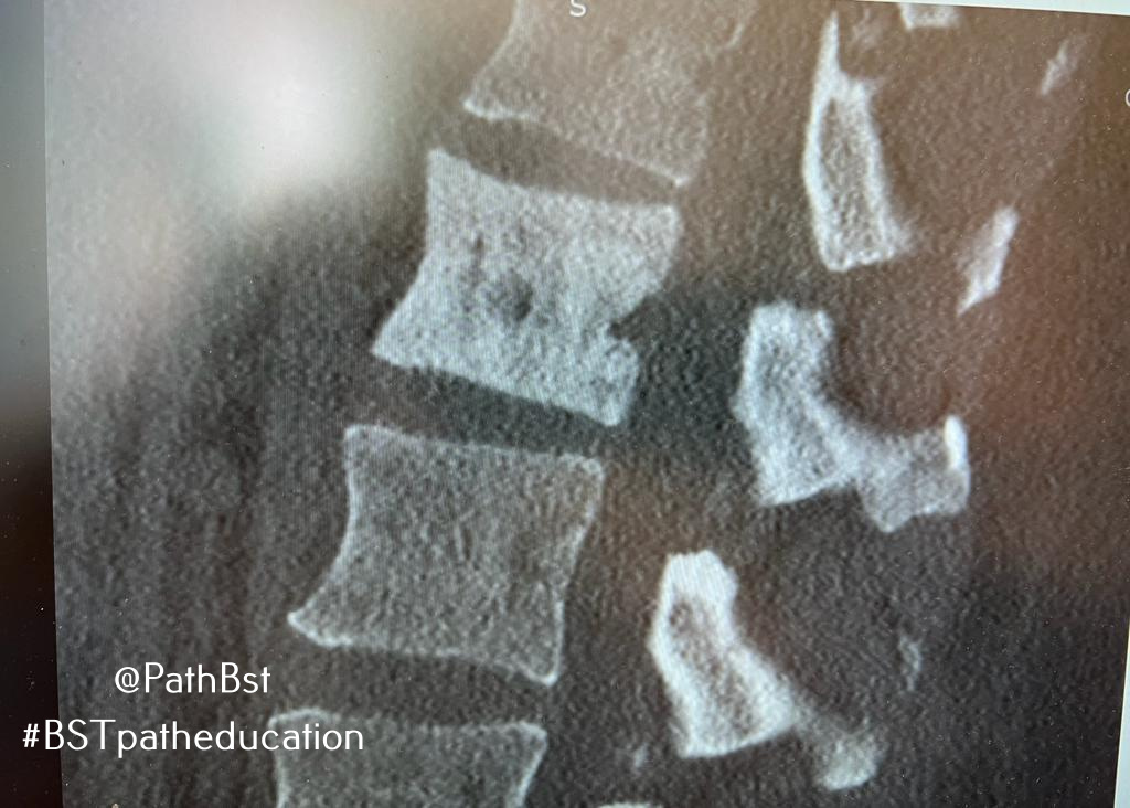
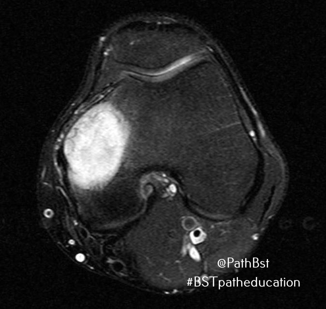
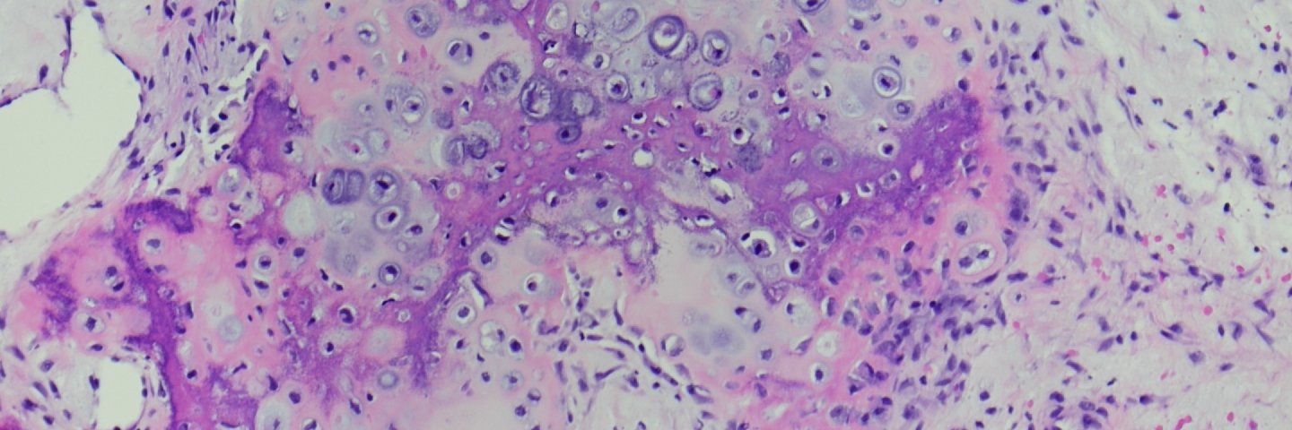
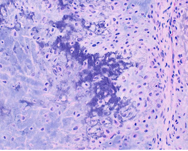
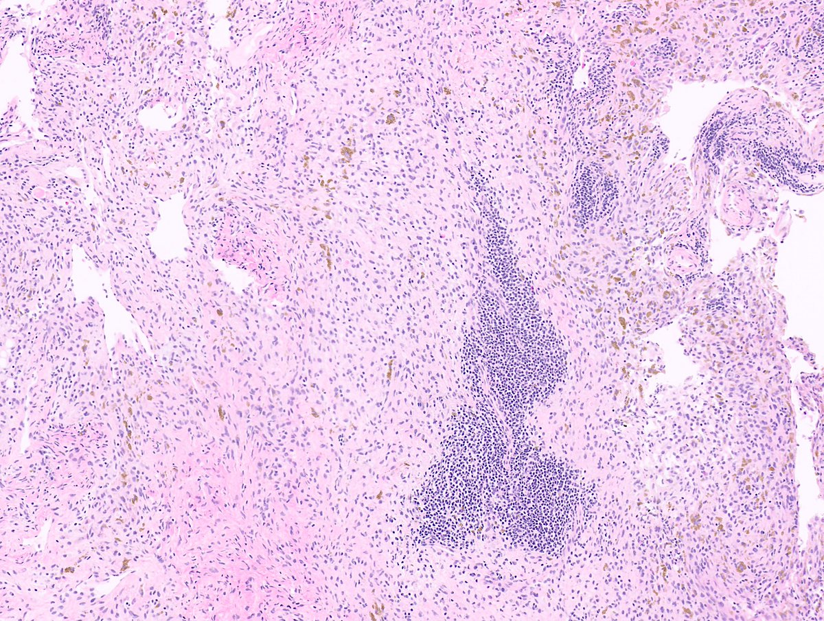
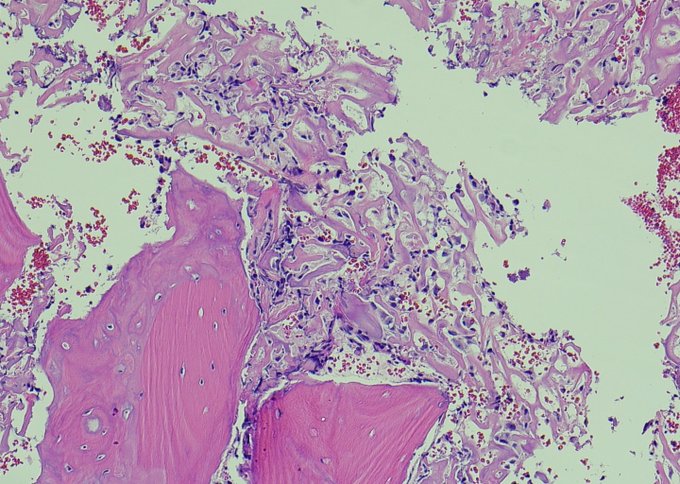
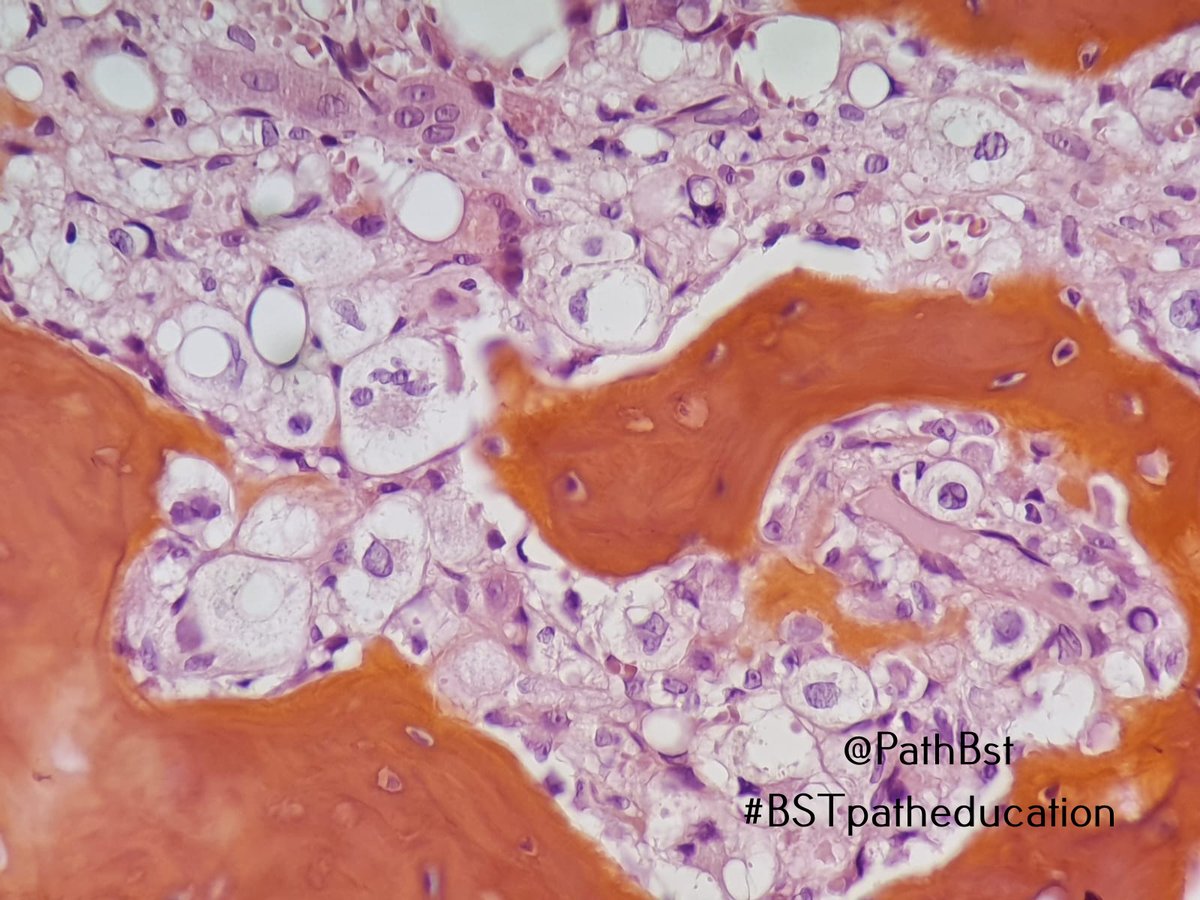
40 year old man. What is the diagnosis? Can you figure out the site? Let's get those #MSKrads and #BSTpath skills to work. #BSTpatheducation
Images courtesy of @kells108