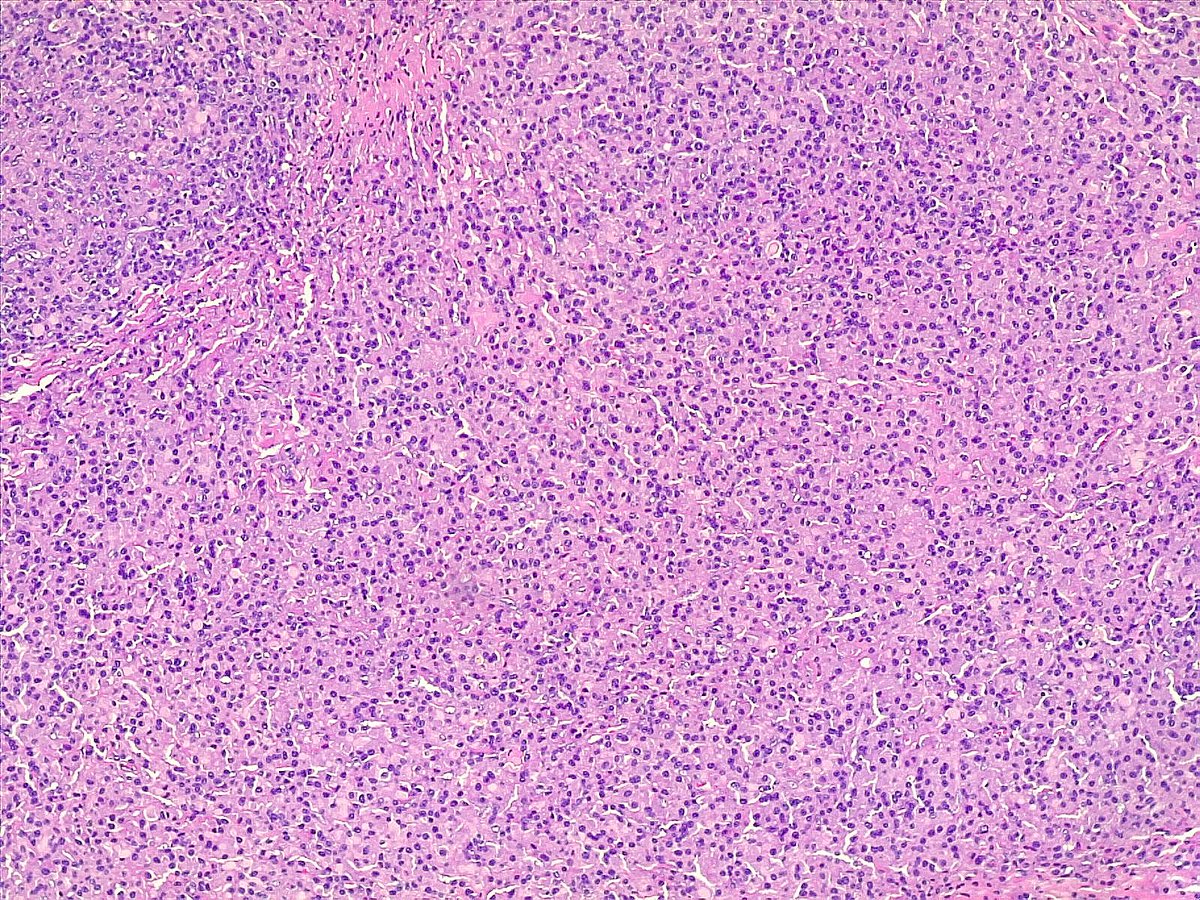
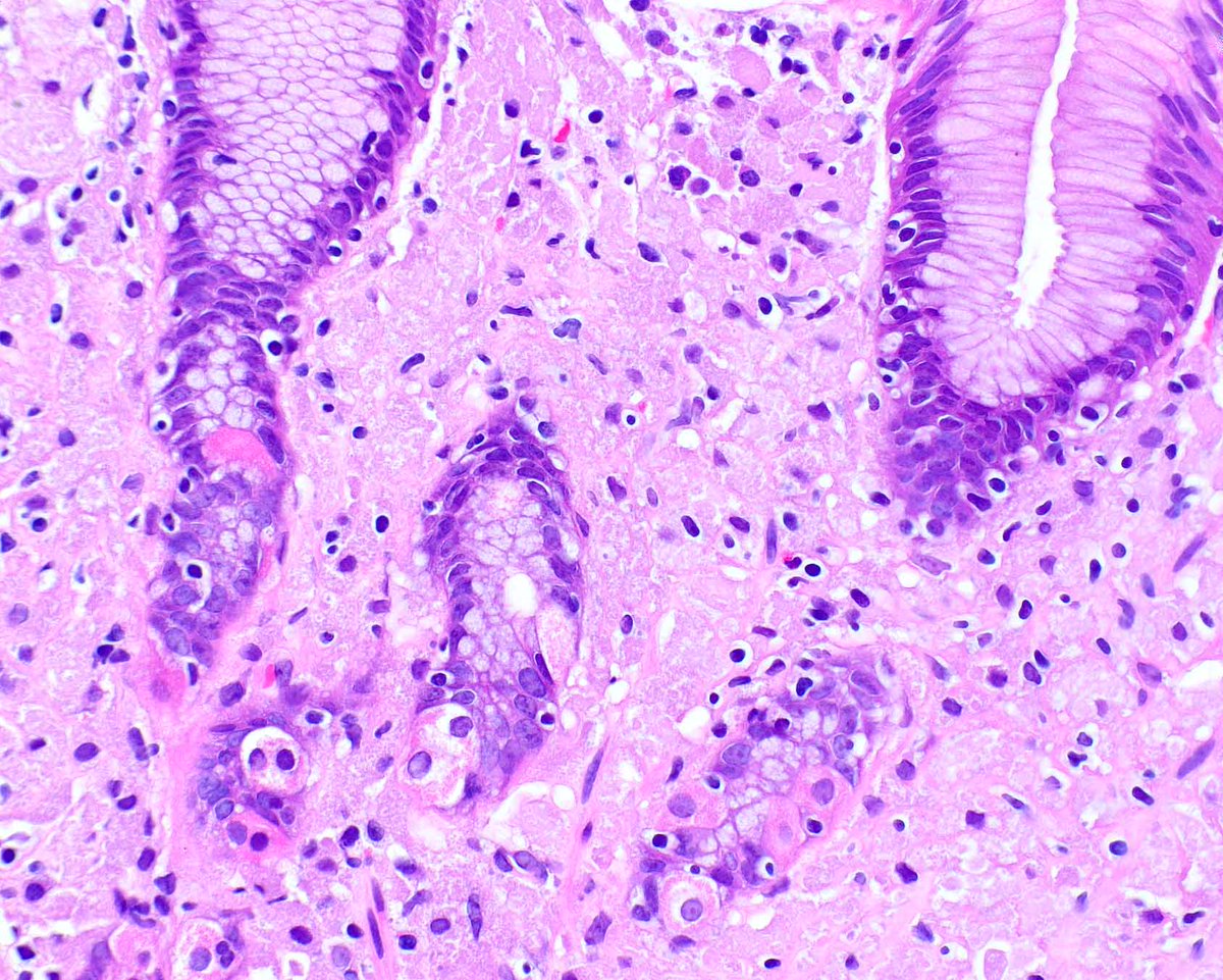
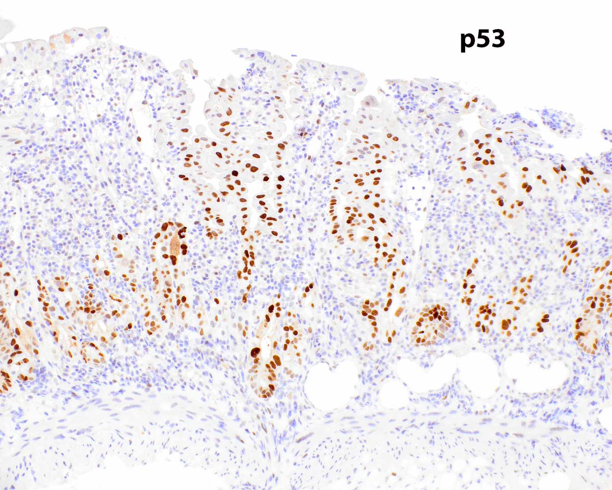
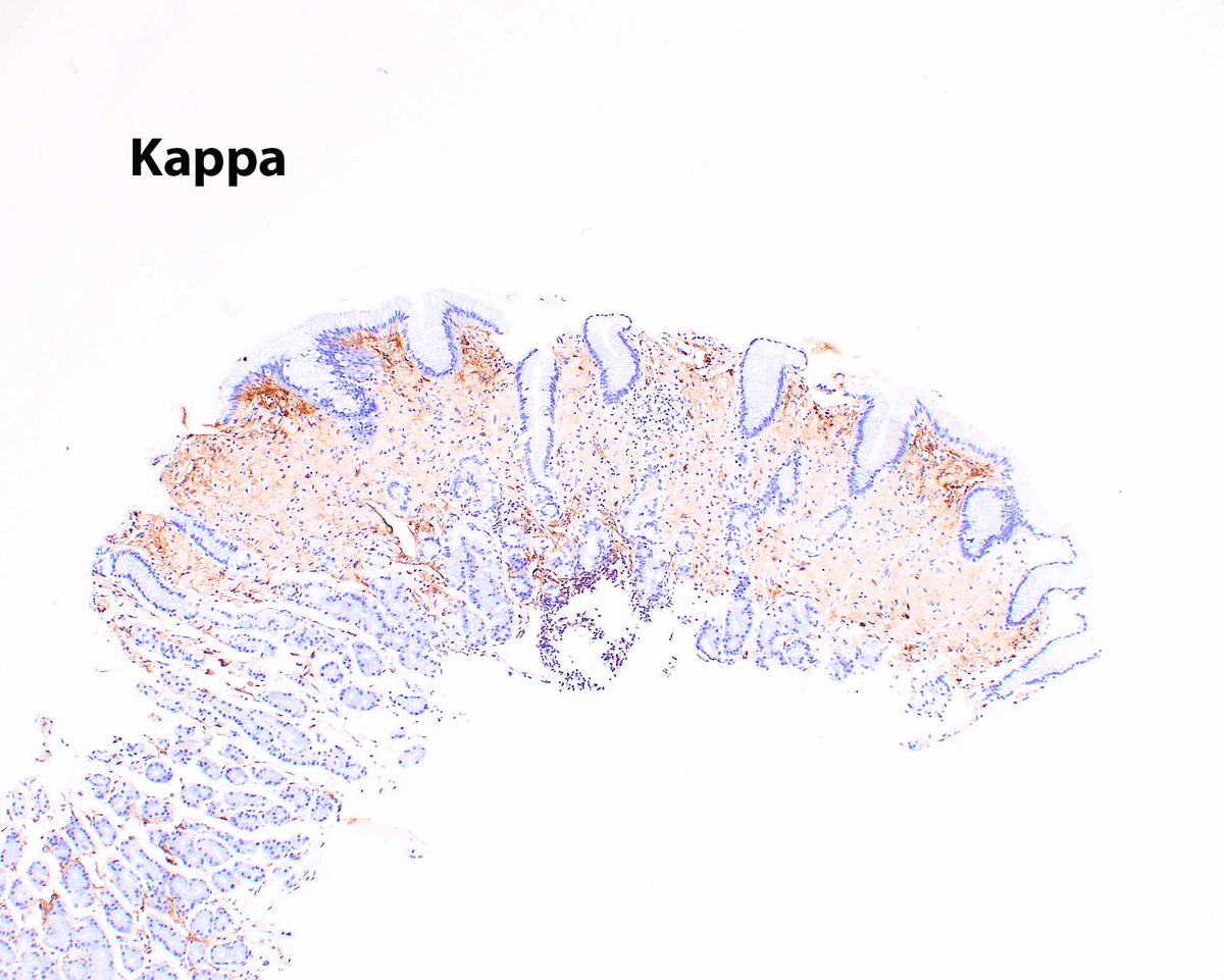
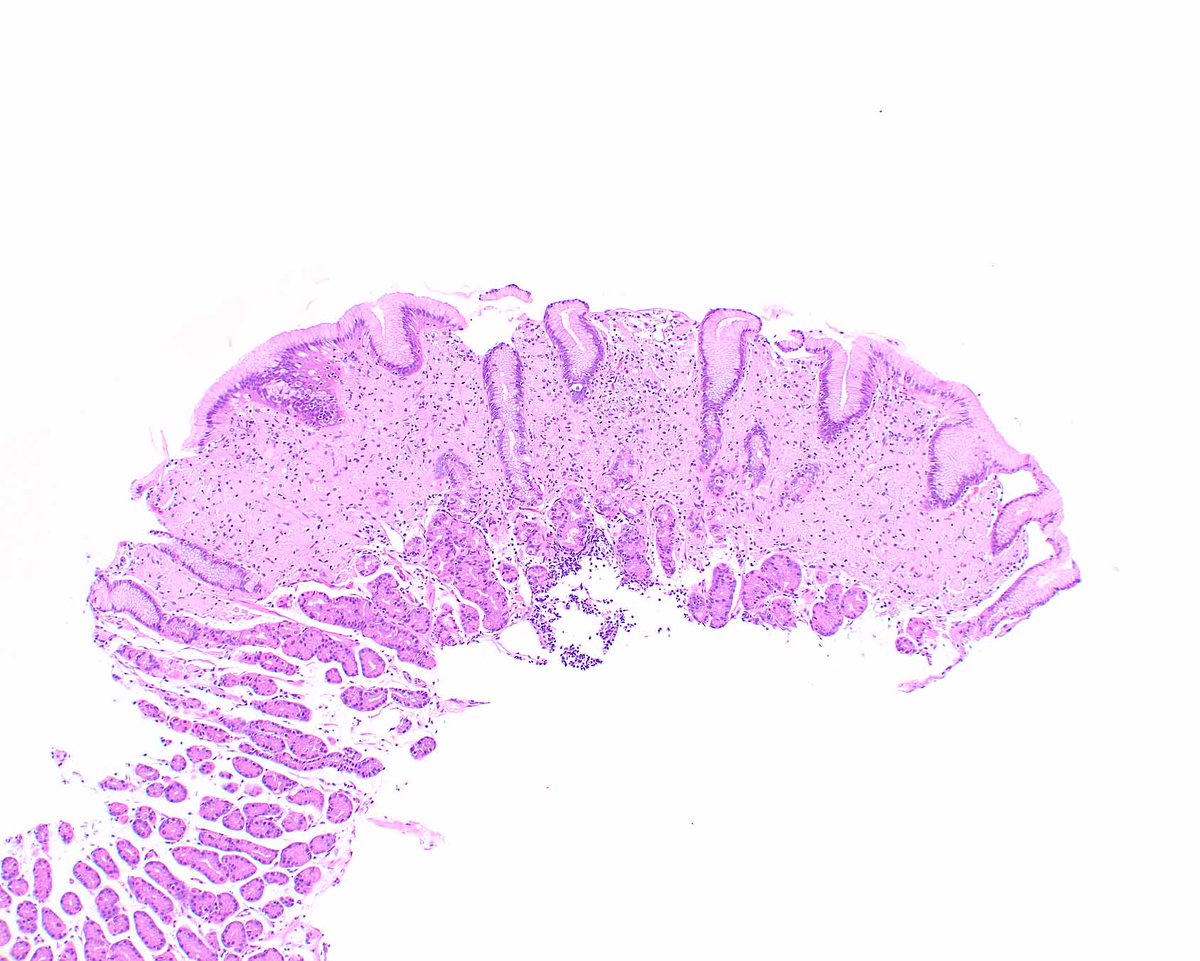
Horrible treatment (radiation) associated epithelial changes in duodenal mucosa. The p53 can be ignored, probably reflecting upregulation rather thana mutational event. The same can be seen in the stomach.
Hutchings DA, et al PMID: 36843539; PMCID: PMC10460459.
A distinguished group of pathologists documents the molecular profile of adenosquamous carcinoma of the gallbladder, demonstrating significant differences between this rare, aggressive tumour and adenocarcinoma. #gipathology#pathtwitter@matteo_fassan
https://t.co/Z6JBJTWyC3
Not everything “benign” behaves that way.
Pancreatic serous neoplasms are usually indolent, but his study is a reminder that some do not follow the script.
This study showed meaningful risk of progression (~28%) on follow-up, without metastasis
.
🔍 What should make us pause:
• Infiltrative growth pattern
• Solid architecture
• Ki-67 higher than expected
Together, they shift the conversation.
💡 Take-home:
Sometimes the goal isn’t to label a case as benign vs malignant,
it’s to recognize when it deserves closer attention and follow-up.
🔓 Open access — worth a read: https://t.co/xIwDIgXpzt
Our Gynecologic Pathology cases @UMiamiPathology are fascinating! Here’s a striking example of pilomatrix-like high-grade endometrial carcinoma (PiMHEC), a recently described entity mostly driven by CTNNB1 mutations. Look for solid basaloid growth with conspicuous central tumor cell necrosis, and pilomatrix-like keratinization with the hallmark ghost cells. This case also had a focal conventional FIGO grade 1 endometrioid component. IHC shows diffuse nuclear β-catenin with loss of PAX8 and ER in the pilomatrix-like component. These tumors are believed to behave aggressively.
📑 #HBV promotes epithelial-mesenchymal transition in #HCC and liver fibrosis through JNK-mediated autophagy❗️
#OpenAccess#LiverX
https://t.co/JAmYQeFYxy
A 3.5cm cervical lymph node, was excised during a total thyroidectomy. This image represents a section from that lymph node, what do you think is the Dx?
What do you think of when you see a granuloma in a vessel wall in the lung? Watch this 51 second YouTube short to learn a quick tip!
Link: https://t.co/OC67nRHHke
I didn't know this was a thing until my first case 2 years ago. Picked up by an eagle-eyed colleague.
Eosinophils everywhere, in keeping with allergic nasal polyp.
Surprisingly hard to see the emperipolesis within dense inflammatory cells.
https://t.co/MjTPAQsuro
This is one of the best examples of lymphocytic #vasculitis I've seen in ages. Here you can see the lymphocytes attacking the right side of the blood vessel, with associated damage to the vessel wall. #pathology#neuropath#pathtwitter
A flare of Behçet disease, a systemic vasculitis, can result in colorectal bleeding. Small blood vessels can be affected, resulting in patchy ischemic changes.
An example of crystal storing histiocytosis; the patient needs to be evaluated for a plasma cell disorder or other lambda-restricted B cell neoplasm.
Arnold CA, et al. Am J Surg Pathol. 2018 Oct;42(10):1317-1324. PMID: 29878935.


![MRB_AI24's tweet photo. 80+ AI tools that replace months of work with minutes.
1. 𝐑𝐞𝐬𝐞𝐚𝐫𝐜𝐡
- ChatGPT
- Copilot
- Gemini
- Abacus
- Perplexity
2. 𝐈𝐦𝐚𝐠𝐞
- Fotor
- Dalle 3
- Stability AI
- Midjourney
- Microsoft Designer
3. 𝐂𝐨𝐩𝐲𝐖𝐫𝐢𝐭𝐢𝐧𝐠
- Rytr
- Copy AI
- Writesonic
- Adcreative AI
4. 𝐖𝐫𝐢𝐭𝐢𝐧𝐠
- Jasper
- HIX AI
- Jenny AI
- Textblaze
- Quillbot
5. 𝐖𝐞𝐛𝐬𝐢𝐭𝐞
- 10Web
- Durable
- Framer
- Style AI
6. 𝐕𝐢𝐝𝐞𝐨
- Klap
- Opus
- Eightify
- InVideo
- HeyGen
- Runway
- ImgCreator AI
- Morphstudio .xyz
7. 𝐌𝐞𝐞𝐭𝐢𝐧𝐠
- Tldv
- Otter
- Noty AI
- Fireflies
8. 𝐒𝐄𝐎
- VidIQ
- Seona AI
- BlogSEO
- Keywrds ai
9. 𝐂𝐡𝐚𝐭𝐛𝐨𝐭
- Droxy
- Chatbase
- Mutual info
- Chatsimple
10. 𝐏𝐫𝐞𝐬𝐞𝐧𝐭𝐚𝐭𝐢𝐨𝐧
- Decktopus
- Slides AI
- Gamma AI
- Designs AI
- Beautiful AI
11. 𝐀𝐮𝐭𝐨𝐦𝐚𝐭𝐢𝐨𝐧
- Make
- Zapier
- Xembly
- Bardeen
12. 𝐏𝐫𝐨𝐦𝐩𝐭𝐬
- FlowGPT
- Alicent AI
- PromptBox
- Promptbase
- Snack Prompt
13. 𝐔𝐈/𝐔𝐗
- Figma
- Uizard
- UiMagic
- Photoshop
14. 𝐃𝐞𝐬𝐢𝐠𝐧
- Canva
- Flair AI
- Designify
- Clipdrop
- Autodraw
- Magician design
15. 𝐋𝐨𝐠𝐨 𝐆𝐞𝐧𝐞𝐫𝐚𝐭𝐨𝐫
- Looka
- Designs AI
- Brandmark
- Stockimg AI
- Namecheap
16. 𝐀𝐮𝐝𝐢𝐨
- Lovo ai
- Eleven labs
- Songburst AI
- Adobe Podcast
17. 𝐏𝐫𝐨𝐝𝐮𝐜𝐭𝐢𝐯𝐢𝐭𝐲
- Merlin
- Tinywow
- Notion AI
- Adobe Sensei
- Personal AI
18. 𝐒𝐨𝐜𝐢𝐚𝐥 𝐦𝐞𝐝𝐢𝐚 𝐦𝐚𝐧𝐚𝐠𝐞𝐦𝐞𝐧𝐭
- Tapilo
- Typefully
- Hypefury
- TweetHunter
[Bookmark this post🔖]
Smart creators use smart tools — follow @MRB_AI24
for more 🪄👇](https://pbs.twimg.com/media/HJAvvabbAAA2dmp.jpg)