our own Dr. Kozakowski participated in this Expert-Publication on <Renal Pathology Society/International Kidney and Monoclonal Gammopathy Research Group Consensus on Pathologic Definitions and Terminology of Monoclonal Gammopathy Associated Kidney Lesions> excited to share~
Fighting systemic viral infections! Dr Hermann Salmhofer explores new ways to minimize damage & improve treatments. https://t.co/q2HrqHBtNV more: https://t.co/9espokQPMR #Virology#MedicalResearch
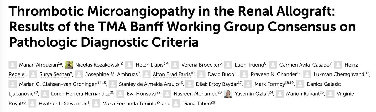
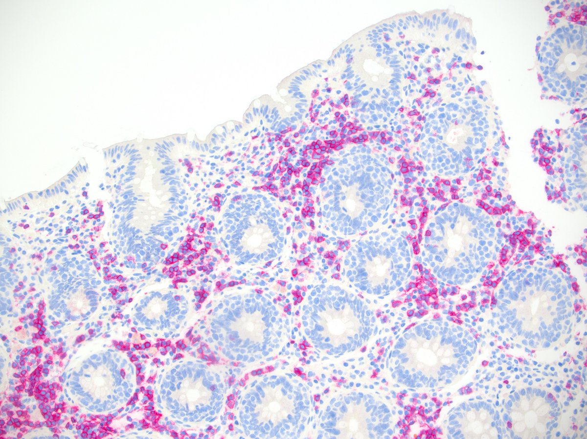
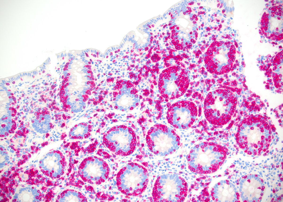
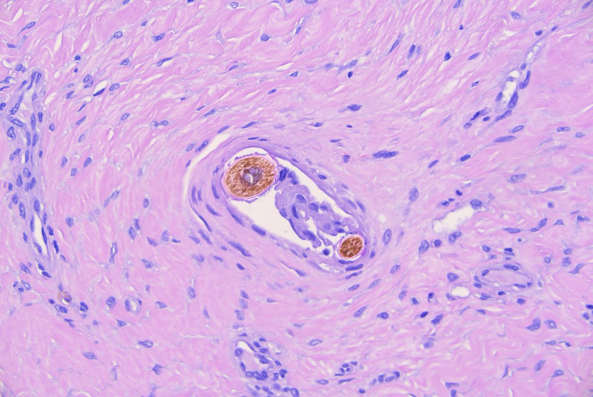
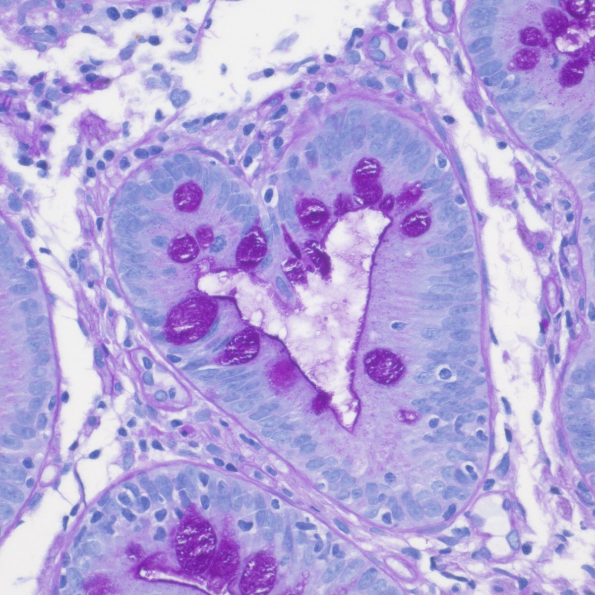
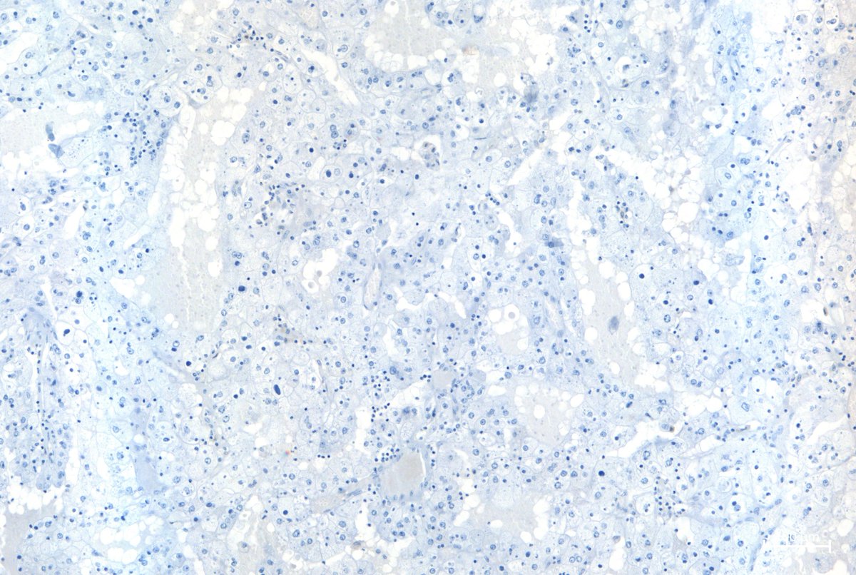
What is the renal pathology here?
If you want to know it, well then, look up the following publication from our renal pathology team:
Kozakowski N, et al. Kidney Int Rep. 2024;10(1):256-259.
doi:10.1016/j.ekir.2024.10.014
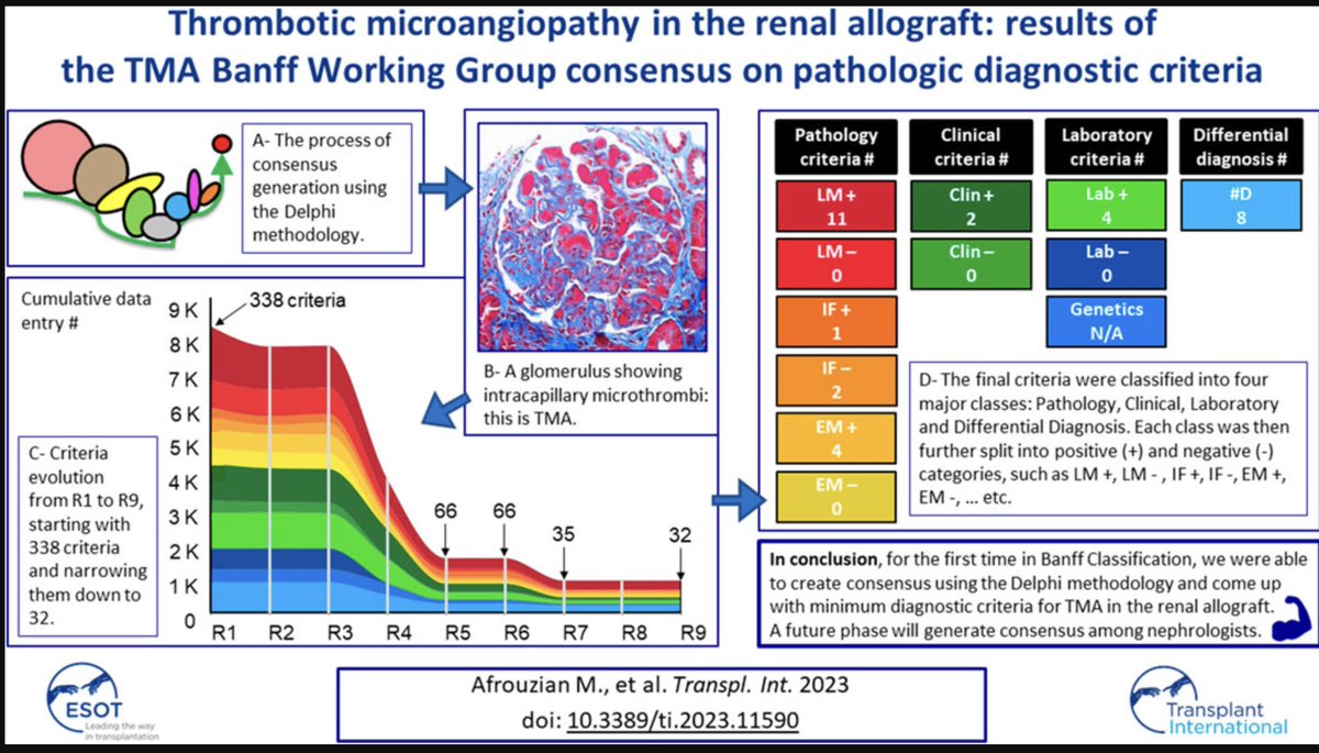
Interested in spatial transcriptomics? Consider reading our pilot study on spatial profiling of glomeruli in pauci-immune focal necrotizing glomerulonephritis!
https://t.co/X6bJlaGOUO
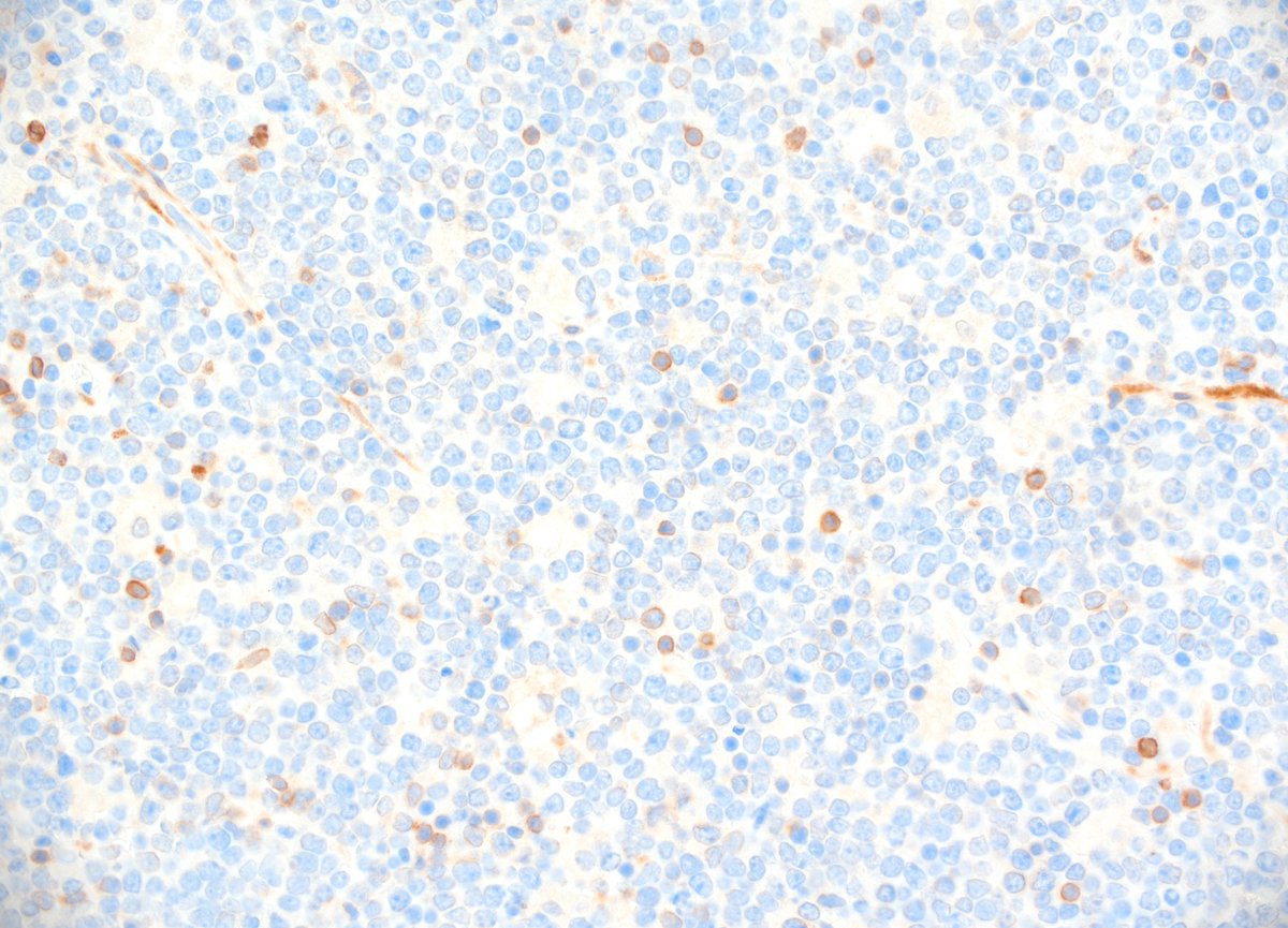
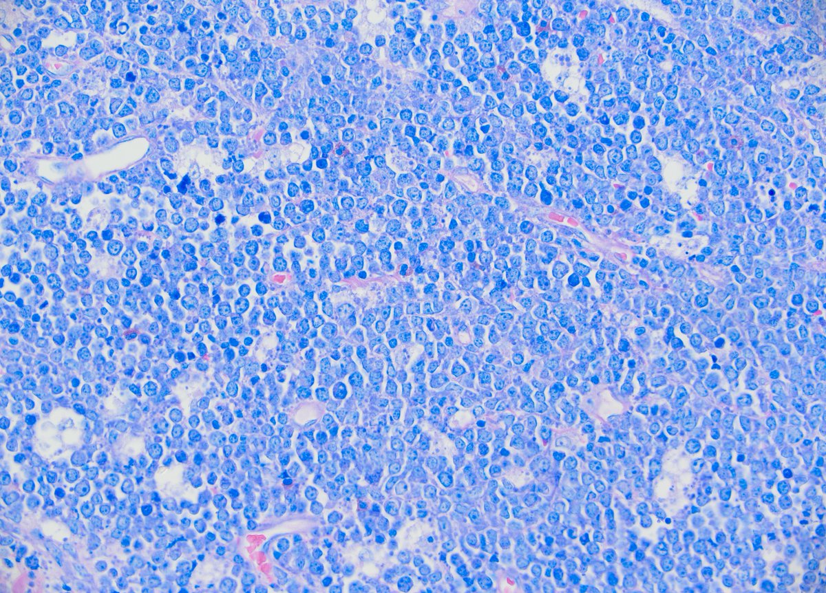
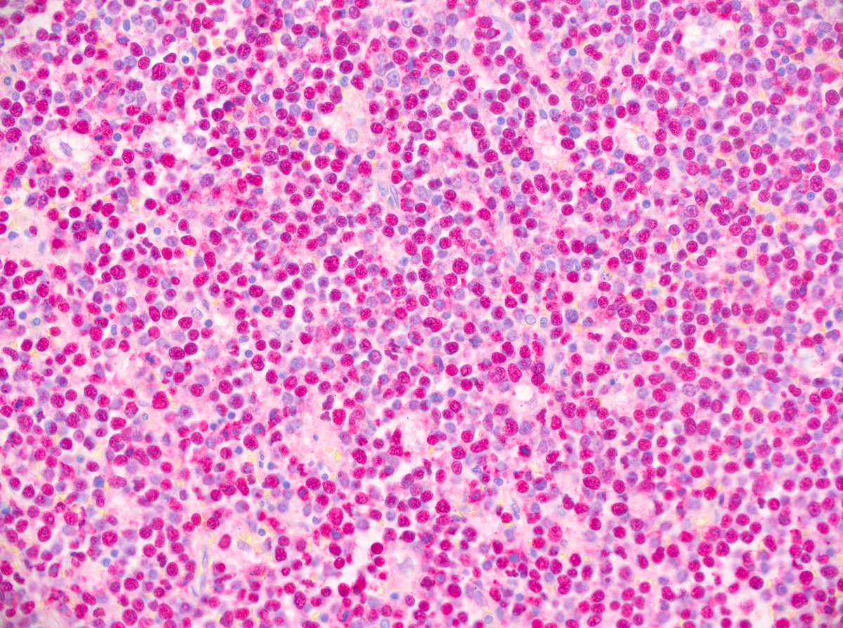
While gazing up at the night's sky hematopathologists don't search for the brightest star. They're looking for Burkitt's lymphoma. Classical starry sky pattern, medium sized, monomorphic tumor cells, EBV associated case (ileocecal resection, 3a, m), Giemsa, bcl6+, blc2-, EBER+
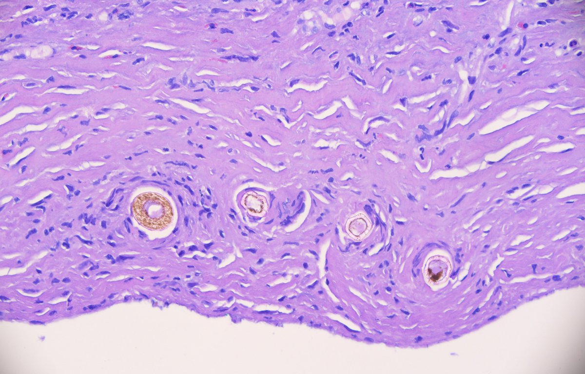
57a, w, colon biopsy, Giemsa, CD3+, CD5-, CD56+, patient with monomorphic epitheliotropic intestinal T-cell lymphoma (MEITL). The neoplastic cells are medium in size, uniform and show a prominent epitheliotropism.
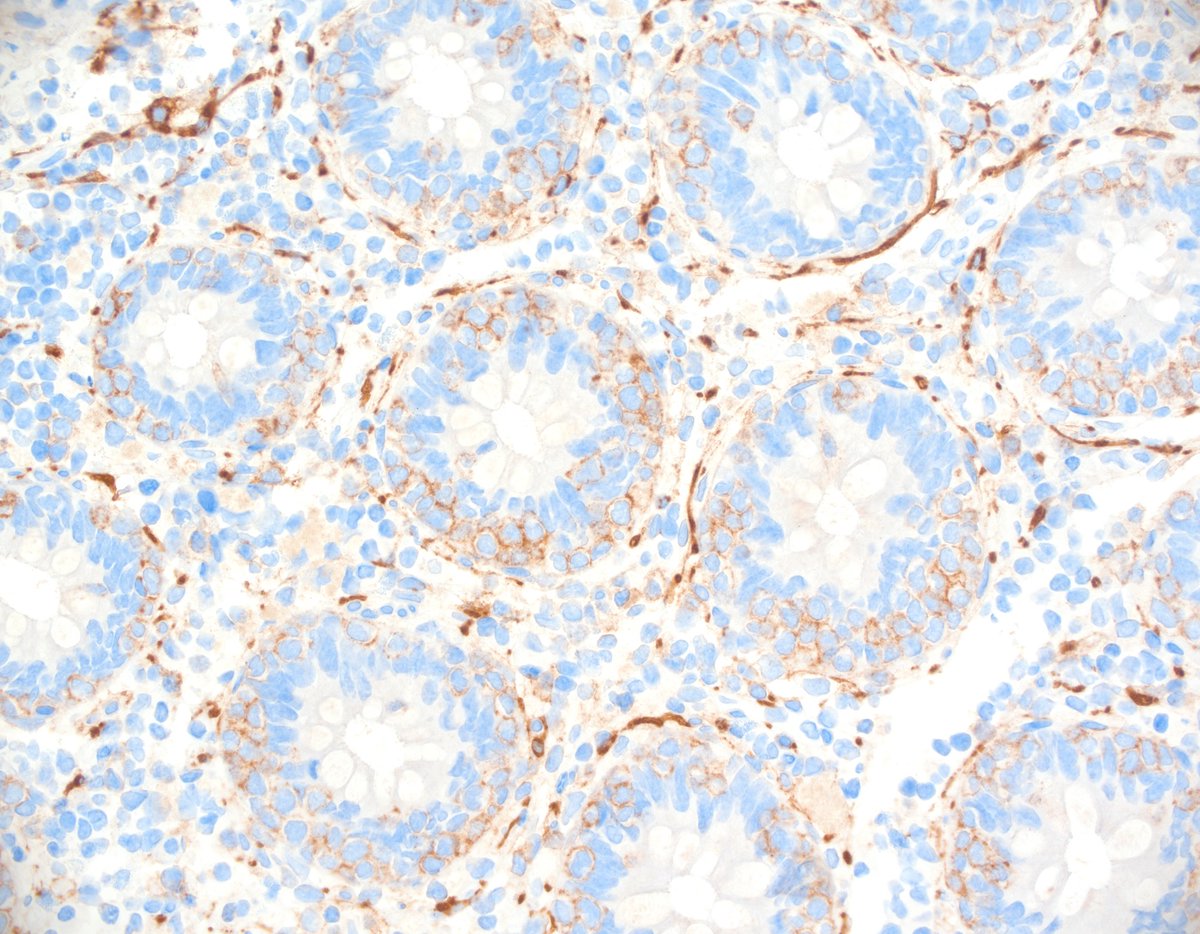
Clear cell renal carcinoma with rhabdoid features - could be challenging at first sight but shows quite typical ccRCC nuclei and positive CAIX (bottom left), negative CK7 (bottom right). #pathology
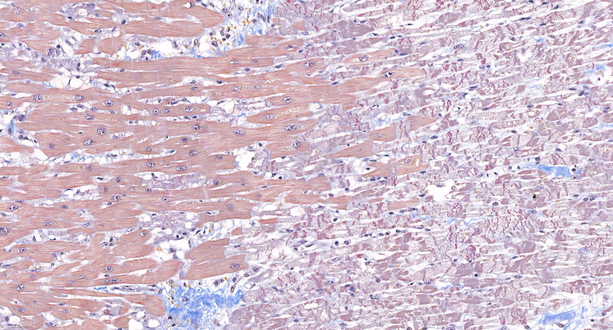
@goziemnweke@AmyHDeekenMD Acid Fuchsine Orange G - one of our favorites for renal pathology because it highlights connective tissue in blue and fibrin in bright red.