Here to drop all the eye-candy from our latest preprint on actin defect mediated Hydra morphogenesis @RouxLab@PearceDJG We'd like to thank the @BrigitteGalliot lab for their support and the Keren lab for their valuable feedback on this work! (1/6)
https://t.co/U6VFKqYimE
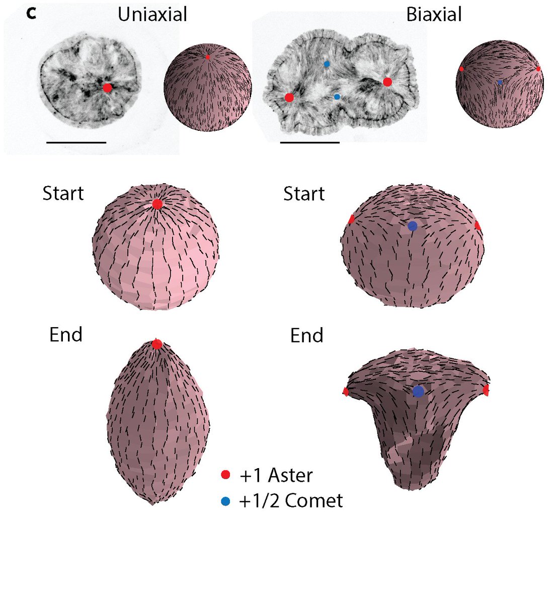
In our new bioRXiv, @yamini_ravichan shows that soft compression of head-regenerating tissues from Hydra induces bicephalous regeneration. During soft compression, a second actin nematic defects appears on the wound.
https://t.co/dJA6KQ7zCj
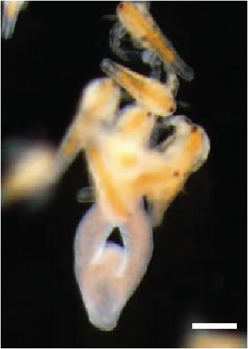
Finally, to test whether the topology change or the lack of actin nematic defects was blocking regeneration, @yamini_ravichan generated toroidal tissues with defects, and they regenerated a head and a foot... and even became adult animals.
https://t.co/dJA6KQ71ML
This shows that actin defects are essential for head morphogenesis. Simulations by @PearceDJG and Karsten Kruse show that the defects organise the actin stresses to shape the Hydra tissue into a head and a foot... or two heads and a foot.
https://t.co/dJA6KQ71ML
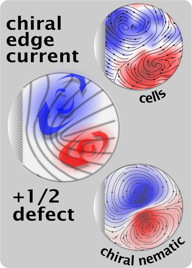
Removing phase difference between a pair of topological defects by creating and annihilating more defects. Read about it in our new manuscript in Soft Matter https://t.co/UgSadI1WuB
Proposed a 100 yrs ago, we show that folding of an epithelium can spontaneously occur by buckling! Congrats to Anastasiya, a wonderful collaboration with the Kruse & Chopard Labs, @sciences_UNIGE, @unige_en, @NCCR_ChemBio
https://t.co/gm1l27NL6L
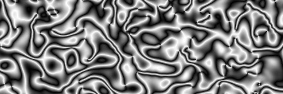
New plotting method for nematic fields. I usually show to show the Schlieren texture, but this is much easier to understand and makes it look far more like experiments!
@shaevitz @dsseara Currently it is tailored to work with the field that comes from my simulation. How are your director fields stored? It should be easy to generalize.
@MarkusDeserno@natxopago Yes, in my simulation the director field exists on a grid, so you can just check each grid point. This representation makes it much easier to identify the charge and orientation of defects by eye though.