I am truly delighted to share with you our recently published book
Behind every page in this book is a child, a family, a challenging diagnosis, and a team that chose to keep searching for answers and striving for better care.
#ISSVA#radiology
https://t.co/rzXkTsodAK
1/Have MULTIPLE questions about the new criteria for MULTIPLE sclerosis?
Do you know the NEW McDonald criteria for multiple sclerosis?
This month’s @theAJNR SCANtastic has the lastest you need to know!
https://t.co/7QhXYeQP4n
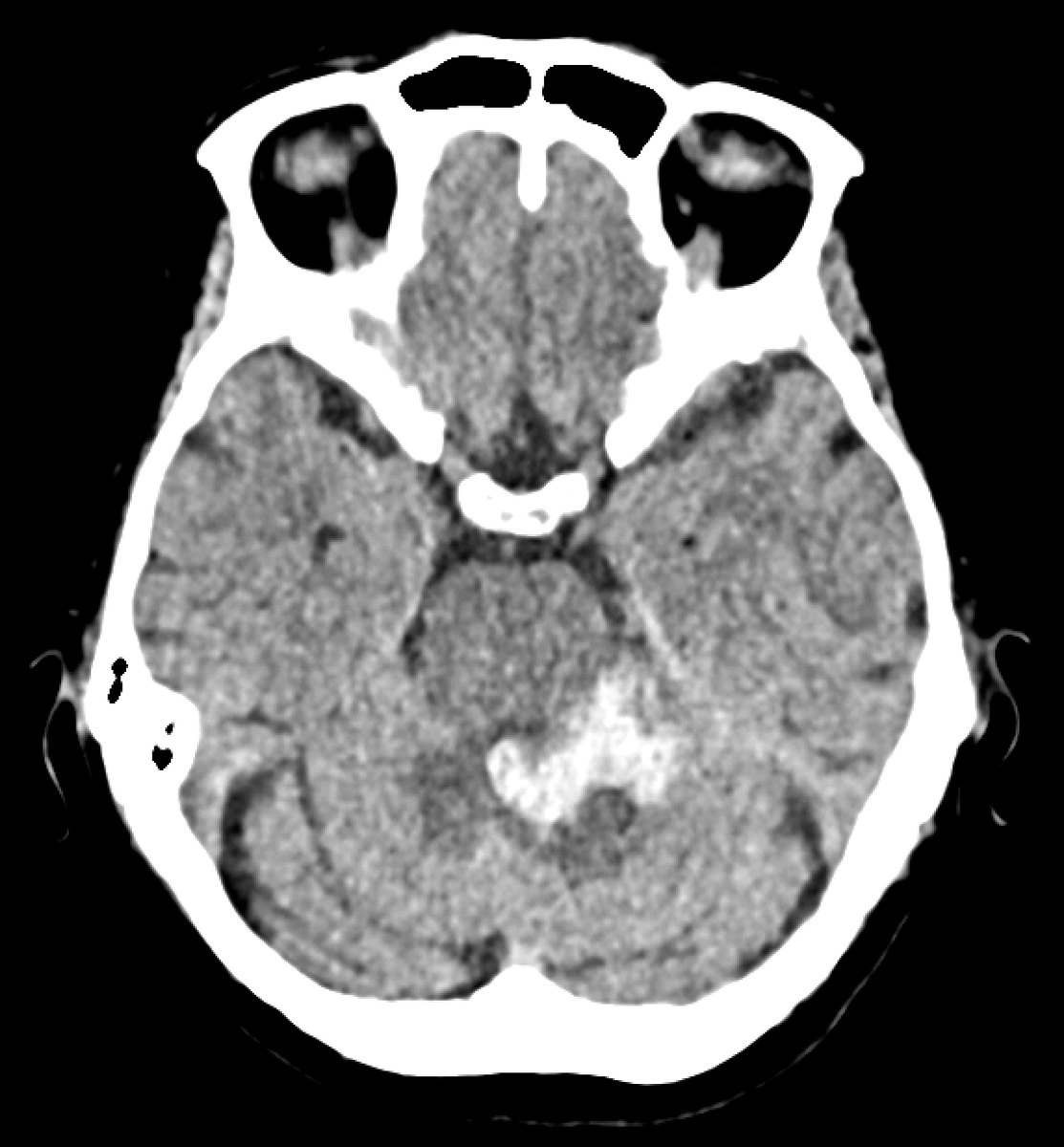
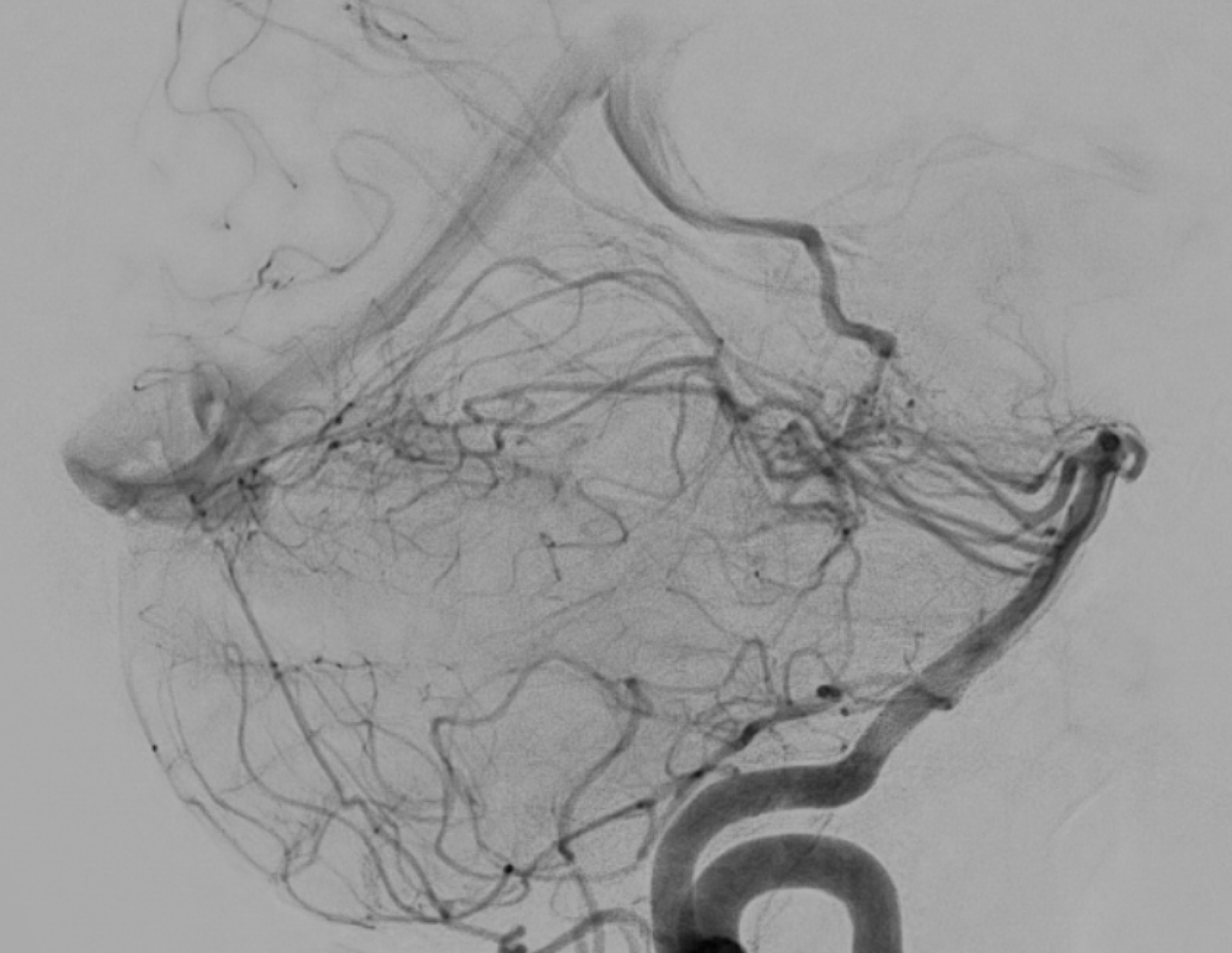
Acute non-traumatic cerebellar haemorrhage in a young patient. Nothing seen on the CT angiogram or the post-contrast MRI, but look at the ASL...
There's high signal in the occipital sinus; this is not normal. If veins light up on ASL, that means labelled arterial blood got there too fast, which usually means it skipped the capillary bed. This implies arteriovenous shunting!
Lo and behold, the DSA shows a left perimesencephalic arteriovenous malformation supplied by basilar perforators and branches from the left superior cerebellar artery, draining into the left basal vein, vein of Galen, and straight sinus.
A fetal spinal defect is identified.
The next question is not simply, “Which dysraphism is this?”
It is:
🔹 Is it open or closed?
🔹 Is there a sac?
🔹 Where is the neural placode?
This #SpinTweetorialWednesday, a practical approach to fetal spinal dysraphism 🧵
🩺Clinical & neuroimaging 🧠 clues to differentiate 🧩autoimmune encephalitis & its mimics🔎
Current opinions in Neurology 📖
10.1097/WCO.0000000000001489.
This week's SPIN-POV: A child with recurrent ataxia, cerebellar signs and atrophy needs CACNA1A sequencing — the same gene causes episodic ataxia type 2, hemiplegic migraine, and SCA6. EA2 is uniquely treatable; acetazolamide can stop the attacks.
Spin Tweetorial Wednesday 🧠🌀
A 4-year-old child with neurodevelopmental regression, refractory myoclonic seizures, ataxia, and language regression.
MRI shows:
Central hypomyelination + cerebellar atrophy + basal ganglia/subcortical calcifications.
What should you think of?
🧠🧵 SPIN Tweetorial Wednesday
A child with recent COVID-19 infection presents with fever, seizures, and encephalopathy.
The MRI patterns here are not random.
#SPINTweetorialWednesday
Acute encephalopathy with biphasic seizures and late reduced diffusion (AESD variant)
The one where MRI lags behind the brain.
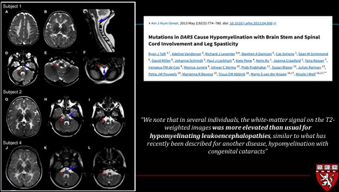
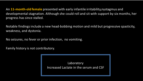
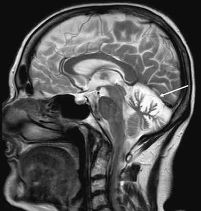
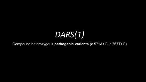
Dr. Cesar Alves (@cesaralvesneuro) shows illustrative examples of DARS mutation related HBSL and LBSL. Sparing of intraxial segmental trigeminal nerve common with DARS1 but rare in DARS2. #PediNeuroRad#radres#pedsrad